Cancer Research and Molecular Mechanisms-Sci Forschen

Full Text

Mini Review
Cell Differentiation and Checkpoint

  Sara Cuesta Sancho      Toru Ouchi   

Department of Cancer Genetics, Roswell Park Cancer Institute, Buffalo, NY14263, USA

*Corresponding author: Toru Ouchi, Department of Cancer Genetics Roswell Park Cancer Institute Elm and Carlton Streets Buffalo, NY 14263, USA, Tel: 716-845-7173; E-mail: Toru.Ouchi@RoswellPark.org

Article Information

Aritcle Type: Mini Review

Citation: Cuesta Sancho S, Ouchi T (2015) Cell Differentiation and Checkpoint. Int J Cancer Res Mol Mech 1(2): doi http://dx.doi.org/10.16966/2381-3318.107

Copyright: © 2015 Ouchi T, et al. This is an open-access article distributed under the terms of the Creative Commons Attribution License, which permits unrestricted use, distribution, and reproduction in any medium, provided the original author and source are credited.

Publication history: 

  • Received date: 19 June 2015

  • Accepted date: 28 June 2015

  • Published date: 02 July 2015
  •  

    Abstract

    DNA damage is induced in many types of cells by internal and external cell stress. When DNA is damaged, DNA Damage Response (DDR) programs are activated to repair the DNA lesions in order to preserve genomic integrity and suppress subsequent malignant transformation. Among these programs is cell cycle checkpoint that ensures cell cycle arrest and subsequent repair of the damaged DNA, apoptosis and senescence in various phases of the cell cycle. Moreover, recent studies have established the cell differentiation checkpoint, the other type of the checkpoint that is specifically activated in the course of differentiation. We will discuss the evidences that support the link between DNA damage proteins and C2C12 cell differentiation.

    Keywords

    Differentiation; Checkpoints; DNA damage response; C2C12

    Introduction

    Genomic integrity is primordial to any organisms. It has been well illustrated that diverse stress from both intrinsic (ex. Reactive Oxygen Species (ROS)) and extrinsic (ex. ionizing radiation (IR), UV light, chemical) environment cause DNA lesions [1]. When DNA damage checkpoint is activated, proliferating cells arrest the cell cycle, allowing the damaged DNA to repair. This process is initiated by recruiting the MRN complex (MRE11-RAD50-NBS1) to DNA Double Strand Breaks (DSBs) and Single Strands Breaks (SSBs), followed by activation of AtaxiaTelangiectasia Mutated (ATM) and Ataxia-Telangiectasia and RAD3- Related (ATR), respectively. ATM and ATR phosphorylate a variety of their substrates, those including p53, MDM2, CHK2, 9-1-1- complex (RAD9, RAD1, HUS1), CHK1, etc [2-7].

    Differentiation is the process in which cells become specialized from the precursor cells to specific cell type, such as neurons, lymphocytes and muscle through differentiation. A global reprogramming of gene expression and withdrawal from the cell cycle are required for the differentiation process [8]. Although it is not well understood how differentiation program proceeds under conditions of DNA damage, it is considered that it could not be completed without the repair of the DNA lesions. Therefore, it is assumed that if cells start the differentiation program prior the DNA was restored, it could lead to abnormally differentiated cells with compromised functions [9].

    C2C12 cells have been widely used as an in vitro model to study myogenic differentiation process. These cells are derived from the mouse skeletal muscle C2 cell line, and they have similar characteristics to those of isolated human skeletal muscle cells [10,11]. Myogenic differentiation consists of a multistep processes that involves two major mechanisms. The first one consists of the induction of the muscle-specific genes expression by Myogenic Regulatory Factors (MRFs). MRFs induce the expression of, for example, Myf-5, MyoD, MRF4 and Myogenin. MyoD and Myf-5 which are primarily expressed in proliferating, undifferentiated myoblasts, allowing the differentiation program start, acting as a determination genes, while Myogenin expression is induced as a result of muscle differentiation (Figure 1) [12-14]. Transcriptional pathways regulated by multiple groups of muscle-specific transcription factors initiate the de novo synthesis of various muscle-specific proteins [15]. The second step in differentiation process is to make a commitment of myogenic cells to irreversible withdrawal from the cell cycle leading permanent G1 phase [16-18]. Withdrawal from the cell cycle causes morphological changes, mononucleated myoblasts alignment, and fusion of their membranes to form multinucleated myotubes, leading to the mature muscle fibers. Accomplishment of these two phases is essential for multinucleated myotubes formation.

    It has been shown that during differentiation DNA Double Strand Breaks (DSBs) occur. For example, development of B lymphocytes requires the induction and consequent repair of DSBs during rearrangement of the antigen receptor genes [19]. Interestingly, there are some biochemical experiments indicating the link between modification of the DDR proteins and neuronal stem cell differentiation. IR-induced DSBs induce acetylation of p53 Lys320 in the Central Nervous System (CNS) [20,21], and acetylated p53 Lys320 promotes neurite outgrowth in vitro and axon regeneration in vivo [22]. Of note, while these results show that DSBs promote cell differentiation of B lymphocyte and neurons, DDR-regulated differentiation checkpoint has been implicated by C2C12 myoblasts, which prevents the appearance of abnormally differentiated cells [9]. Thus, it detains the progression of differentiation until DNA is repaired during muscle differentiation under conditions of genotoxic stress. After serum withdrawal when C2C12 cells were exposed to genotoxic agents, like etoposide and IR, myotube formation is blocked by cell cycle arrest followed by c-Abl-dependent inhibition of MyoD activation. This inhibition of MyoD under genotoxic stress is independent of p53 and c-Jun. c-Abl can phosphorylate MyoD at N-terminal tyrosine (Tyr30) localized within the transactivation domain. Mutations on Tyr30 and Tyr212 to Phe do not interfere with MyoD functionality but mutants become resistant to inhibition of MyoD by DNA damage. Of note, removal of these agents from C2C12 cell culture leads to repair DNA and re-induction differentiation of C2C12 cells into myotubes, indicating that differentiation checkpoint is reversible.

    Figure 1: Myogenic differentiation. Satellite cells (muscle precursor cells) upon stimuli start to proliferate and differentiate into myoblasts (mononuclear cells). The myoblasts proliferate and fuse together to create myotubes over the course of several days. Additional myoblasts fuse to the existing myotubes in the late fusion step to produce larger myotubes. The differentiation process is regulated by many factors, differentiation markers changes during the course of differentiation expressing MyoD and Myf5 at the early steps of the process and Myogenin, MRF4 and pRb when the fusion already start.

    Not only c-Abl, but also several DDR proteins have been implicated to be involved in the differentiation checkpoint, including ATM [23- 25] and NBS1 [26,27], etc. Upon DNA stress, ATM autophosphorylates its own Ser1981, leading to dimer dissociation and, subsequently, phosphorylation of H2AX and a number of transducers and effectors of DNA damage-activated pathways [28,29]. For example, ATM activates CHK2 kinase by phosphorylating its Thr68 [30-32]. Activated CHK2 coordinates a number of cellular processes by phosphorylating downstream effectors, such as CDC25A, CDC25C, BRCA1, PML1, and bRYEFp53, resulting in cell cycle arrest or apoptosis [32-34]. On the other hand, ATM directly phosphorylates p53 at Ser15, causing inhibition of p53-MDM2 interaction and promoting p53-dependent gene expression [35-37].

    Larsen et al. demonstrated that this DDR pathway is activated at the early stages of differentiation of C2C12 cells [38]. Phospho H2AX (g(γ)H2AX) co-localizes to the actual site of DSBs, recruiting and/or stabilizing multiple protein complexes involved in DNA damage signaling [28-30,39,40]. They have demonstrated that, when differentiation is induced, H2AX foci appear in 12h, but most of the signals disappear in 48h. These results indicate that differentiation signals damage DNA during myoblast differentiation. C2C12 myoblasts express wild-type p53 (wtp53) protein, and it has been shown that p53 is activated during differentiation in these cells, suggesting the potential role of the protein in muscle differentiation [41-44]. This model has been supported by the results using immortal and primary myoblasts. Thus, expression of dominant negative p53 (DNp53) proteins in those cells inhibits terminal differentiation. Although it is well documented that spontaneous apoptosis occurs at myogenic differentiation, the mechanism is largely unclear [39,45-48]. Interestingly, DNp53 expression does not affect the cell cycle withdrawal and apoptotic death associated with differentiation process [41,42]. Other studies have also illustrated the possible link of p53 to muscle differentiation. Porrelo et al. have shown that p53 activated in response to DNA damage is rapidly stabilized, binding DNA to the Rb promoter, increasing its expression and inducing muscle differentiation but it is really dependent on the cell differentiation status [42]. In early differentiation (when cells are myoblasts) pRb inhibits DNA synthesis by binding to E2F resulting in repression of cyclin E/cdk2, cyclin D/cdk4 and 6, and cyclin A/cdk2 complexes [49-53]. On the other hand, when cells are in the late differentiation state (myotubes), pRb can promote differentiation binding to MyoD inducing the expression of differentiation makers like Myogenin and MRF4 [54-58] (Figure 2). These results indicate that p53 playing roles in not only inducing genes involved in growth arrest, apoptosis and DNA repair, but also regulating genes whose expression are critical for differentiation [59-62].

    In summary, myogenic differentiation consists of multistep, including appearance and repair of DSBs. When DSBs are generated, DDR proteins are activated to properly ensure the DNA repair before proceeding differentiation, guarantying the correct formation of differentiated cells without compromised genome. Of note, our recent findings showed that ATM inactivation. causes insufficient generation of dendritic cells from bone [63]. Taken together, these results provide a notion that inactivation of DDR proteins results in the abrogation of differentiation.
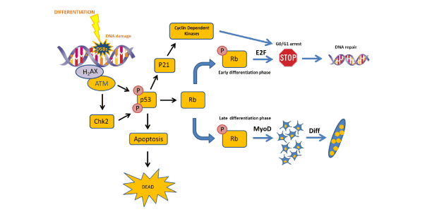
    Figure 2: Schema explaining the role of DDR in myogenic differentiation. Differentiation generates DSBs and the DDR is activated. ATM phosphorylates p53 which could stop the cycle until the DNA damage is repaired activating p21, CDks and generating a G0/G1 arrest. If the DNA damage is not repaired, cells undergo to apoptosis. Finally, p53 plays a role in differentiation through Rb phosphorylation which could activates differentiation or stop it depending on the cell differentiation status.

    Disclosure of potential conflicts of interest

    No potential conflicts of interest were disclosed.

    Acknowledgement

    We thank all the members of the Ouchi Laboratory for helpful discussion. This work is supported by R01CA79892, R01CA90631, and 5P30CA16056 from National Institutes of Health, and Breast Cancer Research Grant from Susan G. Komen Foundation.

    References

    1. Harrison JC, CaH JE (2006) Surviving the breakup: the DNA damage checkpoint. Annual Rev Genet 40: 209-235. [Ref.]
    2. Khanna KK, Lavin MF, Jackson SP, Mulhern TD (2001) ATM, a central controller of cellular responses to DNA damage. Cell Death Differ 8: 1052-1065. [Ref.]
    3. Lavin MF, Khanna KK (1999) ATM: the protein encoded by the gene mutated in the radiosensitive syndrome ataxia-telangiectasia. Int J Radiat Biol 75: 1201-1214. [Ref.]
    4. Shiloh Y (2001) ATM and ATR: networking cellular responses to DNA damage. Curr Opin Genet Dev 11: 71-77. [Ref.]
    5. Tibbetts RS, Brumbaugh KM, Williams JM, Sarkaria JN, Cliby WA, et al. (1999) A role for ATR in the DNA damage-induced phosphorylation of p53. Genes Dev 13: 152-157. [Ref.]
    6. Tibbetts RS, Cortez D, Brumbaugh KM, Scully R, Livingston D, et al. (2000) Functional interactions between BRCA1 and the checkpoint kinase ATR during genotoxic stress. Genes Dev 14: 2989-3002. [Ref.]
    7. Xu X, Tsvetkov LM, Stern DF (2002) Chk2 activation and phosphorylation-dependent oligomerization. Mol Cell Biol 22: 4419- 4432. [Ref.]
    8. Walsh K, Perlman H (1997) Cell cycle exit upon myogenic differentiation. Curr Opin Genet Dev 7: 597-602. [Ref.]
    9. Puri PL, Bhakta K, Wood LD, Costanzo A, Zhu J, et al. (2002) A myogenic differentiation checkpoint activated by genotoxic stress. Nat Genet 32: 585-593. [Ref.]
    10. Gajsek N, Jevsek M, Mars T, Mis K, Pirkmajer S, et al. (2008) Synnaptogenetic mechanisms controlling postsynaptic differentiation of the neuromuscular junctions are nerve-dependent in human nerveindependent and mouse C2C12 muscle cultures. Chem Biol Interact 175: 50-57. [Ref.]
    11. Lee JH, Tachibana H, Morinaga Y, Fujimura Y, Yamada K (2009) Modulation of proliferation and differentiation of C2C12 skeletal muscle cells by fatty acids. Life Sci. 84: 415-420. [Ref.]
    12. Joulia D, Bernardi H, Garandel V, Rabenoelina F, Vernus B, et al. (2003) Mechanisms involved in the inhibition of myoblasts proliferation and differentiation by myostatin. Exp Cell Res 286: 263-275. [Ref.]
    13. Knight JD, Kothary R (2011) The myogenic kinome: protein kinases critical to mammalian skeletal myogenesis. Skelet Muscle 1: 29. [Ref.]
    14. Shen X, Collier JM, Hlaing M, Zhang L, Delshad EH, et al. (2003) Genome-wide examination of myoblast cell cycle withdrawal during differentiation. Dev Dyn 226: 128-138. [Ref.]
    15. Braun T, Gautel M (2011) Transcriptional mechanisms regulating skeletal muscle differentiation, growth and homeostasis. Nat Rev Mol Cell Biol 12: 349-361. [Ref.]
    16. FE S (1992) Molecular mechanism regulating myogenic determination and differentiation. Seminars in developmental biology 154: 284-298.
    17. McKinsey TA, Zhang CL, Olson EN (2001) Control of muscle development by dueling HATs and HDACs. Curr Opin Genet Dev 11: 497-504. [Ref.]
    18. Rescan PY (2001) Regulation and functions of myogenic regulatory factors in lower vertebrates. Comp Biochem Physiol B Biochem Mol Biol 130: 1-12. [Ref.]
    19. Jung D, Alt FW (2004) Unraveling V(D)J recombination; insights into gene regulation. Cell 116: 299-311. [Ref.]
    20. Chao C, Wu Z, Mazur SJ, Borges H, Rossi M, et al. (2006) Acetylation of mouse p53 at lysine 317 negatively regulates p53 apoptotic activities after DNA damage. Mol Cell Biol 26: 6859-6869. [Ref.]
    21. Liu L, Scolnick DM, Trievel RC, Zhang HB, Marmorstein R, et al. (1999) p53 sites acetylated in vitro by PCAF and p300 are acetylated in vivo in response to DNA damage. Mol Cell Biol 19: 1202-1209. [Ref.]
    22. Giovanni SD, Knights CD, Rao M, Yakovlev A, Beers J, et al. (2006) The tumor suppressor protein p53 is required for neurite outgrowth and axon regeneration. EMBO J 25: 4084-4096. [Ref.]
    23. Matsuoka S, Rotman G, Ogawa A, Shiloh Y, Tamai K, et al. (2000) Ataxia telangiectasia-mutated phosphorylates Chk2 in vivo and in vitro. Proc Natl Acad Sci USA 97: 10389-10394. [Ref.]
    24. Matsuoka S, Huang M, Elledge SJ (1998) Linkage of ATM to cell cycle regulation by the Chk2 protein kinase. Science 282: 1893-1897. [Ref.]
    25. Melchionna R, Chen XB, Blasina A, McGowan CH (2000) Threonine 68 is required for radiation-induced phosphorylation and activation of Cds1. Nat Cell Biol 2: 762-765. [Ref.]
    26. Dawson BA, Lough J (1998) Immunocytochemical localization of transient DNA strand breaks in differentiating myotubes using in situ nick-translation. Dev Biol 127: 362-367. [Ref.]
    27. Farzaneh F, Zalin R, Brill D, Shall S (1982) DNA strand breaks and ADP ribosyl transferase activity in eukaryotic differentiation. Nature 300: 362-366.
    28. Bakkenist CJ, Kastan MB (2003) DNA damage activates ATM through intermolecular autophosphorylation and dimer dissociation. Nature 421: 499-506. [Ref.]
    29. Celeste A, Fernandez-Capetillo O, Kruhlak MJ, Pilch DR, Staudt DW, et al. (2003) Histone H2AX phosphorylation is dispensable for the initial recognition of DNA breaks. Nat Cell Biol 5: 675-679. [Ref.]
    30. Ahn JY, Schwarz JK, Piwnica-Worms H, Canman CE (2000) Threonine 68 phosphorylation by ataxia telangiectasia mutated is required for efficient activation of Chk2 in response to ionizing radiation. Cancer Res 60: 5934-5936. [Ref.]
    31. Bartek J, Lukas J (2001) Mammalian G1-and S-phase checkpoints in response to DNA damage. Curr Opin Cell Biol 13: 738-747. [Ref.]
    32. Bartek J, Lukas J (2003) Chk1 and Chk2 kinases in checkpoint control and cancer. Cancer Cell 3: 421-429. [Ref.]
    33. Appella E, Anderson CW (2001) Post-translational modifications and activation of p53 by genotoxic stresses. Eur J Biochem 268: 2764- 2772. [Ref.]
    34. Falck J, Mailand N, Syljuåsen RG, Bartek J, Lukas J (2001) The ATMChk2-Cdc25A checkpoint pathway guards against radioresistant DNA synthesis. Nature 410: 842-847. [Ref.]
    35. Ashcroft M, Kubbutat MHG, Vousden KH (1999) Regulation of p53 function and stability by phosphorylation. Mol Cell Biol 19: 1751-1758. [Ref.]
    36. Rich T, Allen RL, Wyllie AH (2000) Defying death after DNA damage. Nature 407: 777-783. [Ref.]
    37. Shiloh Y (2003) ATM and related protein kinases: safeguarding genome integrity. Nat Rev Cancer 3: 155-368. [Ref.]
    38. Larsen BD, Rampalli S, Burns LE, Brunette S, Dilworth FJ, et al. (2010) Caspase 3/caspase-activated DNase promote cell differentiation by inducing DNA strand breaks. Proc Natl Acad Sci USA 107: 4230-4235. [Ref.]
    39. Huppertz B, Tews DS, Kaufmann P (2001) Apoptosis and syncytial fusion in human placental trophoblast and skeletal muscle. Int Rev Cytol 205: 215-253. [Ref.]
    40. Paull TT, Rogakou EP, Yamazaki V, Kirchgessner CU, Gellert M, et al. (2000) A critical role for histone H2AX in recruitment of repair factors to nuclear foci after DNA damage. Curr Biol 10: 886-895. [Ref.]
    41. Mazzaro G, Bossi G, Coen S, Sacchi A, Soddu S (1999) The role of wild type p53 in the differentiation of primary hemopoietic and muscle cells. Oncogene 18: 5831-5835. [Ref.]
    42. Porrello A, Cerone MA, Coen S, Gurtner A, Fontemaggi G, et al. (2000) p53 regulates Myogenesis by Triggering the Differentiation Activity of pRb. J Cell Biol 151: 1295-1303. [Ref.]
    43. Soddu S, Blandino G, Scardigli R, Coen S, Marchetti A, et al. (1996) Interference with p53 protein inhibits hemopoietic and muscle differentiation. J Cell Biol 134: 193-204. [Ref.]
    44. Tamir Y, Bengal E (1998) p53 protein is activated during muscle differentiation and participates with MyoD in the transcription of muscle creatine kinase gene. Oncogene 17: 347-356. [Ref.]
    45. Kamradt MC, Chen F, Sam S, Cryns VL (2002) The small heat shock protein alpha B-crystallin negatively regulates apoptosis during myogenic differentiation by inhibiting caspase-3 activation. J Biol Chem 277: 38731-38736. [Ref.]
    46. Murray TV, McMahon JM, Howley BA, Stanley A, Ritter T, et al. (2008) A non-apoptotic role for caspase-9 in muscle differentiation. J Cell Sci 121: 3786-3793. [Ref.]
    47. Wang J, Walsh K (1996) Resistance to apoptosis conferred by Cdk inhibitors during myocyte differentiation. Science 273: 359-361. [Ref.]
    48. Zhang K, Sha J, Harter ML (2010) Activation of Cdc6 by MyoD is associated with the expansion of quiescent myogenic satellite cells. J Cell Biol 188: 39-48. [Ref.]
    49. Guo K, Walsh K (1997) Inhibition of myogenesis by multiple cyclin-Cdk complexes. Coordinate regulation of myogenesis and cell cycle activity at the level of E2F. J Biol Chem 272: 791-797. [Ref.]
    50. Skapek SX, Rhee J, Kim PS, Novitch BG, Lassar AB (1996) Cyclinmediated inhibition of muscle gene expression via a mechanism that is independent of pRB hyperphosphorylation. Mol Cell Biol 16:7043-7053. [Ref.]
    51. De Falco G, Comes F, Simone C (2006) pRb: master of differentiation. Coupling irreversible cell cycle withdrawal with induction of musclespecific transcription. Oncogene 25: 5244-5249. [Ref.]
    52. Schneider JW, Gu W, Zhu L, Mahdavi V, Nadal-Ginard B (1994) Reversal of terminal differentiation mediated by p107 in Rb-/- muscle cells. Science 264: 1467-1471. [Ref.]
    53. Weinberg RA (1995) The retinoblastoma protein and the cell cycle control. Cell 81: 323-330. [Ref.]
    54. Banin S, Moyal L, Shieh S, Taya Y, Anderson CW, et al. (1998) Enhanced phosphorylation of p53 by ATM in response to DNA damage. Science 281: 1674-1647. [Ref.]
    55. Chen PL, Riley DJ, Chen-Kiang S, Lee WH (1996) Retinoblastoma protein directly interacts with and activates the transcription factor NFIL6. Proc Natl Acad Sci USA 93: 465-469. [Ref.]
    56. Gu W, Schneider JW, Condorelli G, Kaushal S, Mahdavi V (1993) Interaction of myogenic factors and the retinoblastoma protein mediates muscle cell commitment and differentiation. Cell 72: 309-324. [Ref.]
    57. Novitch BG, Mulligan GJ, Jacks T, Lassar AB (1996) Skeletal muscle cells lacking the retinoblastoma protein display defects in muscle gene expression and accumulate in S and G2 phases of the cell cycle. J Cell Biol 135: 441-456. [Ref.]
    58. Novitch BG, Spicer DB, Kim PS, Cheung WL, Lassar AB (1999) pRb is required for MEF2-dependent gene expression as well as cell-cycle arrest during skeletal muscle differentiation. Curr Biol 9: 449-459. [Ref.]
    59. Barlow C, Hirotsune S, Paylor R, Liyanage M, Eckhaus M, et al. (1996) Atm-deficient mice: a paradigm of ataxia telangiectasia. Cell 86: 159- 171. [Ref.]
    60. Canman CE, Lim LD, Cimprich KA, Taya Y, Tamai K, et al. (1998) Activation of the ATM kinase by ionizing radiation and phosphorylation of p53. Science 281: 1677-1679. [Ref.]
    61. Jhappan C, Morse HC 3rd, Fleischmann RD, Gottesman MM, Merlino G (1997) DNA-PKcs: a T-cell tumour suppressor encoded at the mouse scid locus. Nat Genet 17: 483-486. [Ref.]
    62. Kruse JP, Gu W (2009) Modes of p53 regulation. Cell 137: 609-622. [Ref.]
    63. So EY, Ouchi T (2014) Translational initiation regulated by ATM in dendritic cells development. Cell Death Dis 5: e1418. [Ref.]

    Download Provisional pdf here