Introduction
Small intestinal arteriovenous malformations represent up to 30 to 40% of inexplicable chronic gastrointestinal bleeding and are the most common aetiology in elderly patients. The correct categorization of gastrointestinal vascular anomalies has been inconsistent and a source of confusion [1]. Angiodysplasia gastrointestinal lesions are pathological communications between capillaries of the dilated mucosa and submucosal veins. Angioectasia, vascular ectasia and vascular malformations are the most common vascular lesions diagnosed in the gastrointestinal tract. These aberrant vessels we first described as a clinical entity and a potential bleeding cause in 1839. During the 60s and 70s, with the endoscopic optical fiber arrival, angiodysplasia were identified and described, with the term of “angiodysplasia” established in 1974 [2].
Gastrointestinal Angiodysplasia (GIAD) are generally diagnosed during endoscopy by direct visualization of a mucosa lesion, shaped as a star or fern, it varies from 1mm to 5 cm in any portion of the gastrointestinal tract. Histological it is found a mucosa layer and abnormal dilated, thinned and tortous vessels that do not have a smooth muscle layer, which causes them to be weak and prone to bleeding [3]. Despite of having little known physiopathology there are several hypothesis regarding GIAD formation, associated in most of the times with hypoxia and ageing inside intestinal wall [4]. Initial hypothesis by Boley SJ, et al. [5] and Baum S, et al. [6] suggest that GIAD development it’s preceded by chronic hypoxia and increased intestinal wall pressure. These theories were initially accepted as the probable cause due to the idea that most GIAD were located at ascending colon, where the intestinal wall is usually thinner. Recently Junquera F, et al. [7] established the possible Vascular Endothelial Growing-Factor (VEGF) and other factors role, and its impact in the colon angiodysplasia formation by evaluating the factors levels in different tissue samples of colon GIAD [6].
In general terms, GIAD represents 5% of the causes of gastrointestinal bleeding [7], and approximately up to 50% of all obscure gastrointestinal bleeding, defined as bleeding after a negative superior and inferior endoscopy [6,7]. It is estimated that GIAD is responsible for 4 to 7% of non-varicose upper gastrointestinal bleeding, 35 to 50% of overall gastrointestinal bleeding and 3 to 4% of lower gastrointestinal bleeding [8]. Lesions can be commonly observed during conventional endoscopy at colon (80%) and stomach (5%) where are easier diagnosed and treated [9]. It is important to point that describing the correct distribution of GIAD is difficult due to most endoscopic evaluation do not include the small intestine portion, and are present at the small intestine in 15% of the cases [9]. A study realized by Bollinger E, et al. [10] evaluated 127 patients with bleeding GIAD, and examined the complete gastrointestinal tract using conventional endoscopy and small intestine endoscopic capsule [11], describing that GIAD were present in jejunum in 80%, at duodenum in 51%, at stomach in 23%, at ascending colon in 11.4% and at ileum at 5.7% [12].
Case Report
We present the case of a 69 years old male, resident of Mexico City, with medical history high-blood pressure well controlled with Losartan twice a day and being previous smoker. Personal medical history of surgical procedures such as appendectomy and cholecystectomy. 3 years ago he had an event of lower intestinal bleeding that required hospital admission without an etiology confirmation.
Symptoms initiated 7 days prior to hospital admission with asthenia, dyspnea, abdominal intermittent pain and nausea without vomit, afterwards with abundant melena evacuations. At hospital admission found with mild abdominal pain, without signs of acute abdomen and rectal examination with recent blood finding. Blood test with hemoglobin of 5.7 g/dL and hematocrit of 16.6%, with blood coagulation test being normal so a blood transfusion was started, requiring up to 6 globular packages to achieve normal hemoglobin. Simple and contrasted abdominal scans showed a cecum thickening, anterior to mesenteric vessels and pancreas lymph nodes; and endoscopy reported angiodysplasia located at tongue base with finding of chronic gastritis with no finding of upper gastrointestinal tract bleeding (Figures 1-3).
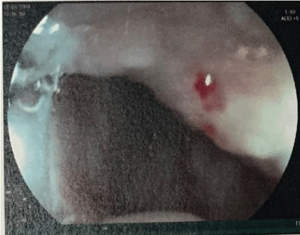
Figure 1: Endoscopic images showing angiodysplasia at tongue base, and no upper gastrointestinal bleeding.
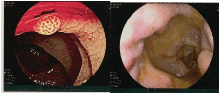
Figure 2: Colonic polyp and mucosa.

Figure 3: Cecum mucosa with telangiectasias and scarce bleeding.
After an initial examination, the patient continued to present important gastrointestinal bleeding and again haemoglobin declining levels, so an obscure gastrointestinal bleeding diagnosis was established. A flexible anterograde and retrograde endoscopy was performed, which reported the presence of jejunum angiectasia and varicose vessels in its final third and the medical team decision was to instill methylene blue 4 cms before those vessels (Figures 4,5) to highlight the intestinal segment in order to facilitate surgical procedure.
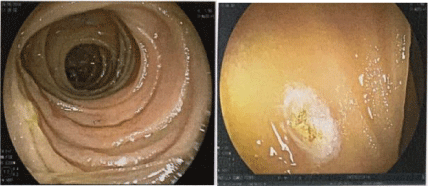
Figure 4: Jejunum angioectasia.
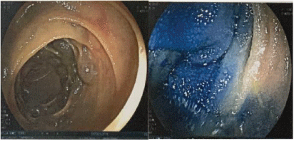
Figure 5: Jejunum varicose vessels instilled with methylene blue.
Due to persistent gastrointestinal bleeding and having already located the intestinal segment with angioectasia, a surgical approach was offered. Initiating with a minimal invasion approach by using a trans umbilical laparoscopic 12 mm port and two 5 mm ports at right and left abdominal flanks, to perform an initial laparoscopic assessment and then locating the methylene blue dye at the jejunum varicose vessels (Figures 6-9).
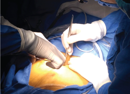
Figure 6: Trans umbilical 12 mm port for lens access.
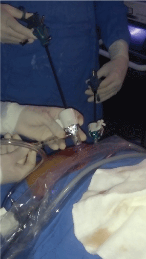
Figure 7: 12 mm and 5 mm ports location.
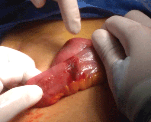
Figure 8: Jejunum with methylene blue dye and angiodysplasia location.
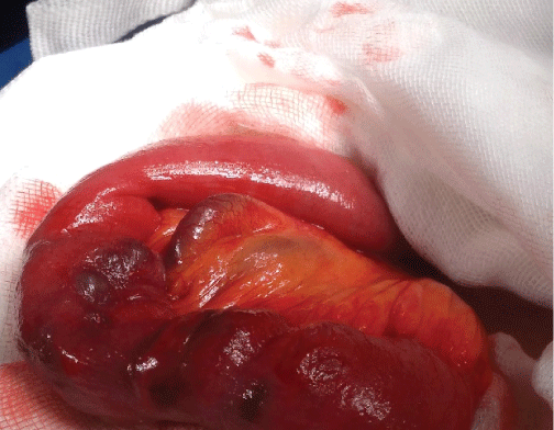
Figure 9: Jejunum with methylene blue dye at varicose vessels.
After locating jejunum and methylene blue dye vessels it is exteriorized at the umbilical port to perform a 15 cms intestinal resection that included the dyed segment with angiodysplasia; stapledmechanical anastomosis was completed and it was reviewed again inside the abdominal cavity, afterwards with the habitual ports closure (Figures 10,11).
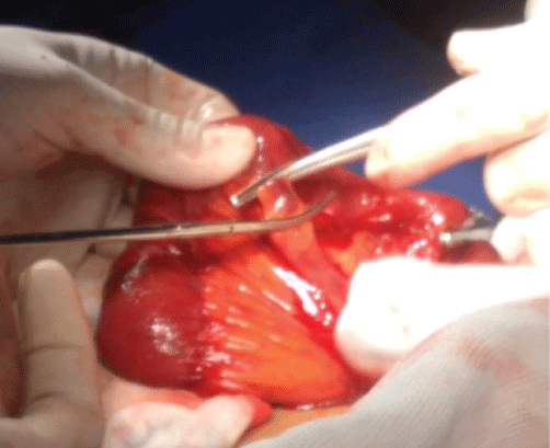
Figure 10: Jejunum varicose vessels are controlled.
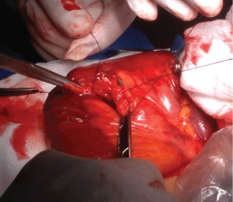
Figure 11: Jejunum varicose vessels are ligated.
After surgery, the patient completed 72 hours of fasting and after the third day, a liquid diet was reestablished, with good initial tolerance, good diet progression tolerance, without abdominal pain and having normal gastrointestinal movement and function, so the patient is discharged from hospital (Figures 12-14).
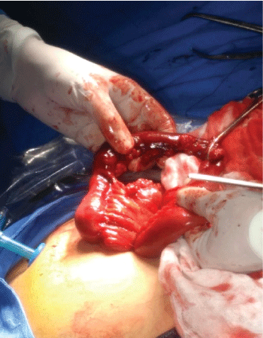
Figure 12: Resected intestinal segment.
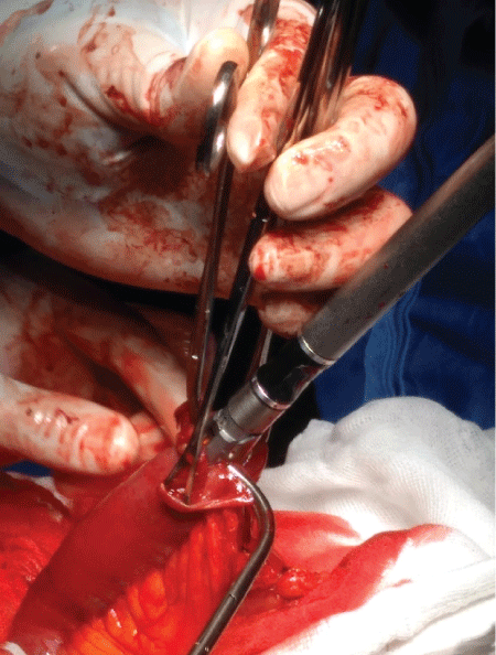
Figure 13: Stapled-mechanic intestinal anastomosis.
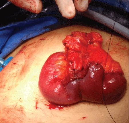
Figure 14: Intestinal segment after completed anastomosis.
Discussion
Obscure gastrointestinal bleeding can be a challenge to correctly diagnose and manage. In this patient, we could adequately locate, diagnose and mark an intestinal segment with angiodysplasia, located at the jejunum that was most likely the etiology for the previous event of gastrointestinal bleeding 7 years before. Having in mind that in these cases of obscure gastrointestinal bleeding endoscopic techniques may be insufficient, with a high rate of bleeding recurrence, up to 36% of the cases [6-9]. Although endoscopic studies have made it easier for surgical teams to correctly study all gastrointestinal bleeding entities and also by enhancing therapeutic options, a surgical approach is still needed in most cases.
Surgical definitive treatment for intestinal angiodysplasia includes a range of major surgery, intestinal resection, intestinal anastomosis or stoma, with higher rates of complications and mortality. Minimal invasion surgery has shown better outcomes, minor surgical trauma, less recovery time and better surgical site infection rates [3,5].
In this case, we describe a complete diagnosis protocol of a patient with obscure gastrointestinal bleeding, and a definite treatment by minimally invasive surgery, with good patient outcome [3].
Conclusion
Surgical management of small intestine angiodysplasia is indicated when endoscopic procedures are insufficient to stop intestinal bleeding, when precise bleeding is not located or when mayor vascular ectasia is found. If it is not possible to locate by an endoscopic approach we recommend an open abdominal approach by laparotomy with trans surgical endoscopy. When precise lesion site is located, usually an intestinal resection and anastomosis is required, this can be done by laparoscopic approach or video-assisted approach too in order to reduce complications and associated mortality
Article Information
Article Type: CASE REPORT
Citation: Jiménez-Canet AA, García-Chávez J, Bizueto-Rosas H, Pérez-González HA, Magaña-Salcedo JR, et al. (2019) Treatment of Jejunal Angiodysplasia by Video-Assisted Intestinal Resection, a Case Report and Literature Review. J Surg Open Access 5(1): dx.doi. org/10.16966/2470-0991.179
Copyright: © 2019 Jiménez-Canet AA, et al. This is an open-access article distributed under the terms of the Creative Commons Attribution License, which permits unrestricted use, distribution, and reproduction in any medium, provided the original author and source are credited.
Publication history:
Received date: 12 Apr, 2019
Accepted date: 25 Apr, 2019
Published date: 30 Apr, 2019
