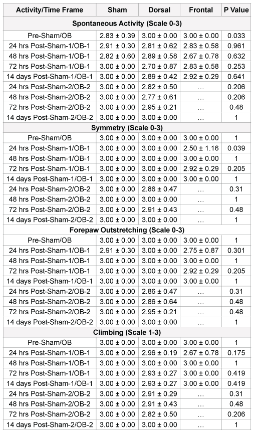
Table 1: Somatomotor evaluation after OB injury in sprague-dawley rats

Somatomotor and Behavioral Changes Following Traumatic Brain Injury
Sherry Adams 1 Jillian A. Condrey 1 Hsiu-Wen Tsai 1 Stanislav I. Svetlov 2 Victor Prima 2 Paul W. Davenport 1**Corresponding author: Dr. Paul W. Davenport, Department of Physiological Sciences,
University of Florida, Gainesville, FL 32603, USA, Tel: 352-294-4025; Fax: 352-392-5145; E-mail:
pdavenpo@ufl.edu
Article Type: Research Article
Citation: Citation: Adams S, Condrey JA, Wen Tsai H, Svetlov SI, Prima V, et al. (2015) Somatomotor and Behavioral Changes Following Traumatic Brain Injury. J Neurol Neurobiol, 1(2): http://dx.doi.org/10.16966/2379-7150.104
Copyright: © 2015 Adams S et al. This is an open-access article distributed under the terms of the Creative Commons Attribution License, which permits unrestricted use, distribution, and reproduction in any medium, provided the original author and source are credited.
Publication history:
Overpressurization blast (OB) exposure used to induce traumatic brain injury (TBI) in the rodent model can result in somatomotor and behavioral changes. Increased anxiety is evidenced after OB TBI in Dorsal and Frontal blast-wave exposed injured animals. Sustained impaired somatosensory functions occur after multiple OB injuries in Dorsal animals. Somatomotor function is impaired acutely but partially recovers after OB injury in Frontal animals. The Dorsal Group had reduction in risk-taking behavior in Group 1 (low pressure OB) and in total exploration in Group 2 (high pressure OB). The critical time point for somatosensory and somatomotor function impairment occurs at 24 hours post-OB injury with Group 1 demonstrating somatosensory and Group 2 demonstrating somatomotor deficits. The results suggest orientation and pressure magnitude have a significant impact on behavioral outcome measures following OB injuries as well as cumulative effects of repeated OB.
Overpressurization blast; Traumatic brain injury; Elevated Plus Maze (EPM); Garcia test; Behavior
Some common behavioral and neurological symptoms following overpressurization blast (OB) traumatic brain injury (TBI) include amnesia, headache, confusion, difficulty concentrating, mood disturbance, sleep pattern alterations and anxiety [1]. The relationship between severity of OB injury and persistence of these symptoms is not known and the effect of repeated OB exposures has not been systematically investigated. The OB directed dorsally over the somatosensory cortex may result in changes in sensory function and result in behavioral status changes after single and repeat OB injuries and may differ from OB injuries directed towards other head areas such as Frontal TBI. Thus, the magnitude of the OB, repeated OB and the force direction may interact to determine the behavioral effects of OB TBI.
TBI affected 1.7 million people annually in the United States [2]. The CDC reported that TBI rates are higher for males than females in every age group. Mild traumatic brain injury (mTBI) can be classified in 70- 90% of all the severity groups of TBI [3]. A cross-sectional study of 119 participants found increased levels of post-traumatic stress disorder (PTSD), depression, anxiety and cognitive failure associated with mTBI and post-concussion syndrome (PCS) [4]. In military personnel returning from Iraq, loss of consciousness (LOC) with mTBI is associated with higher rates of PTSD and depression [5]. Once a person sustains an mTBI, they are more likely to have a greater response to a second event and these multiple events are associated with longer recovery periods [6]. Currently the main clinical focus is treating the mechanical injuries caused by mTBI. When the patient does not show obvious signs of a mechanical injury upon presentation to a critical care center, the patient was often released from the facility without further follow-up. This likely resulted in unreported and underreported cases of behavioral and neurological symptoms resulting from mTBI. The long-term effects of mTBI on behavioral and neurological changes are not known.
Soldiers in combat are most susceptible to sustain a TBI as a result of an OB wave from an improvised explosive device (IED). These injuries are classified as primary blast injuries if the injury is sustained by the pressure wave. OB injuries caused damage to fluid-filled organs moving in a confined structure (such as the brain within the skull), air-filled organs and air-fluid interfaces due to the interaction between the stress wave and shear wave [7]. TBI from head injury with body protection was known to result in observational apneic periods in rodent models [8-12].
Closed-head OB injuries send shearing and stressing forces throughout the brain and brainstem resulting in disruptions in breathing [12]. Disruption in breathing could be associated with loss of consciousness. The specific effects of the forces applied to the head by the OB may be a function of the force vectors within the fluid-filled brain. The impact of the OB injury to the dorsal surface of the closed-head produce a primary force vector from the dorsal brain to the ventral brain and a rostral-caudal lateral spread of the force wave, likely resulting in loss of consciousness and potentially damage the somatosensory area. A Frontal force orientation will produce a rostral-caudal primary force vector with a dorsal-ventral lateral spread of the force wave, likely resulting in prefrontal cortical and brainstem effects with less damage the somatosensory area. Our primary goal in this investigation is to study the somatosensory and somatomotor changes following Dorsal and Frontal OB injury resulting in a TBI.
The OB wave was experimentally produced by a shock tube driven by compressed air. An OB wave directed at the skull of a rodent resulted in an OB TBI if the pressure is of sufficient magnitude. We hypothesized that the somatosensory cortex is directly under OB waves directed at the dorsal skull of a rat between bregma and lambda resulting in somatosensory changes immediately after recovery from the OB TBI. The OB shock tube generated a controlled pressure wave which can produce a range of OB pressures and replicated under experimental conditions [13]. There are two sections of the shock tube separated by a metal diaphragm. The two sections include the high pressure from the driver section and the low pressure from the driven section. The peak and duration of the OB was determined by the driver/driven ratio, thickness of the diaphragm and type of diaphragm material. Stainless steel diaphragms 0.05 mm thick with driver/driven ratio of 15 to 1 have been used to produce the shock wave [13]. An internal cutter was used to initialize the rupture of the diaphragm so the low pressure air mixes with the high- pressure gas resulting in a shock wave. The OB pressure waveform [13] has peak overpressure and gas venting phases. The gas venting phase was believed to cause the most damage due to the prolonged time spent in this phase. Previously reported OB wave venting phase durations were 10 msec at variable psi levels measured at the shock tube [13].
This on-composite (directed at the skull) OB injury resulted in neurodegeneration in the rostral and caudal diencaphalon and mesencephalon [13]. OB injury also resulted in intracranial hematomas as well as brain swelling [13]. Upon autopsy of the animals that succumbed to OB injury, hematomas were found on the dorsal aspect of the brain between bregma and lambda along with evidence of a disruption of ventral vascular supply within the circle of Willis. The kinetic force of the OB on the superficial and interior portion of the brain was evident post- mortem.
The OB blast injury was, however, reproducible and causes significant damage throughout the brain [13]. Computational modeling of OB from anterior, posterior and lateral position on humans showed the brainstem, orbit frontal cortex and cerebellum were predicted to receive the greatest amount of damage from the OB shear stress [14]. In animal OB models, the brainstem has been shown to be susceptible for damage [15-17]. In addition, repeated exposure to OB in a rat model has elicited PTSD-like symptoms [18].
The OB pressure wave generated by a single-driver shock tube system can also be oriented experimentally towards the nose for producing frontal brain OB injury. A frontal OB system was designed for reproducible and reliable frontal OB 19. The pneumatic tube system also consisted of driver (pressurized) and driven (ambient) sections. Before each OB injury, a diaphragm was inserted between the two sections. This created a closed volume driver section. The driver section was then pressurized with helium gas until the difference in pressures between the driven and driver section exceeds the strength of the diaphragm (Mylar sheets). Once the diaphragm ruptures, a shock wave was propagated down the shock tube toward the animal’s frontal skull. The eyes and nose were protected using a nose cone and eye shield. These were protected to prevent the air pressure shock wave from transferring into the lungs via the nasal passages and upper airways. The shock tube design has a 4-inch inner diameter shock tube that is approximately 7-feet in length. The fixture positioned the animal at 53 cm (20.87’) upstream from the driven section opening [20]. Mice with mild and moderate frontal OB injury showed changes in motor performance, cognitive performance and behavior with the alterations extending beyond 30 days in the moderate OB TBI group [19]. A rat model was shown to result in increased anxiety, enhanced contextual fear conditioning and altered response to a predator scent following three consecutive days of frontal OB injury [18]. These changes are suggestive of PTSD-like symptoms in animals experiencing mTBI.
In 1980, the Diagnostic and Statistical Manual of Mental Disorders first referenced the PTSD diagnosis [21]. The diagnosis was revised several times, however, and remains classified as an anxiety disorder [21]. PTSD and anxiety have been seen in humans sustaining an mTBI. However, anxiety and somatosensory changes following Dorsal and Frontal OB injury remains unknown. Specifically, it is unknown if Dorsal and Frontal OB resulted in similar changes of anxiety and/or somatosensory function. Rats exposed to multiple OB injuries (one per day for three consecutive days) developed chronic neurological and behavioral sequelae including cognitive impairment, PTSD and depression [18]. Behavior differences and variability occur in animals due to the heterogeneity in lesions following multiple OB injuries [22]. Lesion in the motor cortex as a result of a frontal OB in a rat resulted in less time spent in the center of the open field test [22]. When rats are exposed to an OB directed at high-intensity (147 kPa) to one side of the body with or without body protection (Kevlar vest), neurotrauma occurs; however, at lower intensity (126-kPa), the body protection (Kevlar vest) protects the brains from fiber degeneration [23]. Soldiers wearing protective Kevlar helmets also remained susceptible to non- penetrating damage such as coup-contrecoup, torsion and the trauma from OB injury [24]. Patients that have sustained an OB have shown neurological and neurobehavioral alterations including physical, affective, behavioral and memory problems in the absence of structural changes within the central nervous system (CNS) [25]. Despite otherwise normal external appearance after an OB injury, there may be long-lasting effects to behavioral and somatomotor function.
In humans, exposure to stress that is traumatic or life-threatening can result in PTSD. The combination of chronic stressful situations and an OB injury may result in increased cases of PTSD as a result of allostatic load (wear and tear on the body and brain) resulting from chronic over activity or inactivity of physiological systems that are normally involved in adaptation to an environmental challenge [26,27]. Approximately 14% of soldiers are reported to suffer from PTSD- like symptoms [28]. mTBI without loss of consciousness results in problems with memory, anxiety and mood disorders in military personnel [29,30]. Stressful situations combined with injuries that may go unreported because of lack of evidence of external damage may result in PTSD-like symptoms that could incapacitate soldiers. It is important to understand changes in behavioral and somatosensory status following an OB injury in regard to OB pressure, OB orientation and repeated OB exposure.
We hypothesized that somatosensory and behavioral changes will occur after one OB and the severity of these changes increased if the animal is exposed to two OB incidents. Further, we hypothesized that somatosensation and behavior will change as a function of the OB pressure and orientation. We hypothesized that somatosensory and behavioral changes will persist over time and not return to pre-OB levels. To test these hypotheses, Dorsal and Frontal OB injuries were induced in an animal model with neurological function and anxiety assessed pre-OB and post-OB.
Animals
These experiments were performed on male Sprague-Dawley rats weighing 250-300 grams. The animals were housed in the University of Florida animal care facility. They were exposed to a 12-hour light/12-hour dark cycle with food and water ad libitum. The experimental protocol was reviewed and approved by the Institutional Animal Care and Use Committee of the University of Florida.
OB Injury
The animals were randomized into three groups for the first OB exposure (OB-1): (1) Dorsal (n=29); (2) Frontal (n=8); and (3) Sham for a control group (n=11). All the animals from Specific Aim 1 were used in the Dorsal group. Not all the Dorsal animal group had pre-testing for the EPM. The differences in group sizes for the Dorsal group was a result of Banyan Biomarkers closing their facilities prior to completion of the experiment. On the day of the OB- 1 procedure for the Dorsal group, the animals were anesthetized with isoflurane. The anesthetized animals were placed onto a flexible mesh surface with the dorsal surface of head positioned underneath the air nozzle. Body was protected with a plexiglass shield placed over the entire body leaving only the head exposed 13. Then a 10-msec duration 68.0-102.6 psi OB was performed. The animals were then removed from anesthesia and returned to their cages. On the day of the OB-1 for the Frontal group, the animals were anesthetized with isoflurane. The anesthetized animals were placed onto the animal holder with the nose facing the shock tube. The head was laid on a flexible mesh surface. The neck, torso and abdomen of the animal were fixed to the animal holder to avoid movement during the blast. The eyes were covered. A cone was placed over the nose to prevent OB air from entering the nasal passages. Then a 50-65 psi OB was performed. The animals were removed from the chamber and returned to their cages. On the day of the sham blast for the Sham group, the animals were anesthetized with isoflurane. The anesthetized animals were placed in prone position similar to both the Dorsal and Frontal OB groups. Then a balloon was popped near the left external meatus creating a loud noise (dB 89.18 ± 1.83) equal to the OB. The animals were removed from anesthesia and returned to their cages.
The second OB (OB-2) was presented 14 days post-OB-1 with only Dorsal (n=23) or Sham (n=11) exposure, in the OB-1 animals. On the day of the OB-2 procedure, the animals were anesthetized with isoflurane. The anesthetized animals were placed onto a flexible mesh surface with the dorsal surface of head positioned underneath the air nozzle. The body was protected with a plexiglass shield placed over the entire body leaving only the head exposed [13].
Then a 10 msec duration 71.2-98.0 psi OB was performed. The animals were removed from anesthesia and returned to their cage. For the OB-2 Sham group, the animals were anesthetized with isoflurane. The anesthetized animals were placed in prone position then a balloon was popped near the left external meatus creating a loud noise (dB 89.60 ± 0.84) equal to the OB sound. The animals were removed from anesthesia and returned to their cages.
Behavioral tests
All behavioral measures were collected by a trained laboratory researcher in a sound- attenuated room. These tests were randomly recorded and scored by a trained observer who was unaware of each animal’s level of injury. For all testing procedures, rats were brought into the room 30 minutes prior to testing and left undisturbed to habituate to the environment. The testing room was ventilated and maintained at a temperature of 21 ± 2˚C. After the behavioral tests of each animal, the devices were cleaned with 70% alcohol and dried to prevent olfactory cues. All behavioral tests were performed at the same hours of the day (9:00a.m.-12:00 p.m.).
Elevated plus maze
The elevated plus maze (EPM) is a “+”-shaped maze that is elevated 50 cm above the floor with four 50 cm long arms. Two of the arms are open (without walls) and two are closed (with walls) and an open middle area is between the arms. The experimental procedure was initiated by placement of the rat on the middle area with head facing an open arm. The rats were allowed to roam the maze without any visual or audio distractions for 5 minutes (300 seconds). The EPM exposure was video-recorded and behavioral patterns analyzed by the video tracker software, including the amount of time spent in closed, open and middle areas and total distance traveled. The EPM was performed four days pre-OB-1/Sham-OB-1, three days post-OB-1/Sham- OB-1 and three days post-OB-2/Sham-OB-2.
Somatomotor testing
Somatomotor examinations were carried out one day prior to surgery, 24 hours, 48 hours, 72 hours and 14 days following OB/sham exposure (OB-1 and OB-2). The somatomotor tests consisted of the following six tests.
Spontaneous activity: The animal was observed for 5 minutes in its normal environment (cage). The rat’s activity was assessed by its ability to approach all four walls of the cage. Scores were: 3) rat moved around, explored the environment, and approached at least three walls of the cage; 2) slightly affected rat moved about in the cage but did not approach all sides and hesitated to move, although it eventually reached at least one upper rim of the cage; 1) severely affected rat did not rise up at all and barely moved in the cage; and 0) rat did not move at all.
Symmetry in the movement of four limbs: The rat was held in the air by the tail to observe symmetry in the movement of the four limbs. Scores were: 3) all four limbs extend symmetrically; 2) limbs on one side extended less or more slowly than those on the opposite side; 1) limbs on one side showed minimal movement; and 0) forelimb on one side did not move at all.
Forepaw outstretching: The rat was brought up to the edge of the table and allowed to walk on forelimbs while being held by the tail. Symmetry in the outstretching of both forelimbs was observed while the rat reached the table and the hind limbs were kept in the air. Scores were: 3) both forelimbs were outstretched, and the rat walked symmetrically on forepaws; 2) one side outstretched less than the opposite side, and forepaw walking was impaired; 1) one forelimb moved minimal; and 0) one forelimb did not move.
Climbing: The rat was placed on the wall of a wire cage. Normally the rat uses all four limbs to climb up the wall. When the rat was removed from the wire cage by pulling the tail, the strength of attachment was noted. Scores were: 3) rat climbed easily and gripped tightly to the wire; 2) one side was impaired while climbing or did not grip as hard as the opposite side; 1) rat failed to climb or tended to circle instead of climbing.
Body proprioception: The rat was touched with a blunt stick on each side of the body, and the reaction to stimulus was recorded. Scores were: 3) rat reacted by turning head and was equally startled by the stimulus on both sides; 2) rat reacted slowly to stimulus on one side; and 1) rat did not respond to the stimulus placed on either side.
Response to vibrissae touch: A blunt stick was brushed caudal to cranial against the vibrissae on each side. Scores were: 3) rat reacted by turning head or was equally startled by the stimulus on both sides; 2) rat reacted slowly to stimulus on one side; and 1) rat did not respond to stimulus on either side.
All EPM parameters were represented as mean ± SD (See Appendix A). The Shapiro-Wilk test was used to assess normality of the variables. Changes in the EPM variables were analyzed between groups and within groups using one-way analysis of variance (ANOVA) for Sham, Dorsal and Frontal groups. The Dorsal group was further broken down into Group 1 (low pressure) and Group 2 (high pressure) and between group and within groups measurements were analyzed same as above. Post hoc comparisons were performed with Tukey HSD test. A p ≤ 0.05 was considered statistically significant.
The nonparametric somaotomotor scores for each experimental group were averaged to obtain a mean ± SD (Tables 3-7). Changes in the somatomotor scores for each test were analyzed between groups and within groups using one-way analysis of variance (ANOVA) for Sham, Dorsal and Frontal groups. The Dorsal group was further broken down into Group 1 (low pressure) and Group 2 (high pressure) and between group and within groups measurements were analyzed same as above. Post hoc comparisons were performed with Tukey HSD test. A p≤0.05 was considered statistically significant.
Dorsal OB group (n=29) did not result in significant (p=0.240) group differences in Psi between OB-1 (83.2 ± 10.2) and OB-2 (78.5 ± 10.2) for somatomotor testing. Somatomotor breakdown of Dorsal OB-1 group found significant (p=1.48E-11) difference between Group 1 (low pressure) 69.86 ± 0.79 Psi and Group 2 (high pressure) 90.77 ± 5.55 Psi. Somatomotor breakdown of Dorsal OB-2 group found significant (p=8.74E-12) between Group 1 (low pressure) 69.94 ± 1.96 Psi and Group 2 (high pressure) 87.26 ± 3.63 Psi. Dorsal OB group (n=23) did not result in significant (p=0.223) group differences in Psi between OB-1 (80.5 ± 9.8) and OB-2 (76.7 ± 9.2) for EPM. EPM breakdown of Dorsal OB-1 group found significant (p=4.56 E-12) difference between Group 1 (low pressure) 70.15 ± 1.28 Psi (n=10) and Group 2 (high pressure) 90.64 ± 5.42 Psi (n=19). EPM breakdown of Dorsal OB-2 group found significant (p=1.295 E-12) difference between Group 1 (low pressure) 69.63 ± 2.09 Psi (n=14) and Group 2 (high pressure) 87.26 ± 3.62 Psi (n=9). Four animals did not receive EPM testing and were excluded.
Frontal OB group somatomotor testing (n=12) had OB Psi = 46.7 ± 4.9. The EPM (n=7) had OB Psi=65.0 ± 0.0. The EPM testing protocol resulted in exclusion of three Frontal group animals.
The distance traveled for the Sham group (n=11) was Pre-OB=3.51 ± 3.05, Post-Sham OB-1=4.62 ± 2.68 and Post-Sham OB-2=3.48 ± 2.25 and were not significantly different (Table 2). The Dorsal OB group Pre- OB (n=11) distance travelled was 8.35 ± 2.00 and significantly greater for Pre-OB than the Sham and Frontal groups. The Dorsal OB group distance travelled significantly decreased from their Pre-OB for both Post-OB-1 (n=29)=6.54 ± 2.01 and Post-OB- 2 (n=22)=5.15 ± 2.55. The Frontal OB group Pre-OB (n=8) distance travelled was 6.19 ± 1.22 and not significantly different than the Sham group. The Frontal group distance travelled Post-OB-1 (n=7) was 4.70 ± 3.34 and not significantly different from their Pre-OB distance. Three Frontal group animals fell off the EPM during the Pre-OB trial and were excluded from the analysis. The Sham group animals did not explore as much as the other two groups. The Dorsal group was significantly more active as evidenced by increased distanced traveled in EPM Pre- and Post-OB-1 than the other two groups, however, by Post-OB-2 the activity level normalized to the Sham group suggesting a cumulative effect as a result of a multiple OB injury. The Dorsal group also showed a progressive decline in activity after each OB injury.
Table 3 showed the Dorsal group distance traveled in the EPM between group 1 70.15 ± 1.28 psi (n=10)=6.22 ± 2.15 meters and Group 2 90.64 ± 5.42 psi (n=19)=6.71 ± 2.06 meters for their OB-1. The distance traveled in the EPM for OB-2 for Group 1 69.63 ± 2.09 psi (n=14)=5.50 ± 2.78 meters and Group 2 87.26 ± 3.62 psi (n=9)=4.17 ± 2.02 meters. There was no significant difference in the distance travelled between Group 1 and Group 2 for OB-1 (p=0.556) and OB-2 (p=0.229). There was also no OB-1 and OB-2 significant difference in the distance travelled for Group 1 (p=0.502). However, there was an OB-1 and OB-2 significant decrease in the distance traveled for Group 2 (p=0.010).
Table 2 showed the amount of time spent in the open arms. The times spent in the open arms for the Sham group (n=11) were Pre-Sham- OB-1=7.41 ± 25.06, Post-Sham OB-1=5.41 ± 11.65 and Post-Sham- OB-2=0.09 ± 0.28 and significantly decreased from Pre-Sham-OB-1. The times spent in the open arms for the Dorsal OB group were Pre- OB (n=11)=24.75 ± 23.36, Post-OB-1 (n=29)=16.01 ± 27.98 and Post- OB-2 (n=22)=4.30 ± 8.57 with a progressive significant decrease. The times spent in the open arms for the Frontal OB group were Pre-OB (n=8)=39.58 ± 29.16 and Post-OB-1 (n=7)=5.63 ± 10.76 and the Post- OB-1 was significantly decreased. Significance (p=0.0017) was reached between the Sham and Dorsal group for Post- Sham-2/OB-2 time period with the Sham group habituating to the EPM and barely exploring the open arms. Pre-OB (p=0.112) and Post-Sham-1/OB-1 (p=0.194) were not significantly different between groups. All three groups spent significantly less time in the open arms of the EPM with each subsequent trial. There was an overall significant decrease in the duration of the time spent in the open arms with each subsequent trial only after multiple OB injury suggesting a cumulative effect occurs with OB injuries.
Table 3 showed the Dorsal group duration of time spent in the open arms of the EPM for Group 1=27.65 ± 34.28 and Group 2=9.88 ± 22.69 for OB-1 and OB-2 for Group 1=2.22 ± 3.51 and Group 2=4.17 ± 2.02. There was a significant difference (p=0.030) as evidenced by decreased time spent in open arms for Group 2 compared to Group 1post-OB-1 but no significance was seen for OB-2 (p=1.000). Significance was also reached with Group 1 (p=0.010) as evidenced by decreased time spent in open arms OB-2 compared to OB-1 but not for Group 2 (p=0.940). The higher pressure OB (Group 2) spent significantly less time in the open arms after OB-1, however, the lower pressure OB (Group 1) showed a cumulative effect by spending less time in the open arms after OB-2. Group 1 and Group 2 had significant changes in psi (p<0.001) for OB-1 and OB-2 and amount of time spent in the open arms of the EPM (p=0.030) for OB-1 but not for OB-2 (p=0.730). There appeared to be an effect of psi on exploration in the EPM.

Table 1: Somatomotor evaluation after OB injury in sprague-dawley rats
Table 3-2 showed the amount of time spent in the closed arms. The times spent in the closed arms for the Sham group (n=11) were Pre-OB = 208.51±79.90, Post-Sham OB-1=256.82 ± 30.53 and Post-Sham OB- 2=273.39 ± 28.64. The times spent in the closed arms for the Dorsal OB group were Pre-OB (n=11)=182.64 ± 51.86, Post-OB-1 (n=29)= 235.21 ± 47.26 and Post-OB-2 (n=22)=235.61 ± 81.16. The times spent in the closed arms for the Frontal OB group were Pre-OB (n=8)=185.28 ± 33.50 and Post-OB-1 (n=7)=253.21 ± 52.09 Sham (p=0.021), Dorsal (p=0.004) and Frontal (p=0.009) all were significantly different as evidenced by increased time spent in the closed arms in post time periods. However, significance was not found between groups at Pre-OB (p=0.163), Post-Sham-1/OB-1 Op=0.294) and Post-Sham-2/OB- 2 (p=0.290). All three groups increased time spent in the closed arms following Pre-OB.
Table 3 presented the Dorsal group duration of time spent in the closed arms of the EPM with OB-1 for Group 1= 220.40 ± 37.56 and Group 2=243.00 ± 50.82 and with OB-2 for Group 1=227.05 ± 95.94 and Group 2=255.84 ± 50.89. There were no significant differences between OB-1 (p=0.090) and OB-2 (p=0.780) or Group 1 (p=0.110) and Group 2 (p=0.250). Regardless of injury or no-injury, the animals spend more time in the closed arms of the EPM after first trial.
Table 2 showed the amount of time spent in the middle arms for the Sham group (n=11) Pre-OB=74.08 ± 79.90, Post-Sham OB-1=37.77 ± 34.61 and Post-Sham OB-2=26.53 ± 28.72; Dorsal OB injury group Pre-OB (n=11)= 92.61 ± 31.18, Post-OB-1 (n=29)=48.44 ± 34.69 and Post-OB-2 (n=22)= 60.08 ± 77.06 and Frontal OB injury group Pre-OB (n=8)=75.18 ± 25.22 and Post-OB-1 (n=7)=41.17 ± 47.91. Significance was reached as evidenced by decreased time spent in the middle sections in the Dorsal group (p=0.004) over time periods but not in the Sham (p=0.245) and Frontal (p=0.103) groups. No significance was reached between treatment groups for Pre-OB (p=0.132), Post-Sham-1/OB-1 (p=0.469) and Post- Sham-2/OB-2 (p+0.225). The Dorsal group had significant differences in time spent in the middle section between the trials with a slight increase in time post-OB-2.
Table 3 showed the duration of time spent in the middle of the EPM for OB-1 for Group 1=51.96 ± 35.02 and Group 2=46.59 ± 35.33 and OB-2 for Group 1=70.73 ± 92.60 and Group 2=37.09 ± 40.23. No significance was reached for OB-1 (p=0.700) or OB-2 (p=0.730) between groups. No significance was reached for Group 1 (p=0.560) or Group 2 (p=0.300) between OB-1 and OB-2. The time spent in the middle arm does not appear to be affected by either psi or repeat OB injury.
Garcia somatomotor tests spontaneous activity
Table 4 showed the spontaneous activities for the Sham group (n=11), Dorsal group (n=27) and Frontal group (n=12). No significant differences were found between time points for Sham (p=0.336), Dorsal (p=0.166) or Frontal (p=0.673). There were significant differences for Pre-OB (p=0.033) between groups as evidenced by decreased responsiveness in the Sham group, but no significance was reached for the rest of the time points. Spontaneous activity was only significantly different between the group’s pre-OB injury with activity remaining the same at all the time points following injury and sham injury. This suggests OB injury does not affect spontaneous activity.
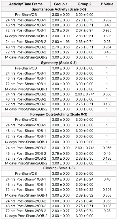
Table 5 showed the spontaneous activity with OB-1 between Group 1 = 69.86±0.79 (n=9) and Group 2=90.77 ± 5.55 (n=18). There were no significant differences 24 hours to 14 days post-OB between Group 1 and Group 2. There were also no significant differences between OB-1 and OB-2 for Group 1 and for Group 2 24 hours to 14 days post-OB. There were no significant differences in spontaneous activity between Group 1 and Group 2 and two OB injuries in Group 1 suggesting OB pressure does not have an effect on activity.
Table 4 showed the symmetry for the Sham, Dorsal and Frontal groups. There were no significant differences between groups. There was a significant difference between treatment groups at 24 hours post- Sham-1/OB-1 as evidenced by decreased responsiveness in the Frontal group (p=0.039). There were no significant differences between treatment groups for pre- Sham/OB-1 at 48 hours to 14 days post-Sham-1/OB-1. It appears that symmetry was affected only 24 hours following frontal injury.
Table 5 showed the symmetry between Group 1 and Group 2 for OB-1 and OB-2. Symmetry between OB-1 and OB-2 for Group 1 was not significantly different. Symmetry between OB-1 and OB-2 for Group 2 was significantly different as evidenced by decreased responsiveness at only 24 hours post-OB-2 (p=0.031). OB Psi appears to affect symmetry only at 24-hour post-OB-2 suggesting that higher Psi along with a second OB injury transiently affects motor control.

Table 2: EPM evaluation after OB injury in sprague-dawley rats
Data expressed as mean ± SD. One-way ANOVA test followed by post hoc analysis by Tukey HSD method. *Denotes statistically significant change of values within group. Intergroup comparison one-way ANOVA test followed by post hoc analysis by Tukey HSD method.

Table 3: EPM evaluation after dorsal OB injury in sprague-dawley rats
Data expressed as mean ± SD. One-way ANOVA test followed by post hoc analysis by Tukey HSD method. *Denotes statistically significant change of values within group. Intergroup comparison one-way ANOVA test followed by post hoc analysis by Tukey HSD method.

Table 4: Somatomotor tests for sham, dorsal and frontal groups
Data expressed as mean ± SD. One-way ANOVA test followed by post hoc analysis by Tukey HSD method. Intergroup comparison one-way ANOVA test followed by post hoc analysis by Tukey HSD method.
Forepaw outstretching
Table 4 showed the forepaw outstretching for the Sham, Dorsal and Frontal groups. There were no significant differences between time points for Sham (p=0.425), Dorsal (p=0.183) and Frontal (p=0.535) groups. Forepaw outstretching does not appear to be affected by Dorsal or Frontal OB injuries.
Table 3-5 showed the forepaw outstretching between Group 1 and Group 2 for OB-1 and OB-2. Group 1 and Group 2 animals with OB-1 and OB-2 were significantly different as evidenced by decreased responsiveness at 24 hours for the Group 2, OB-2 condition. There was a deficit in forepaw outstretching only at the first 24 hours following a second OB injury as a result of high pressure OB Psi.
Climbing
Table 4 showed the climbing for the Sham, Dorsal and Frontal groups. There were no significant differences between group or OB-1 and OB-2. There were also no significant differences between Group 1 and Group 2 for either OB-1 or OB-2 (Table 3-5). Climbing does not appear to be affected by OB orientation, Psi or number of OB injuries.
Proprioception
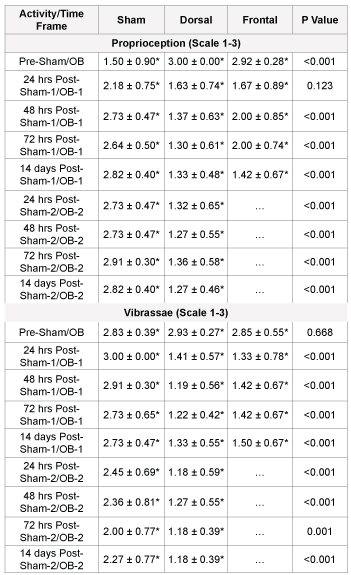
Table 6 showed the proprioception for the Sham, Dorsal and Frontal groups. There were significant differences between treatment groups (p<0.001) and time points for Sham (p<0.001), Dorsal (p<0.001) and Frontal for Pre-OB injury (p<0.001); 48 hours Post-OB-1 (p<0.001); 72 hours Post-OB-1 (p<0.001); 14 days Post-OB-1 (p<0.001); 24 hours Post-OB-2 (p<0.001); 48 hours Post-OB-2; Sham (n=11)=2.73 ± 0.47 and Dorsal (n=24)=1.27 ± 0.55 (p<0.001); 72 hours Post-OB-2 (p<0.001); and 14 days Post-OB-2 (p<0.001). Proprioception seemed to be affected in all groups regardless of injury. The OB injured groups have a decrease in response to proprioception following the OB, whereas, the Sham group had an increase in response to proprioception following the initial pre- OB testing. This suggested proprioception is decreased as a result of OB injury regardless of OB orientation. The Dorsal and Frontal OB injuries resulted in a decreased response to the proprioception stimuli that could have been due to habituation, however, this habituation was not seen in the Sham group.
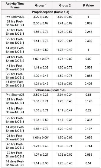
Table 3-7 showed the proprioception between Group 1 and Group 2 for OB-1 and OB-2. Comparing OB-1 and OB-2 in Group 1 animals resulted in significant differences with decreased responsiveness 24 hours post- OB-2 (p=0.002). Comparing 24 hours Post-OB-2 resulted in significant differences as evidenced by decreased responsiveness in Group 1 compared to Group 2 (p=0.020). Proprioception appeared to be sensitive to Psi as evidenced by an almost total lack of response in Group 1 (low pressure) 24 hours following the second OB injury. There was a cumulative effect of injury evidenced by the lack of response to proprioception following OB-2 at the 24 hour time point.
Vibrissae
Table 7 showed the vibrissae test response for the Sham, Dorsal and Frontal groups. There was a significant group by time effect for Sham (p=0.001), Dorsal (p<0.001) and Frontal (p<0.001) animals. The significant differences between groups (Sham, Dorsal and Frontal) were 24 hours Post-OB-1 (p<0.001); 48 hours Post-OB-1 (p<0.001); 72 hours Post-OB-1 (p<0.001); 14 days Post-OB-1 (p<0.001); 24 hours Post-OB-2 (p<0.001); 48 hours Post-OB-2 (p<0.001); 72 hours Post-OB-2 (p=0.001) and 14 days Post-OB-2 (p<0.001). All three groups showed a within group significantly decreased responsiveness to vibrissae stimuli following sham/ OB injuries, however, the Dorsal and Frontal OB injured animals showed a greater decline in responsiveness. The decreased responsiveness in the Sham group is likely due to habituation, whereas, both habituation and OB TBI may affect the Dorsal and Frontal groups. There was a significant difference between treatment groups (p<0.001) as evidenced by decreased responsiveness in the Dorsal and Frontal groups compared to the Sham group. The responsiveness to vibrissae was significantly different between the groups at every time point following the OB injury suggesting that both Dorsal and Frontal OB injuries affect somatosensory function.
Table 7 showed the vibrissae between Group 1 and Group 2 pre-OB-1 for OB-1 and OB-2. Comparison of OB-1 and OB-2 for Group 1 resulted in a significant difference at 24 hours post-OB (p=0.002) as evidenced by decreased responsiveness post-OB-2. However there were no significant OB-1 and OB-2 differences for Group 2 at all time-points. Although no significance was seen in response to vibrissae post-OB injury, there was a complete absence of response in Group 1 (low pressure) 24 hours following the second injury. Group 1 animals experienced a complete lack of response to vibrissae stimuli 24 hours after OB-2 suggesting a cumulative damage. The vibrissae response was not affected as a result of higher pressures and multiple injuries.
Behavior and cognitive performance have been the topic of multiple TBI studies using fluid percussion [31], impactor tip [32] and weight-drop [33,34]. The TBIs in these studies were induced either every day or every other day for a week. Two injuries within 7 days of each other resulted in increased damage to hippocampal neurons in the area of CA1 [35] and neuronal damage in the cortex and hypothalamus following single and repetitive concussive brain injury [32]. The multiple injury weightdrop models did not find axonal injury in the brainstem [34]. Although no brainstem neuronal loss was seen with immunohistochemistry in the weight-drop model, a disruption in the neuronal network is possible with the OB model having kinetic forces transmitted throughout the brain. Most studies focused on short-term survival ranging from hours to days with rare instances of one month beyond the TBI [36]. Longterm behavioral deficits were more commonly seen in cognition than in sensorimotor function in rodent models of TBI [37-39].
Anxiety and PTSD are commonly associated with mTBI in soldiers. Anxiety in animals is commonly measured using the EPM. Studies found that anxiety levels were not altered after frontal blast exposure [40] and weight-drop [41]. A controlled cortical impact (CCI) in a rat model used enrichment and non-enrichment 15 days prior to TBI found that EPM open arm time increased following injury suggesting reduced anxiety [42]. The authors attributed this to prefrontal lobe damage which actually resulted in increased risk-taking behavior. Another CCI model that performed EPM testing 21 days after mild, moderate or severe injury found increased time spent in the open arms [43]. Mice underwent a weight drop and were found to have increased levels of anxiety in the mTBI group as evidenced by decreased distance traveled and decreased time spent in the open arms [44]. Anxiety-like behavior appeared to be ambiguous and variable in these prior studies.
Our results suggested there is increased anxiety experienced after OB TBI. We found that the Dorsal OB injury group decreased distance traveled, decreased time spent in open arms, decreased time spent in the middle and increased time spent in closed arms. The Frontal OB injury group had decreased time spent in open arms and increased time spent in closed arms. The Sham group also showed a decreased time spent in open arms and increased time spent in the closed arms. When comparing all groups, there were differences between the distances traveled pre-injury with the Sham group being more sedentary. There were also differences seen between groups post-OB-1. These results suggested there was a habituation that occurs with all three groups over testing time. However, this habituation did not account for the changes in all categories for the Dorsal OB injured group.
The Dorsal group, due to the extent of the global brain insult, may be at greatest risk for developing anxiety. In the Dorsal group, we found that there were differences based on lower (Group 1) and higher OB pressures (Group 2). In the first injury (OB-1), Group 2 spent significantly less time in the open arms as compared to Group 1. When comparing OB-1 and OB-2 in Group 1, we found that the animals spent basically no time in the open arms after the OB-2 injury. Thus, higher OB pressure resulted in a greater effect on EPM measures of anxiety. The Group 2 animals traveled half the distance after OB-2. These results suggested a cumulative effect occurs especially in the Group 1 (lower pressure) animals with less time spent in the open arms as compared to Group 2 on first injury but then significantly less time spent in the open arms when comparing OB-1 and OB-2 within these Group 1 animals. The Group 2 animals were found to also have a cumulative effect based on the decreased distance traveled following OB-2. Although both Group 1 and Group 2 had the same injury orientation but different pressures elicit differences in OB-2 outcomes. The Group 1 animals appeared to decrease their risk-taking behavior by not exploring the open arms. The Group 2 animals, maybe due to more severe injury, did not explore (move) even in the closed arm area.

Table 5: Somatomotor tests for dorsal groups 1 and 2
Data expressed as mean ± SD. One-way ANOVA test followed by post hoc analysis by Tukey HSD method. *Denotes statistically significant change from time value within group. Intergroup comparison one-way ANOVA test followed by post hoc analysis by Tukey HSD method.

Table 6: Somatosensory tests for sham, dorsal and frontal groups
Data expressed as mean ± SD. One-way ANOVA test followed by post hoc analysis by Tukey HSD method. *Denotes statistically significant change of values within group. Intergroup comparison one-way ANOVA test followed by post hoc analysis by Tukey HSD method.
Somatomotor testing measured both somatosensory and somatomotor changes following OB injuries. It was hypothesized that the Dorsal OB injury would result in more somatosensory changes due to the orientation of the OB impact force towards the dorsal somatosensory cortex (blast overpressure directed between bregma and lambda) and the Frontal OB injury would result in more somatomotor change due to orientation of the impact force towards the pre-motor cortex (pre-frontal cortex area). To our knowledge, there were no studies that compared dorsal and frontal somatomotor changes using the Garcia somatomotor tests. When all the groups (Sham, Dorsal and Frontal) were compared, we found there were changes in spontaneous activity pre- OB injury with the Sham group less active or exploratory. The comparison of groups only found significant changes in symmetry 24 hours post-OB-1 with the Frontal group affected and the Sham and Dorsal groups remaining at pre-OB injury scores. This suggested that motor function in the form of symmetry of movements was disrupted acutely in the Frontal group.
The somatosensory and motor deficits that persisted throughout the time points were with proprioception and vibrissae. All three groups showed significant changes in proprioception persisting from pre-OB to post-OB-1and/or post-OB-2. However, the Sham group followed an opposite trend where the reaction was blunted pre-OB and then gradually returned 48 hours post- OB-1. The Dorsal group had full response to proprioception pre-OB but following OB-1 was blunted and continued to remain blunted throughout the entire period post-OB-1 and post OB- 2. The Frontal group had almost full response to proprioception pre-OB injury but then had nearly half the response at the 24 hour post OB-1 time point with a little recovery for the 48 and 72 hour periods and then a further decrease to the lowest level at 14 days post OB-1. When groups were compared, significance was seen pre-OB, post OB-1 (48 and 72 hours and 14 days) and post OB- 2 (24, 48, and 72 hours and 14 days). This suggested the Sham group was not as anxious or responsive to begin with probably due to lack of startle but eventually had almost full response to proprioception. Both the Dorsal and Frontal groups started with responsiveness to proprioception but their response precipitously dropped following OB injury. This does not appear to be habituation since the Sham group’s responses were opposite of the Dorsal and Frontal groups. Thus, OB TBI reduces proprioception responsiveness; however, OB orientation was not a factor in proprioception.
The vibrissae response was significantly different over the time points in all three groups (Sham, Dorsal and Frontal). The Sham group had attentuaion in the response to vibrissae stimulation over all time periods. This could be due to habituation to the stimulus paradigm. Although attenuation did occur, the responses were maintained on at least one side of the body. In the Dorsal group, the responses to vibrissae were nearly abolished on both sides 24 hours post OB-1 and remained absent for 14 days post OB-2. The Frontal group was similar to the Dorsal group with diminished responses from 24 hours post OB-1 through 14 days post OB-1. When comparing groups during the time points, there was a significant difference between Sham and both OB injured groups at all times points. The Sham groups maintained responsiveness to vibrissae even though attenuation did occur, whereas, both the Dorsal and Frontal groups did not respond to vibrassae stimulation post-OB throughout the entire time period following either OB- 1 or OB-2. This would suggest that somatosensory and/or somatomotor functions in response to vibrassae stimulation were affected following both a Dorsal and Frontal OB injury. This also could be related to the brain injury and only a more painful stimulus would elicit a response from the animals.

Table 7: Somatosensory tests for dorsal groups 1 and 2
Data expressed as mean ± SD. One-way ANOVA test followed by post hoc analysis by Tukey HSD method. *Denotes statistically significant change from time value within group. Intergroup comparison one-way ANOVA test followed by post hoc analysis by Tukey HSD method.
When comparing only the Dorsal OB injured rats to see if pressure affected somatomotor function, we found slightly different responses. The OB-1 injured rats did not have any differences between Group 1 (low pressure) and Group 2 (high pressure) somatomotor functioning, however repeat injury OB-2 resulted in changes between Group 1 and Group 2. In forepaw outstretching and climbing, Group 2 (higher pressure) animals had diminished functioning which approached significance 24 hours post OB-2 injury. Whereas, proprioception and vibrissae were more affected in the Group 1 (lower pressure) animals 24 hours post OB-2 injury. Thus, proprioception and vibrissae responses were non-existent in the Group 1 animals with some response seen in the Group 2 animals. When we looked at the differences in OB-1 and OB-2 in Groups 1 and 2, we found that the critical time point was 24 hours. Group 1 had a small response following OB-1 in both proprioception and vibrassae but 24 hours after OB-2 there was almost no response. This suggested a cumulative effect of the injuries or lack of responsiveness due to other confounding problems where startle was not enough of a sensation to elicit a response. For the Group 2 animals, symmetry and forepaw outstretching were effected 24 hours post OB-2. The decreased response was not as pronounced as the Group 1 animals but there was a slight attenuation in these motor activities following OB-2. This suggested that these somatomotor changes are primarily a result of the cumulative effects of injuries within a short time period (two weeks) of one another.
The results of this study are relevant to what is found in humans with mild TBI injuries. Human studies have shown greater changes with psychological or anxiety-related measures similar to the EPM and startleeliciting stimuli in our Group 1 animals. The primary dysfunction in the Group 2 animals appeared to be more motor related as evidenced by decreased time moving in the EPM and changes in forepaw outstretching and symmetry of movement. The Frontal group showed the biggest impairment in symmetry of motion at the 24 hour post OB-1 time point which suggests the motor functioning is transiently impaired. In a frontal blast mouse model 7 and 14 days after injury, histochemistry revealed degeneration in axons in the deep cerebellar white matter (arbor vitae) and peduncles, corticospinal tract and visual pathways [40]. In the present study, the Dorsal group animals showed the greatest impairments in proprioception and vibrassae at the 48- and 72-hour post OB-1. The Dorsal group’s responsiveness to these somatosensory tests were obliterated almost entirely, whereas, the Frontal group had an impaired yet present response (responsiveness on at least one side). Spontaneous recovery occurred within days after experimental TBI induced with weight drop in mice [45]. A weight-drop TBI between lambda and bregma resulted in decreased spontaneous activity in an open field 7 days post injury [46], whereas, no changes in spontaneous activity are seen in a right parietal cortex CCI [47] or fluid percussion impact that is lateral and posterior to bregma [48]. These results suggested orientation and pressure may have a large impact on behavioral outcome measures following OB injuries as well as the cumulative effects of OB suggesting specific measures are needed before the OB TBI individual is returned to duty.
Download Provisional pdf here
All Sci Forschen Journals are Open Access