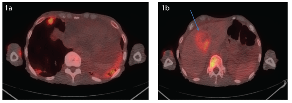
Figure 1a and 1b: FDG avid right lung and hepatic metastases (arrows) on fused axial PET/CT images in an 85 year old female with invasive, poorly differentiated squamous cell cancer of oropharynx.

Usha A Joseph1* Ron J Karni2 David Q Wan1 Isis W Gayed1
1Department of Diagnostic and Interventional Imaging/Nuclear Medicine, University of Texas McGovern Medical School at Houston, Texas, USA*Corresponding author: Joseph UA, Department of Radiology, University of Texas- McGovern Medical School at Houston, 6431Fannin, Suite 2.130b, Houston, Texas 77030, USA, Tel: 713 566 4562; Fax: 713 566 4135; E-mail: Usha.A.Joseph@uth.tmc.edu
Introduction: Fluorine-18 Fluorodeoxyglucose (FDG) Positron emission tomography/Computed tomographic (PET/CT) imaging is used to identify metabolically active primary and metastatic cancers. In head and neck cancers distant metastasis to lung is most common followed by bone and liver. Metastases to heart, spleen and gastrointestinal tract may also be seen.
Objective: Identify FDG avid distant metastasis on PET/CT scans in head and neck cancer patients to determine whether abdomen and pelvis imaging is warranted on all follow up PET/CT scans or limited PET/CT of neck and chest is an effective alternative option in patients with favorable tumor characteristics.
Methods: FDG PET/CT scans and electronic medical records of 68 consecutive head and neck cancer patients between January, 2015 and November, 2015 at our hospital were retrospectively reviewed.
Results: PET/CT scans in the 68 consecutive head and neck cancer patients of all types, sites and ages between 20-86 years were included. Lung metastasis was the most common distant metastatic site and was seen in 11/68 Patients (16.2%). 3/68 patients (4.4%) (2 males, 1 female) showed FDG avid liver metastases with concomitant lung and ipsilateral cervical nodal metastases. Below the diaphragm distant metastasis was not seen in the absence of lung metastasis. However, one oropharyngeal cancer patient without lung metastasis and evidence of a mildly avid right upper lung mycetoma had multiple FDG avid bone metastases in the clavicle, ribs, thoracic spine, left ilium and right ischium.
Conclusions: Limited PET/CT from top of the head to the waist level may be sufficient in follow up of early stage head and neck cancer patients in the absence of pulmonary, lymph node or bony metastasis in the chest on initial staging scan due to the unlikely possibility of missing distant below the diaphragm metastasis.
Limited PET/CT; Head and neck cancer follow-up; Lung metastasis; Below diaphragm distant metastasis
Head and neck cancers comprise 5% of all malignancies [1]. The incidence of distant metastasis in head and neck cancer is relatively small compared to other malignancies, but once discovered their long term survival is poor [2]. Some head and neck cancers are seen with advanced stage disease at initial presentation. Lung is the most frequent site of distant metastasis (66%) in squamous cell cancer (SCC) of head and neck; other sites are bone 22%, and liver 10%. Occasionally skin, mediastinum and bone marrow metastasis may be seen [2,3]. Majority of distant metastasis occur within the first 2 years of initial diagnosis [4] and is usually associated with poorly differentiated cancers.
Lymphatic spread is most common in head and neck cancers while hematogenous spread occurs later. High risk factors for developing distant metastasis include bilateral or 3 or more lymph nodal metastases, large lymph nodes 6 centimeters or greater in diameter (N3), low jugular lymph node metastasis, regional nodal tumor recurrence, development of second primary tumor, advanced or stage 4 disease and extensive, or bulky lymph nodal metastases [2,3,5,6]. Incidence of lung metastasis is high with bilateral nodal (N2c) disease [2]. Kotwall et al in their study showed that cancer of hypopharynx had highest incidence of distant metastasis (60%) followed by base of tongue (53%), and anterior tongue (49%) at time of death [7].
Multiple organ involvement was seen in 57% of head and neck cancer patients [8]. Distant metastases to bone (48.5%), lung (30.3%), and liver (29.3%) developed in 99/352 (28.1%) patients with nasopharyngeal cancer [9]. Probert et al., found that 65% of patients manifesting distant metastasis had no evidence of disease locally or in cervical lymph nodes, suggesting a significant proportion of such patients may have occult metastasis at initial diagnosis [10]. Troell et al., reported that liver metastasis rarely occurs in the absence of other distant metastasis and suggested that liver ultrasound may be sufficient in those with elevated liver function tests and normal chest x-ray as liver metastasis rarely occurs in the absence of other distant metastasis particularly lung metastases [4]. De Bree et al., suggested that only high risk patients with significant lymph node metastases and certain molecular characteristics of the primary tumor should be subjected to intense screening using FDG-PET/MRI [11]. Thus it would appear that in well differentiated early stage head and neck cancer patients, demonstrating only unilateral lymph nodal metastasis, the likelihood of distant metastasis is small and many remain distant metastasis free for long time periods after complete cancer control is achieved following initial therapy.
Thus in follow up of early stage head and neck cancer patients repeated whole body PET/CT scans may be unjustifiable due to low yield, additional unnecessary radiation to patient, less than optimal use of scanner time, higher cost to patient and insurance carrier and increased patient discomfort. For all these reasons our group investigated the incidence of distant metastasis in patients with variety of head and neck tumors to evaluate the reliability of a novel modified limited PET/CT imaging protocol limited to the head, neck and chest in patients with favorable tumor characteristics. Our study objective was to see if limited PET/CT can be a practical, feasible and effective imaging option without compromising patient care.
A retrospective review was done of PET/CT scans in a total of 68 consecutive head and neck cancer patients of various types and locations imaged at our hospital between January and November 2015. PET /CT scans and electronic medical records of patients were reviewed for demographics, medical history, primary malignancy, pathology, relevant CT, MRI, and treatment history. PET CT scans and /or reports were reviewed for primary tumor site, nodal and distant metastasis, second malignancy if present, and included metabolic activity as measured by standard uptake value (SUV) in the primary and metastatic malignancy. PET/CT in our 68 head and neck cancer patients was comprised of 19 patients undergoing initial staging scans and restaging PET /CT scans in 49 patients done from 2- 48 months after completion of treatment.
Our PET /CT camera is a GE Discovery ST PET/CT System. The16- slice CT unit has diagnostic capability. The PET camera has Bismuth Germanate (BGO) Crystals and can acquire images in both 2D and 3D mode with a 3.75mm slice thickness which is matched by the CT for image co-registration.
Patients with head and neck cancers visiting our facility for the first time receive a 2 part FDG PET/CT examination. After the 60 min post-injection uptake phase, an initial two bed image (in 2D mode) is taken of the head and neck region with the arms in the down position for optimal visualization of the head and neck region. After this first scan is completed, it is immediately followed with a 2D body image extending from the vertex to the upper thighs with the arms in the up position in order to optimize imaging of the thorax. On subsequent imaging only the vertex to upper thigh PET/CT scanning is performed.
The total scan duration is dependent upon the length of a patient’s torso. PET images are acquired in ~8inch bed positions lasting 4 minutes each. An eye to thigh scan for the average sized patient will take 6 bed positions and a total scan time of generally 26 minutes. The scan time can increase to 30 min when a 7 beds is used for 6 feet tall patient and less total scan time (5 bed positions, 22 min scan time) for someone 5 feet tall (5 beds, 22 min scan). The 7 bed scan would also apply to Head/Neck cancer patients where we image from vertex to mid thighs. There is no modulation of scan time per bed position or depending on body region. When PET/CT imaging is limited to only through the thorax (above diaphragm only) on subsequent scans, the average total scan time could be shortened by 8-10 minutes.
PET/CT tumor imaging is done 60 min after the intravenous injection 12-15m Ci of F18 FDG in adult patients while patients are seated comfortably in a quiet dimly lit room, avoiding physical activity after radiotracer injection. Patient is injected after fasting overnight or 4 hours prior to radiotracer injection and are instructed to avoid heavy physical activity 1 day pre scan. Spot blood glucose level obtained pre radiotracer injection should be < 250mg/dl. Low dose non contrast CT imaging is done in the supine position with arm elevated and patient breathing normally. PET imaging is done in the supine position starting from the base of skull to below pelvis. The reconstructed CT and PET images are reviewed separately and as fused PET/CT images. Any abnormality identified on PET scan is correlated with CT findings and maximum metabolic activity recorded as SUV max. A separate head and neck view is obtained on the initial PET/CT for purposes of radiation treatment planning.
Analysis of the results using descriptive statistics was used and reported as percentages, mean and median as well as ranges.
A retrospective review was performed of a total of 68 consecutive head and neck cancer patients, 56 males, 12 females, average age of 21-84 years. The majority 89.7% (61/68 patients) had squamous cell cancer, and the other 10.3% (7/68 patients) included of one patient each of biopsy proven invasive squamous cell carcinoma of the left pre auricular skin and concurrent follicular lymphoma of the left nasopharynx and multiple left cervical lymph nodes, lymphoepithelial cancer, parotid cancer, parotid carcino sarcoma, unknown primary, high grade papillary thyroid cancer and olfactory esthesioneuroblastoma. Follow up scans were performed at an average interval of 16.3 months and a median of 11 months after therapy (range 2-48 months).
Ipsilateral or bilateral cervical lymph nodal metastases was seen in 28/68 (41.2%) patients. Local extension to skull base was seen in 3 patients, orbital invasion in 3 patients and intracranial extension in 3 patients.
Lung was the most common site for distant metastasis, seen in 11/68 (16.2%) head and neck cancer patients. Three patients (4.4%) (2 males, 1 female, age range of 41-77 years) showed FDG avid liver metastases with concomitant lung and ipsilateral cervical nodal metastases (Table 1). This group included a patient with locally invasive poorly differentiated squamous cell cancer (SCC) of oropharynx with ulcerating mass in the right tonsil/base of tongue (Figure 1a,1b). The second patient had invasive poorly differentiated squamous cell cancer (SCC) in a supraglottic mass involving larynx/hypopharynx with vocal cord fixation, false vocal cord involvement and obliteration of the airway (Figure 2a-2d). The third patient had undifferentiated sinonasal cancer of the naso-ethmoid sinus with lung and ipsilateral cervical nodes, liver, brain and inferior pericardial nodal metastases (Figure 3).
Figure 1a and 1b: FDG avid right lung and hepatic metastases (arrows) on fused axial PET/CT images in an 85 year old female with invasive, poorly differentiated squamous cell cancer of oropharynx.
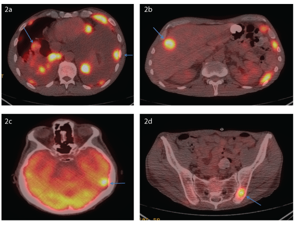
Figure 2a, 2b, 2c, 2d: FDG avid metastases in lung, pleura, left temporal lobe, liver and left ischium (arrows) on fused axial PET/CT images in 52 year-old male with invasive poorly differentiated squamous cell cancer of the supraglottis/larynx.
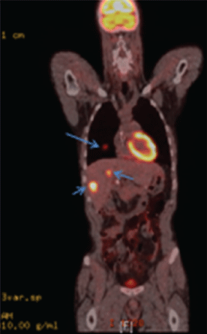
Figure 3: FDG avid right lung and liver metastases (arrows) on fused coronal PET/CT image in a 40 year-old male with undifferentiated sinonasal carcinoma in a large locally invasive left naso-ethmoid, paranasal and maxillary sinus cancer.
| Patient Number | Cancer | CLN | Lung | Liver | Brain | Bone | Other Mets | Other Nodes | Pathology |
| 1 | N-P | No | Bilat | No | No | Lt rib | No | H,MS | SCC-PD |
| 2 | O-P | Ipsi | RLL | No | No | T1 | No | No | SCC |
| 3 | Larynx | Ipsi | Bilat | No | No | No | SC | No | ISCC |
| 4 | Paranasal | Ipsi | RUL | No | No | No | RH | No | LE |
| 5 | Nasal Cavity | Ipsi | LUL | No | No | No | RH | No | SCC |
| 6 | U Primary | Bilat | RUL | No | No | No | No | No | SCC |
| 7 | Parotid | No | Multi | No | Yes | No | No | No | CS |
| 8 | O-P | Ipsi | RLL | No | No | No | No | No | ISCC |
| 9 | O-P | Ipsi | Yes | Yes | No | No | No | No | ISCC-PD |
| 10 | Supraglottis | Ipsi | Bilat | Yes | Temp | rib | Pl, perit | H,Ms, | SCC-PD |
| 11 | NES | Ipsi | Yes | Yes | Yes | L | No | IPLN | SNUC |
Table 1: FDG Avid Lung and below diaphragm metastases in 11/68 head and neck cancer patients.
NP=Nasopharyngeal Cancer; CLN=Cervical Lymph Nodes; Mets=Metastasis; O-P=Oropharyngeal Cancer; Bilat=Bilateral; Ipsi=Ipsilateral; RLL=Right Lower Lobe; SCC=Squamous Cell Cancer; Lt Rib=Left Rib; H=Hilar; Ms=Mediastinal; SCC PD=Squamous Cell Cancer-Poorly Differentiated; ISCC= Invasive Squamous Cell Cancer; RUL=Right Upper Lung; RH=Right Hilar; LE=Lymphoepithelial Cell Cancer; LUL=Left Upper Lung; U Primary=Unknown Primary; Multi=Multiple; CS=Carcino Sarcoma; Temp=Temporal Lobe; Pl/Perit=Pleura/Upper Peritoneal Metastases; ISCC-PD=Invasive Squamous Cell CancerPoorly Differentiated; NES=Naso-Ethmoid Sinus Cancer; L=Local Involvement by Large Mass of Ethmoid Air Cells, Clivus, Sella turcica, and Left Orbit; IPLN=Inferior Pericardial Lymph Nodal Metastasis; SNUC=Undifferentiated Sinonasal Cancer.
Another three (4.4%) patients with lung and brain metastases, included a parotid carcino sarcoma; the second patient with supraglottis cancer had lung, temporal lobe, rib, hilar and mediastinal nodal and pleuro-peritoneal metastases and the third patient had lung, liver and pericardial lymph node metastases in addition to the brain metastases.
Five patients (7.3%) had bony local invasion or bony metastases at different sites. Four of them also had additional lung metastases. Only one patient (1.4%) had wide spread bony metastases involving ribs, clavicle, multiple thoracic vertebrae, right ischium and left ilium without lung metastasis. However, his bony metastases would not have been missed if he had an upper body limited PET-CT scan on follow –up and would have triggered the conversion of the scan to a standard full body PET-CT scan at the time of imaging.
Two additional patients did not have lung metastases but had cervical lymph node metastases. One of them had hilar and mediastinal lymph node metastases and the other one had thyroid metastasis. Thus, both the patients would have still been appropriately restaged on follow up upper body limited PET-CT scan in the absence of lung metastases.
Our study like previous others confirms that lung is the most common site for distant metastasis in head and neck cancers and was seen in 16.2% in our study group. Distant metastasis to liver, bone, brain, was always seen in association with lung metastasis in our PET/ CT scan analysis of 68 consecutive head and neck cancer patients at our hospital over a 11 month period. The only exception was a patient with oropharyngeal cancer and right upper lung mycetoma who had multiple FDG avid bone metastases in the thorax and pelvis in the absence of lung or soft tissue metastasis below the diaphragm. Liver metastases was seen in 3/68(4.4%) with poorly differentiated or undifferentiated head and neck cancers, concomitant lung and ipsilateral cervical nodal metastases. Our findings showed that distant metastasis to brain, bone, mediastinal and hilar lymph nodes did not occur in the absence of lung, lymph node, or bone metastasis in the chest.
Our study showed that if there are no clinical or radiographic evidence of lung metastasis, lung nodules, lymph nodes or bony metastases in the chest, or non FDG avid lung pathology, the likelihood of developing distant metastasis below the diaphragm including liver and bone metastasis is an unlikely occurrence. Our findings parallel those of Senfit et al., who reported that screening for distant metastasis on FDG PET scan is indicated if solid lung lesion or suspicious lung lesions less than 5 mm are seen on CT as these findings on CT are considered positive for distant metastasis regardless of PET findings [1]. Troell et al., also have suggested that liver ultrasound may be sufficient in those with elevated liver function tests and normal chest x-ray as liver metastasis rarely occur in the absence of other distant metastasis particularly lung metastases [4].
Distant metastases were found in 12.3% (96/779) patients in Probert’s series and most commonly involved lungs, bone and liver [10]. Locally extensive and most advanced primary tumors were most likely to develop metastasis; even when the primary tumor site was completely eradicated, 51% developed metastasis suggesting that subclinical metastasis must have been present prior to or at time of initial therapy [6]. Persistent tumor at the primary site is associated with high incidence of distant metastasis, seen in 49% of 99 patients [4,10] Primary, advanced stage tumors in the hypopharynx, oropharynx and oral cavity are associated with the highest incidence of distant metastasis and are influenced by location, initial T and N stage of primary tumor, presence or absence of regional control above clavicles, advanced nodal disease especially with jugular vein invasion or extensive soft tissue disease in neck [2].
De Bree et al., found that clinical and histopathological risk factors are mainly related to the extent of lymph node metastases. They felt that because the yield from examinations for detection of distant metastases is too low to warrant routine screening of all head and neck squamous cell cancers, only patients with high risk factors should be selected for intense screening for distant metastases and advocated more research to develop new screening protocols for distant metastases [11].
Our study found that in follow up of early stage head and neck cancers with none or few small, ipsilateral lymph nodes, absence of pulmonary metastases, lung nodules or other lymph nodes or bony metastasis in the chest, limited repeat PET /CT scans to the waist level may be sufficient due to the unlikely possibility of missing distant below the diaphragm metastasis in the absence of chest metastasis. Arens Al et al., also are proposing the use of a limited FDG PET-CT of the thorax and upper abdomen as adequate imaging modality for patients with non-small cell lung cancer as they found no negative impact on staging or treatment in their study of 1059 patients with suspected or recently proven lung cancer limited to thorax. PET/CT limited to the chest and upper abdomen resulted in correct staging in 98.7% of patients, with identical management as in the 98.8 % patients who underwent full field of view PET. Additional lesions considered malignant was seen in 10.7% of patients leading to a change in management in 1.2% of their lung cancer patients [12].
Deuvorst SE et al., have found high negative predictive value for FDG PET-CT scans in 190 patients with head and neck cancer and high risk factors and no difference in median overall survival from the time distant metastasis was found on initial staging or at follow up restaging scans in their study [13].
Our study is limited by small sample size and retrospective nature and further large scale study is needed to further confirm our findings.
As medicine evolves, older concepts are being challenged and customized medical care tailored for an individual patient is being increasingly advocated. At present diagnostic CT and MRI imaging are routinely limited to head, neck, chest, and/or abdomen and pelvis depending on individual patient’s disease circumstances and needs. Substituting limited PET/CT imaging for routine whole body PET/ CT imaging on follow up PET/CT may be considered a useful option in patients with small initial tumors, confined to none or few small ipsilateral lymph nodes without evidence of lung metastasis or lung abnormality. The advantages of limited PET/CT is shortening of total imaging times by 8-10 min, resulting in improved laboratory efficiency, better patient throughput, less patient discomfort, better patient acceptance, less cumulative radiation exposure, and potential of reduced financial burden to patient and insurers without compromising patient care. These advantages can increase the use and appropriate utilization of FDG PET-CT scans with less cost especially in smaller hospitals and tertiary centers in developing countries. Nair S et al., in their study of 131 patients with various head and neck cancers, found only 3 patients (2.2%) had a change in metastatic status with lung metastases identified in two patients and liver and lung metastasis in one patient. They suggested a more realistic evaluation of cost versus benefit be done since in many developing countries, the use of FDG PET-CT scan is limited owing to the high cost and limited availability [14].
In conclusion, limited PET/CT from top of the head to the waist level may be sufficient in follow up of early stage head and neck cancer patients in the absence of pulmonary, lymph node or bony metastasis in the chest during initial staging PET-CT scan due to the unlikely possibility of missing distant below the diaphragm metastasis.
Download Provisional PDF Here
Article Type: RESEARCH ARTICLE
Citation: Joseph UA, Karni RJ, Wan DQ, Gayed IW (2018) Limited PET/ CT– A New Concept in Follow-up of Head and Neck Cancer Patients. J Mol Med Clin Appl 2(1): dx.doi.org/10.16966/2575-0305.111
Copyright: © 2018 Joseph UA, et al. This is an open-access article distributed under the terms of the Creative Commons Attribution License, which permits unrestricted use, distribution, and reproduction in any medium, provided the original author and source are credited.
Publication history:
All Sci Forschen Journals are Open Access