Abstract
With the rapid development of CT technology, the procedure of Coronary CT Angiography (CCTA) has been increasingly used and widely available for the diagnosis of Coronary Artery Disease (CAD). Other than having its advantages to improve the sensitivity and specificity in the detection of CAD, CCTA also associated with high radiation dose. Several dose-reduction strategies have been introduced to reduce radiation dose during CT procedure. However, one of the technique, namely prospective ECG-triggering CCTA provides tremendous radiation dose reduction to patient. Therefore, this article provides information on the prospective ECG-triggering technique and how it can further reduce the radiation dose to patient.
Keywords
Coronary CT angiography; Coronary artery disease; Electro Cardiogram (ECG)
Prospective ECG-triggering Coronary CT Angiography
(CCTA): How safe is this procedure?
Over the last fifteen years, tremendous developments have taken place
in the CT technology which allows the CT scanner to produce images
of the coronary artery lumen and wall in order to provide analysis on
severity and characteristics of coronary artery disease has definitely made
CCTA a reliable non-invasive diagnostic tool in coronary artery imaging.
This article is written to provide information about CCTA scanning
techniques, radiation dose associated with CCTA procedure and how
prospective ECG-triggering can further reduce the radiation dose to
patient.
The acquisition of the dataset for CCTA consists of three steps including
topogram, determination for the adequate contrast enhancement of
CCTA and the image acquisition for the entire coronary artery tree were
made completed with the contrast enhancement setting either using bolus
tracking or the test bolus techniques. The scan is acquired in a single
breath hold during comfortable inspiration and starts with the injection
of a contrast agent with a high concentration of iodine (300–400 mg/mL)
at a high flow rate (4–6 mL/s). The total volume of contrast agent depends
on the scan length, but typically 60–80 mL is injected, followed by a saline
flush (40–70 mL at 4–6 mL/s). The actual CT scan starts after the delay is
calculated as the contrast material transit time [1].
There are two techniques in CCTA procedures namely prospective
ECG-triggering and retrospective ECG-gating. Both prospective ECGtriggering
and retrospective ECG-gating techniques are characterized
based on its scanning mode, sequential and helical, respectively. In
sequential or known as step-and-shoot scanning mode, a cross-sectional
image is produced by acquiring a series of axial slices of the body from
different angular positions while the X-ray tube and detector rotate 360°
around the patient with the table being stationary. The image is then
reconstructed from the resulting projection data which is like merging
several contiguous stacks of partial cardiac images until a complete cardiac
image is acquired consists of the entire cardiac length within the desired
scanned range. However, if the patient moves during the acquisition, the
data obtained from different angular positions are no longer consistent
and stair-step artifact occurs resulting image degradations and may be of
limited diagnostic value [2].
On the other hand, helical or spiral scanning mode uses a different
scanning principle. Unlike the sequential CT, this mode allows table
to move continuously through the gantry in the z-direction while the
X-ray tube rotates 360° around the patient. The X-ray traces a spiral path
around the patient and produces volume data. The table movement in the
z-direction during the data acquisition will generate inconsistent datasets,
causing reconstructed images being degraded by the artefacts. Thus, some
special reconstruction algorithms using the interpolation principles to
generate planar set of data for each table position producing artefact-free
images [2]. Thus, it is possible to reconstruct individual slices from a large
data volume by overlapping reconstructions as often as required.
Radiation dose issue
Although rapid developments in CT technology have led to improve the
sensitivity and specificity of CCTA procedure in the detection of Coronary
Artery Disease (CAD) in both image quality and diagnostic value, CCTA
runs the potential risk of high radiation dose [3,4]. Previous literature
has shown that radiation dose increases with increasing detector rows
in CT due to narrow detector collimations and long anatomic coverage
[5]. In fact, it is generally agreed that CT is an imaging modality with the
highest radiation dose of all radiological examinations, as it contributes
up to 70 per cent radiation dose of all radiological examinations [6].
Previous research estimated that CT scan causes 800 cancers in woman
and 1300 in man per year in the United Kingdom [7]. Moreover, in the
United States, approximately 500 out of 600,000 children less than 15
years old who are estimated to undergo CT procedure each year will die
of cancer [8]. Moreover, the radiation risks associated with CCTA have
raised serious concerns in the literature and many researchers agreed on
that there is potential risk of radiation-induced malignancy resulting from
CCTA [3,9]. In fact, many researchers were questioning: Does utilization
of CCTA lead to the greatest benefit and is the risk of radiation greater
than the benefit expected from the CT examinations? [3,10].
Therefore, several dose-saving strategies have been introduced to deal
with radiation dose issues, and these techniques include anatomical-based
tube current modulation [11], ECG-controlled tube current modulation
[12], tube voltage reduction [13], iterative reconstruction algorithm
software [14], a high-pitch scanning [15] and prospective ECG-triggered
CCTA [16,17]. This article is written to provide information about
prospective ECG-triggered CCTA technique which is one of the strategies
that could be used to further reduce the radiation dose to patient during
CCTA procedure
Prospectively ECG-triggered coronary CT angiography
Prospectively ECG-triggered technique is a low-radiation dose
scanning method using sequential mode to acquire axial images and an
incrementally moving table to cover the heart with minimal overlap of
axial slices. This technique in cardiac CT is not new and it recognizes
that CT image synchronization with heart diastolic phase was optimal for
heart imaging. However, the results were not being achieved when the
patient heart rate increases. Unlike the principle of helical continuous
scanning in retrospective ECG gating, the principle of data acquisition in
this technique takes place only in the selected cardiac phase (diastolic) by
selectively turning on the X-ray tube when triggered by the ECG signal.
The X-ray tube is remained off for most of the scanning period especially
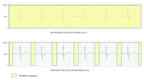
other than diastolic phase in cardiac cycle (Figure 1) [18]. Moreover,
during the image acquisition, the table is stationary and then moves to the
next position for another scan initiated by the subsequent cardiac cycle
(diastolic phase). This results lead to a significant reduction in radiation
dose [19]. It has been widely reported that prospective ECG-triggering
protocol produces low radiation dose with the effective dose ranging from
3.8 to 6.8 mSv which was significantly lower than that of conventional
CCTA method, retrospective ECG-gating technique. This dose reduction
ranging from 76% to 83% [17,20].
In 64-slice scanner system, the scan is prescribed by using 3 to 5
incremental of 64 × 0.625 mm (40 mm) image groups which requires
up to 4 incremental table translations of 35 mm allowing for 5 mm of
overlap. The minimum interscan delay is approximately between 0.6 and
1.0 second which normally requires skipping a cardiac cycle between data
acquisitions which results in one image acquisition per 2 cardiac cycles
[19]. However, the process will be faster with larger detectors (128-, 256-
or 320-slice CT) being used. The detector width determines the number
of steps/scans to cover the entire heart and complete the examination. For
instance, the dual-source 64-slice CT has a narrower detector array (32 ×
2 × 0.6 mm = 38.4 mm per acquisition); thus, it takes more incremental
steps (normally 4-5 cardiac cycles) to cover the heart and complete an
examination than with the 320-row system (320 × 0.5 mm = 160 mm)
which covers the heart in a single acquisition [21].
Prospectively ECG-triggered technique has a limited number of cardiac
phases available for reconstruction. Therefore, mid-diastolic phase (75%
of R-R interval) was normally selected for data acquisition for all subjects.
In addition, by using add-on 'padding' will allow more cardiac phases
for reconstruction. Padding technique is described as prolonging the
acquisition window in order to produce consistent image quality although
scanning a patient with minor heart rate variations (more than ± 5 b.p.m).
Although padding can be described as widening the acquisition window, it
is actually turns the x-ray tube on before and after the minimum or actual
acquisition time (milliseconds) required. Available padding options with
current software ranges from 0 to 200 milliseconds. However, radiation
dose will definitely increase with application of padding window due to
expense of radiation exposure on the particular windows phase [19].
In terms of diagnostic quality and accuracy, prospective ECG-triggering
CCTA was proven to have high sensitivity (>89.3%), specificity (>90.5%),
positive predictive value (>89.8%), negative predictive value (>90%) and
accuracy (>92.3%) in the diagnosis of CAD from a systematic review
based on 23 selected studies using prospective ECG-triggering [22].

Figure 1: Variations in CCTA scanning techniques produced different radiation dose. In standard retrospective gating, the tube current is constantly ‘on’ throughout the acquisition produces high radiation dose while in prospective ECG-triggering, the exposure is ‘on’ at the selective cardiac phase
(diastolic phase) for a short period resulting low radiation dose production.

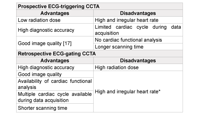
*Depending on types and generations of multidetector CT scanner
Table 1: Summary of advantages and disadvantages of prospective ECG-
triggering versus retrospective ECG-gating CCTA
The main limitations of prospective ECG triggering technique is
unavailability of producing cardiac functional analysis due to limited
access of the cardiac cycle during data acquisition. The cardiac functional
analysis can be provided with multiple phases of cardiac scanning using
helical scanning mode. If the clinical scenario or referring physician
requires information about cardiac function, then retrospective gating
must be undertaken. Heart rate variability is another limitation for the
prospective ECG triggered technique. Heart rate variability of > 5 b.p.m.
is considered not applicable for prospective ECG-triggering with scanner
system which is not equipped with ‘padding window’ software application
especially in 64-slice scanner [19]. A summary of advantages and
disadvantages of prospective ECG-triggering versus retrospective ECGgating
CCTA is shown in Table 1.
In conclusion, coronary CT angiography with prospective ECGtriggered
technique has proven to produce low radiation dose with
high diagnostic quality and accuracy in the diagnosis of CAD. With low
effective dose from 3.8 mSv, it is believed that prospective ECG-triggering
CCTA is a safe procedure to be performed in radiological investigation
procedures as a screening tool for the assessment of coronary arteries.
Article Information
Article Type: Review Article
Citation: Sabarudin A (2015) Prospective ECGtriggering
Coronary CT Angiography (CCTA): How
Safe is this Procedure? J Hear Health, Volume1.1:
http://dx.doi.org/10.16966/2379-769X.104
Copyright: © 2015 Sabarudin A. This is an
open-access article distributed under the terms
of the Creative Commons Attribution License,
which permits unrestricted use, distribution, and
reproduction in any medium, provided the original
author and source are credited.
Publication history:
Received date: 12 March, 2015
Accepted date: 24 March, 2015
Published date: 28 March 2015


