
Figure 1: A. Coronal view of CT scan showing splenic hematoma of 7 × 9 × 10 cm and hemoperitoneum. B. Sagittal view of CT splenic rupture, splenic hematoma with active bleeding.

Mariana Caraballo Angeli Omar Abdel-lah Fernández* Yari Aguilera Molina José Quiñones Sampedro
Department of Surgery, University Hospital of Salamanca, Castilla y León, Spain*Corresponding author: Dr. Omar Abdel-lah Fernández, C/ Muñoz Torrero 1 5B CP: 37007 SALAMANCA, ESPAÑA, Spain, Tel: +34 923261198; E-mail: omarabdellah@gmail.com
Background: Spleen injury is an uncommon life threatening complication after routine colonoscopy with a low incidence of 0.001%-0.004% approximately. There should be a high level of suspicion when patients that underwent a colonoscopy start presenting abdominal pain during the first 24 hours and are hemodynamically unstable.
Method: Present two cases of splenic injury after routine colonoscopy and a brief literature review.
Results: Two women arrived to the Emergency Department (ED) with abdominal pain four days after undergoing routine colonoscopy. Splenectomy was performed in one patient, and the other was admitted into the Intensive Care Unit (ICU), recovering satisfactorily. Both patients were discharged on the 6th day after admission without any further complications.
Conclusion: The real incidence of splenic injury remains unknown, due to the reluctance to publish and the absence of guidelines. Many of splenic injuries don´t present symptoms, so they are not diagnosed and remain unrecognized.
Most of the patients present symptoms in the first 24 hours. In patients with a late presentation, a high level of suspicion is advised in order to make a timely diagnosis.
Diagnose is clinically based and confirmed with a Computer Tomography Scan (CT), considered the gold standard. Treatment should be based on the hemodynamic status of the patient.
Splenic lesion; Colonoscopy
Colonoscopy is considered among the most appealing approaches for early detection and prevention of Colorectal Cancer (CRC) [1]. Colonoscopy is reported to be a safe procedure; complications after a routine colonoscopy are unusual, but it remains an invasive test with a low but real risk for morbidity and mortality. The real number of complications is probably underestimated, mainly because endoscopists self-reported complications are not described6 . The most frequent complications of colonoscopy are bowel perforation (0.34% to 2.14%) and bleeding (1.8% to 2.5%) [2,3]. The rates of complications are higher in patients who undergo biopsy or polypectomy [2]. Infrequent complications include: pneumothorax, pneumomediastinum, volvulus, hernia incarceration, retroperitoneal abscess and emphysema. Other complications include appendicitis and bacteremia with endocarditis [4]. Another underappreciated and life-threatening complication is splenic injury. This complication was first described by Werry and Zhener in 1974 [5]. A total of 103 cases have been described in 75 reports [6].
Two case reports are presented. These cases describe two patients who underwent routine colonoscopy, and afterwards went to the Emergency Department (ED) with symptoms of splenic injury followed by a brief literature review of this unusual complication.
The two patients were women, who underwent outpatient colonoscopy.
A 63-year-old woman, with a history of dyslipidemia, hypertension and previous cholecystectomy presented to the ED for evaluation of abdominal pain four days after having undergone outpatient colonoscopy. The colonoscopy was performed for CRC screening and it revealed a three millimeter sessile polyp, 20 cm from the anal verge. Polypectomy was performed without any complications.
The patient reported mild pain in both upper quadrants of the abdomen, radiating to the back on the left side. The pain worsened with deep inspiration and movements; she denied having any other symptoms.
Initial examination showed, temperature: 36.1°C, heart rate 84 bpm, respirations of 18, blood pressure 104/80 mmHg. She was alert and hemodynamically stable, her abdomen was slightly rounded with diffuse tenderness, no bowel sounds and Kehr sign was positive. She presented with acute abdomen and her laboratory data was: hemoglobin 12.4 g/dl, hematocrit 36%, white blood cell count 10999/mm3 , platelet count 259000/ mm3 . Coagulations studies, routine chemistries and electrocardiogram were normal. A CT of the abdomen and pelvis revealed hypodense lines on the spleen compatible with fissures. Splenic hematoma of 7 × 9 × 10 cm and hemoperitoneum was described (Figure 1). Emergency laparotomy revealed a 2l hemoperitoneum, laceration and decapsulation of the upper pole of the spleen, for that emergency splenectomy was performed without difficulties. The spleen pathology was unremarkable. Postoperatively the patient recovered without complications. The patient was discharged the 6th day after surgery.
Figure 1: A. Coronal view of CT scan showing splenic hematoma of 7 × 9 × 10 cm and hemoperitoneum. B. Sagittal view of CT splenic rupture, splenic hematoma with active bleeding.
A 62-year-old woman with history of hypertension, heart valve replacement surgery, in treatment with acenocumarol switched to lowmolecular-weight heparin due to the programmed colonoscopy for evaluation of occasional rectal bleeding. The colonoscopy revealed diverticulosis, internal hemorrhoids and two polyps that were millimetric in the left colon, which weren’t resected due anticoagulant treatment.
The patient went to the ED for acute diffuse abdominal pain of 48 hours. The pain was radiating to the left anterior thorax, arm and left shoulder. She denied nausea or vomiting, but reported diarrhea. Initial examination showed that the patient had a heart rate of 114bpm and a blood pressure 101/58 mmHg. She was alert and appeared somewhat uncomfortable. Lungs were clear to auscultation without chest wall tenderness. No significant cardiological findings were reported. Abdomen was slightly rounded without bowel sounds. Her laboratory data was: hemoglobin 9.5 g/dl, hematocrit 28.2%, white blood cell count 11250/mm3 , platelet count 234000/mm3 . Coagulations studies, routine chemistries and electrocardiogram were normal. Cardiac markers were carried out do to high risk of acute cardiac pathology. A CT scan of the abdomen and pelvis described multiple diverticulums in the right and left colon. Small quantity of liquid was found in the perihepatic, perisplenic and in the upper right paracolic gutter and pelvis. Patient was left in the emergency observation area. Cardiac markers were normal.
Due to the persistence of the abdominal pain, six hours after the first evaluation a surgical consultant was assessed. The patient reported undergoing a colonoscopy four days before and denied previous trauma. The surgical team’s evaluation, reveled an abdomen that was slightly rounded, abdominal guarding was found and bowel sounds were present. No periumbilical or flank ecchymosis was noted. A new control blood analysis revealed a drop in the hemoglobin of 1.3 points in six hours. After reevaluating the CT scan a subcapsular spleen hematoma without active bleeding signs was noted (Figure 2). Patient was admitted to the ICU for close monitoring; operative intervention was not required. Transfusion of two units of packed red blood cells was needed during her 48 hours stay in the ICU. The patient remained hemodynamically stable, with gradual improvement of the pain, and was discharged the 6th day after her admission. Follow-up CT a month after her discharge showed, remarkable improvement of the hematoma (Figure 3).
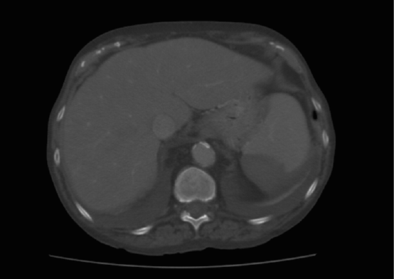
Figure 2: Axial view of CT scan showing subcapsular spleen hematoma.
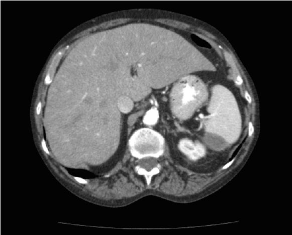
Figure 3: Axial view of follow-up CT scan showing remarkable improvement of the subcapsular spleen hematoma.
Spleen lesions after a colonoscopy are a rare complication, with a low incidence of 0.001% - 0.004% approximately; a higher risk (72-75%) does appear to exist in women [6-9]. This complication was first described in 1974 by Werry and Zhener [5]. A total of 103 cases have been described in 75 reports [6]. Published articles estimate an incidence from 1 x 6,000 cases annually [9-11] to 1- 21 × 100,000 annually [8,12], with a mortality rate of 5.4% [7,9,10,12,13].
Risk factors have been described predisposing spleen injury; those patient-dependent are: splenomegaly, adhesions between spleen and colon from prior surgery, neoplasm, inflammation such as diverticular disease, pancreatitis, inflammatory bowel disease, endometriosis and anticoagulation. Those operator-dependent include: supine position, in experienced operator, biopsy, polypectomy, excess traction, direct injury, excessive torquing. Other risk factors are: difficult colonoscopy, hooking splenic flexure to straighten left colon, external pressure on the left hypochondrium, slide by advancement, alpha maneuver, straightening sigmoid loop [5-7,9,11,14].
The splenic lesion often occurs due to an avulsion of the splenic capsule, with a consequent destruction of the adhered portion of the parenchyma, and the development of sub-capsular or intra-parenchymal hematoma [5,11].
Clinical presentation may vary; 93% of patients usually refer localized abdominal pain in the upper left quadrant with radiated to the left shoulder (positive Kehr´s sign) in the first 24 hours after the colonoscopy procedure [9,14]. Others report mild abdominal discomfort or soreness up to an acute abdomen. Also nausea, vomiting and increased abdominal distention may be present, as well as hemodynamic changes: hypotension, tachycardia, dyspnea or even shock [5,6,9].
Initial diagnosis should be clinically based on previous history of a colonoscopy. Due to the low incidence of complications, delayed diagnosis may result in an adverse outcome. Confirmed with a CT scan, which is the modality of choice, it provides the most sensitive and specific method for definitive diagnosis and also indicates the best management option [7]. Ultrasound can be useful for visualizing the spleen and determining the presence of free fluid, although increasing amount of gas in the bowels after colonoscopy may limit its accuracy. Complete laboratory study should be performed.
The treatment, like in other splenic injuries, depends on the hemodynamic status of the patient. A hemodynamically unstable patient should be taken urgently to the operating room; an exploratory laparotomy should be performed and a splenectomy should be considered. Most of the reports of splenic rupture after colonoscopy describe surgical intervention more than non-surgical management [6]. Two recently published reviews suggest that 60–70% of patients with post-colonoscopy splenic injury underwent splenectomy; mostly because of delayed diagnosis [9,13,15].
However, a hemodynamically stable patient without anticoagulant treatment or a patient who responds well to moderate resuscitation can be closely observed in a monitored setting [6].
The success of non-surgical management has been correlated to the degree of the splenic lesion. Nevertheless, it doesn’t mean that the approach will succeed. Treatment with embolization has been reported to be safe and cost effective, and can potentially preserve splenic function [7].
In the future, more of these complications are to be expected, due to the increase of colonoscopy screening programs. In order to reach early diagnosis and correct treatment, guidelines should be defined, as well as statics, correlations, and risk factors should be revisited
The real incidence of splenic injury remains unknown, due to the reluctance to publish and the absence of guidelines. Many of splenic injuries don´t present symptoms, so they are not diagnosed and remain unrecognized.
Most of the patients present symptoms in the first 24 hours. In patients with a late presentation, a high level of suspicion is advised in order to make a timely diagnosis.
Diagnose is clinically based and confirmed with a Computer Tomography Scan (CT), considered the gold standard. Treatment should be based on the hemodynamic status of the patient.
Download Provisional PDF Here
Aritcle Type: Case Report
Citation: Angeli MC, Fernández OA, Molina YA, Sampedro JQ (2015) Splenic Injury after Colonoscopy: Two Case Reports and Literature Review. J Gastric Disord Ther 1 (1): doi http://dx.doi. org/10.16966/2381-8689.105
Copyright: © 2015 Angeli MC, et al. This is an open-access article distributed under the terms of the Creative Commons Attribution License, which permits unrestricted use, distribution, and reproduction in any medium, provided the original author and source are credited.
Publication history:
All Sci Forschen Journals are Open Access