Drug Research and Development-Sci Forschen

Full Text

Mini Review
Kidney-Type Glutaminase Inhibitors for Treating Cancer. An Overview

  Lee McDermott*   

Department of Pharmaceutical Sciences, University of Pittsburgh, 3501 Terrace Street, 808 Salk Hall, Pittsburgh PA 15261, USA

*Corresponding author: Lee McDermott, Department of Pharmaceutical Sciences, University of Pittsburgh, 3501 Terrace Street, 808 Salk Hall, Pittsburgh PA 15261, USA, Tel: 412-648-9706; E-mail: lam179@pitt.edu


Abstract

A number of tumor cell lines exhibit dependence on glutamine in vitro, a phenomenon termed as ‘glutamine addiction’. The rate limiting step in the processing of glutamine in mitochondria is controlled by glutaminase, an amidohydrolase that converts glutamine to glutamate. In many tumor cell lines the kindney-type glutaminase (KGA) and particularly its C isoform (GAC) are upreglulated. As such, inhibition of KGA/GAC has been viewed as an attractive strategy for exploiting tumor cells’ glutamine addiction for cancer therapy, as it aims to deprive them from their ability to metabolize a nutrient they apparently need for their survival and proliferation. In this very brief review we discuss the progress toward the identification of KGA/GAC inhibitors.

Keywords

Cancer; Glutaminase inhibitors; KGA; GLS1; GAC; LGA; GLS2; BPTES; Compound 968; CB-839


Introduction

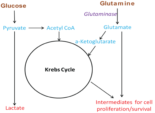
Metabolic differences between normal and cancer cells were observed for the first time about ninety years ago by Otto Warburg. In his 1924 and 1925 papers ‘Ueber den stoffwechel der tumoren’ (About the metabolism of tumors) and ‘Ueber den stoffwechsel der carcinomzelle’ (About the metabolism of the cancer cell) Warburg was the first to show that when compared to normal tissue/cells, tumors exhibit increased rate of glucose uptake and lactic acid production even when ample oxygen was available [1,2]. Thirty-one years after the report of this phenomenon, called aerobic glycolysis or the Warburg effect, in 1955, Harry Eagle in his paper ‘Nutrition needs of mammalian culture cells’ described another interesting observation. HeLa cancer cells have high glutamine requirements in vitro for optimal growth when compared to normal mouse L fibroblasts [3]. Since then, research on the role of glutamine in cancer cell proliferation has revealed that high glutamine utilization and dependence, a property termed as ‘glutamine addiction’, is a metabolic hallmark of many tumor cells in vitro [4,5]. The Warburg effect has been linked, to the dysregulation of a number of pathways known for their involvement in cancer and multiple theories have been advanced for explaining its biological benefit to cancer cells [6,7]. Nevertheless, despite considerable progress in understanding this phenomenon, success in exploiting it for cancer therapy has thus far been limited to tumor imaging (18FDG-PET) [8]. Glutamine addiction, like the Warburg effect, has also been linked to dysregulation of pathways involved in cancer. The biological rationale proposed for it lies on the fact that, as glucose is quickly uptaken and converted to lactate, tumor cells increase their use/uptake of glutamine to produce a-ketoglutarate, to anapleroticaly fuel their Krebs cycle, and to also produce intermediates for the synthesis of lipids, nucleosides and other biomolecules needed for their proliferation and survival (Figure 1) [4,5]. As many cancer cells appear addicted to glutamine, taking advantage of this addiction has been considered as a very attractive strategy for cancer treatment and has drawn much attention, particularly over the last 10 years.

Figure 1: Simplified diagram of glutamine use in Kreb cycle anaplerosis and other intermediate production

Inhibition of KGA/GAC: A Strategy for Exploiting Tumors’ Glutamine Addiction

Glutaminase is an amidohydrolase that converts glutamine to glutamate in the first step of the glutamine processing in mitochondria. The human genome encodes two main glutaminase isoforms, the kidney isoform (KGA/GLS1) and the liver isoform (LGA/GLS2). LGA is encoded by the GLS2 gene in chromosome 12, and it is highly expressed in liver and to a much lower degree in brain and pancreas [9]. KGA is encoded by the GLS gene, in chromosome 2, and has much wider tissue distribution than LGA [9]. Both enzymes are catalytically active as tetramers but have very different kinetic behavior in terms of activation by inorganic phosphate, Km for glutamine, and/or inhibition by glutamate [10]. In the 1969 paper ‘The proportionality of glutaminase content to growth rate and morphology of rat neoplasms’, Knox et al. [11] demonstrated that kidney glutaminase activity is proportional to the growth rate of tumors in rats. Since that paper, cumulative evidence suggests that KGA, and in particular the kidney glutaminase isoform C (GAC) splice variant that has a 71 residue shorter c-terminus, could be an important target for therapy [12-14]. TCGA (The cancer genome atlas) data suggest that there is high GLS gene expression in a number of cancers. Upregulation of KGA, and particularly GAC, has been seen in a number of human tumor cell lines and correlates with increased proliferative rates and in certain instances with tumor progression [5,13,15-19]. The observed KGA/GAC upregulation has also direct links to transcription factors and enzymes/pathways with concrete involvement in cancer such as Myc, STAT-1, c-Jun, Erb2 and the RAF-RAS-MEK-ERK signaling pathways [20-25]. Furthermore, inhibition of KGA expression has been shown to inhibit tumor cell growth [26]. In this context, impairing the ability of tumors to process glutamine by selectively inhibiting KGA/GAC with small molecules has been viewed as an appealing and likely viable strategy for anti-cancer therapy.

KGA/GAC Inhibitors

A review of the literature suggests that DON (Figure 2) and L-2- amino-4-oxo-5-chloropentanoic acid, are the earliest compounds cited as KGA inhibitors [27]. Both compounds act as glutamine mimics and inactivate the enzyme by covalently binding in the active site. Neither is KGA selective however, and both interact with several other targets in addition to glutaminase [28-30]. Among the two, DON also made it to the clinic but its development was halted due to excessive toxicity, apparently the result of its broad enzyme inhibition spectrum [31,32]. A screen of the 1280 member Library of Pharmacologically Active Compounds (LOPAC1280) identified ebselen, apomorphine and chelerytrine as agents that could inhibit KGA/GAC (Figure 3).Unfortunately the utility of these agents in studies/therapy is limited because they interact with other targets and their KGA/GAC vs LGA selectivity is very narrow [33]. In a different screen, thiourea THDP-17 (Figure 3) was identified as an un-competitive KGA inhibitor [34]. KGA/GAC vs LGA selectivity data have not been reported for this agent. Compound 968 (Figure 3) identified as KGA/ GAC inhibitor after protein pull down experiments where lysates from Cdc42-(F28L)-expressing NIH 3T3 cells were incubated with streptavidin beads labeled with the biotinylated N,N-dimethyl-bromophehyl moiety of 968, a key moiety for its activity [35,36]. Studies suggest that 968 acts allosterically and preferentially binds to GAC monomers [37]. The KGA/GAC vs LGA selectivity of this compound has not been reported. However, there are data suggesting that 968 may also bind LGA [38]. As a probe, compound 968 has been featured in a number of biological studies. It has been shown to afford growth inhibition in glutamine-addicted cancer cell lines as well as growth inhibition in vivo in a P493B lymphoma mouse model [35,36,39-43]. The first truly selective KGA/GAC inhibitor reported was BPTES/SNX-1770 (Figure 4). This compound, along with a small list of close derivatives, first appeared in patent application US 2002/0115698 A1. Initial kinetic and biophysical studies showed that the compound is a highly selective allosteric KGA inhibitor that upon binding leads to formation of inactive KGA tetramers [44]. Crystallographic work, using KGA and GAC, confirmed the BPTES allosteric binding mode and showed that the compound binds at the interface between two interacting KGA/GAC dimers, in a 2:4 stoichiometry, and stabilizes an important flexible loop between residues Glu320-Pro327 near the active site (Figure 4) [24,45,46]. In the binding region of BPTES, KGA/GAC and LGA differ in only 2 amino acids. Specifically, Phe318 and Phe322 in KGA/GAC are substituted in LGA by Tyr251 and Ser255 respectively. These differences and particularly the Phe322/Ser255 difference are key for the BPTES binding to KGA/GAC. Studies with KGA/GAC mutants revealed that a Phe318/Tyr Phe322/Ser double mutant or a KGA Phe322/Ser single mutant were unable to bind BPTES but they retained catalytic activity, while a Phe318/Tyr single mutant was catalytically active and also bound BPTES [24,45]. BPTES has been shown to effect cell growth inhibition in glutamine addicted cancer cell lines in vitro and tumor growth inhibition in test animals [19,22,39,47-52]. Despite of its demonstrated activity BPTES did not advance to the clinic because of poor drug-like properties [52]. Preclinical formulation work that aimed to overcome its poor properties was recently published and demonstrated that BPTES encapsulation in poly (lactic-coglycolic acid)-poly (ethylene glycol) (PLGA-PEG) nanoparticles improved compound delivery and tumor uptake and afforded improved tumor growth inhibition than un-encapsulated BPTES in a JH090 pancreatic tumor model [52]. Medicinal chemistry efforts aiming to expand the SAR (structure activity relationships) around BPTES and also produce BPTES derivatives or analogs/mimics with improved potency and drug-like properties have been reported by our laboratory and others and selected compounds from these efforts are shown in (Figure 5) [53-56]. BPTES derivatives and BPTES analogs/mimics have also been claimed in recently published patent applications [57-65]. Among the broader class of BPTES-like compounds, the most advanced is CB-839 (Figure 6) [57,66]. This compound is a more potent KGA/GAC inhibitor than BPTES. It binds in a similar fashion, was shown to have activity against various cancer cell lines and to effect tumor growth inhibition in the CTG-0052, JIMT-1, Caki-1 and other tumor models [18,56,66-69]. CB-839 is currently in Phase I/II clinical trials in combination with other agents (NCT02071927, NCT02071888, NCT02861300, NCT02771626, NCT02071862, NCT03047993 and NCT03057600). As a single agent CB-839 appears to exhibit an acceptable safety profile despite the relatively high, 600 mg twice a day with food, dose regimen required for maintenance of sustained therapeutic plasma levels and for the alleviation of its high absorption variability [70,71]. Early clinical reports suggest that the CB-839/azacytine combination for acute myeloid leukemia (AML), the CB-839 combination with everolimus for renal cell carcinoma (RCC), the CB-839/pomalidomide and dexamethasone combination for multiple myeloma (MM) and the CB-839/paclitaxel combination for triple negative breast cancer, warrant further study [72-75].
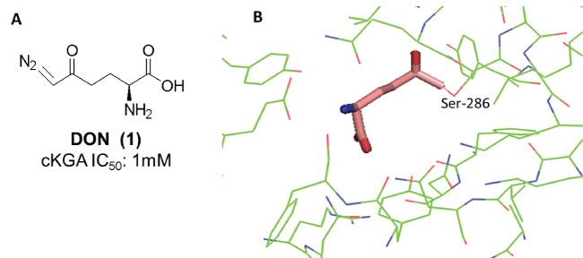
Figure 2: A: Structure of DON and its activity against the catalytic domain of KGA (cKGA) [30]. B: cKGA-DON co-crystal (PDB: 3VOY) showing covalent attachment to Ser 286 in the active site
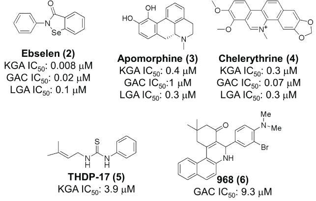
Figure 3: Structures of ebselen, apomorphine, chelerythrine , THDP-17 and 968 and their reported activities [33,34 and 36 respectively].
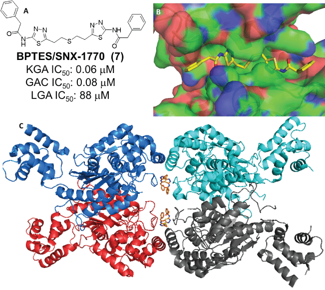
Figure 4: A: Structure of BPTES and its glutaminase selectivity profile [45]. B & C: Crystal structure of BPTES in complex with GAC (PDB: 3UO9)
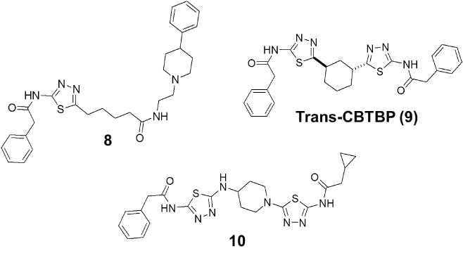
Figure 5: Selected examples from reported medicinal chemistry efforts aimed to improve upon BPTES
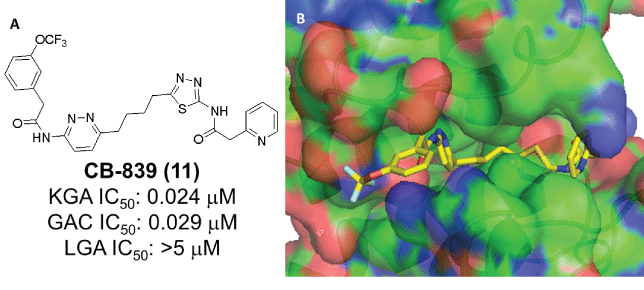
Figure 6: A: Structure of CB-839 and its glutaminase selectivity profile [52,53]. B: Crystal structure of CB-839 in complex with GAC (PDB: 5HL1)

Discussion

The ability of selective KGA/GAC inhibitors to potently inhibit the growth of glutamine addicted cancer cells and afford tumor growth inhibition in vivo is well documented. The preclinical in vivo experience with these compounds, however, suggests that, as single agents, they can only achieve tumor growth inhibition and not tumor regression or stasis, as one would expect given the attributed importance of glutamine in Krebs cycle anaplerosis, the synthesis of nucleosides and other intermediates important for cell survival and proliferation [35,44,53,62,66,69,76,77]. The inability of KGA/GAC inhibitors to afford tumor stasis or regression as single agents may be accounted for by the fact that tumors are heterogeneous, they can use alternative fuels, such as acetate and lipids, and recent findings which show that the metabolic behavior of tumor cells in vitro and in vivo can be vastly different [78- 83]. Taking all of this into consideration it seems likely that, in cancer therapy, KGA/GAC inhibitors will be most useful in combination with other agents rather than as a stand-alone treatment. Preclinical exploration of combination regimens of KGA/GAC inhibitors with established chemotherapeutic agents and/or inhibitors of complementary pathways suggests that synergy is indeed possible [41,43,62,66,69,84-92]. For instance, the available preclinical in vivo data with CB-839 show that combinations with everolimus, paclitaxel, pomalidomide, 5-FU, 5-AZA, and anti-PD-L1/anti-PD-1 antibodies have enhanced efficacy and can produce tumor stasis/regression [66,69,85,90,92,93]. This offers hope that such combinations may translate to the clinic. Early clinical reports in that front appear encouraging [72-75]. However, it is too early to proclaim success. Ongoing and future clinical trials are expected to shed more light on the utility of these regimens and the utility of KGA/GAC inhibitors in general for cancer treatment.


References

  1. Warburg O (1924) Ueber den stoffwechel der tumoren. Biochem Zeitschrift 152: 319-344.
  2. Warburg O, Posener K, Negelein E (1925) Ueber den stoffwechesel der carcinomcezelle. Klinische Wochenschrift 4: 534-536. [Ref.]
  3. Eagle H (1955) Nutrition needs of mammalian cells in tissue culture. Science 122: 501-514. [Ref.]
  4. Wise DR, Thompson CB (2010) Glutamine addiction: a new therapeutic target in cancer. Trends Biochem Sci 35: 427-433. [Ref.]
  5. Altman BJ, Stine ZE, Dang CV (2016) From Krebs to clinic: glutamine metabolism to cancer therapy. Nat Rev Cancer 16: 619-634. [Ref.]
  6. Kroemer G, Pouyssegur J (2008) Tumor cell metabolism: cancer’s Achilles’ heel. Cancer Cell 13: 472-482. [Ref.]
  7. Liberti MV, Locasale JW (2016) The Warburg Effect: How Does it Benefit Cancer Cells? Trends Biochem Sci 41: 211-218. [Ref.]
  8. Mankoff DA, Eary JF, Eary JM, Eary M, Rajendran JG, et al. (2007) Tumor-specific positron emission tomography imaging in patients: [18F] fluorodeoxyglucose and beyond. Clin Cancer Res 13: 3460-3469. [Ref.]
  9. Aledo JC, Gómez-Fabre PM, Olalla L, Márquez J (2000) Identification of two human glutaminase loci and tissue-specific expression of the two related genes. Mamm Genome 11: 1107-1110. [Ref.]
  10. Curthoys NP, Watford M (1995) Regulation of glutaminase activity and glutamine metabolism. Annu Rev Nutr 15: 133-159. [Ref.]
  11. Knox WE, Horowitz ML, Friedell GH (1969) The proportionality of glutaminase content to growth rate and morphology of rat neoplasms. Cancer Res 29: 669-680. [Ref.]
  12. Elgadi KM, Meguid RA, Qian M, Souba WW, Abcouwer SF (1999) Cloning and analysis of unique human glutaminase isoforms generated by tissue-specific alternative splicing. Physiol Genomics 1: 51-62. [Ref.]
  13. Perez-Gomez C, Campos-Sandoval JA, Alonso FJ, Segura JA, Manzanares E, et al. (2005) Co-expression of glutaminase K and L isoenzymes in human tumour cells. Biochem J 386: 535-542. [Ref.]
  14. Cassago A, Ferreira AP, Ferreira IM, Fornezari C, Gomes ER, et al. (2012) Mitochondrial localization and structure-based phosphate activation mechanism of Glutaminase C with implications for cancer metabolism. Proc Natl Acad Sci USA 109: 1092-1097. [Ref.]
  15. Szeliga M, Obara-Michlewska M (2009) Glutamine in neoplastic cells: focus on the expression and roles of glutaminases. Neurochem Int 55: 71-75. [Ref.]
  16. Huang F, Zhang Q, Ma H, Lv Q, Zhang T (2014) Expression of glutaminase is upregulated in colorectal cancer and of clinical significance. Int J Clin Exp Pathol 7: 1093-1100. [Ref.]
  17. Pan T, Gao L, Wu G, Shen G, Xie S, et al. (2015) Elevated expression of glutaminase confers glucose utilization via glutaminolysis in prostate cancer. Biochem Biophys Res Commun 456: 452-458. [Ref.]
  18. Matre P, Velez J, Jacamo R, Qi Y, Su X, et al. (2016) Inhibiting glutaminase in acute myeloid leukemia: metabolic dependency of selected AML subtypes. Oncotarget 7: 79722-79735. [Ref.]
  19. Xiang Y, Stine ZE, Xia J, Lu Y, O’Connor RS, et al. (2015) Targeted inhibition of tumor-specific glutaminase diminishes cell-autonomous tumorigenesis. J Clin Invest 125: 2293-2306. [Ref.]
  20. Gao P, Tchernyshyov I, Chang TC, Lee YS, Kita K, et al. (2009) c-Myc suppression of miR-23a/b enhances mitochondrial glutaminase expression and glutamine metabolism. Nature 458: 762-765. [Ref.]
  21. Zhao L, Huang Y, Tian C, Taylor L, Curthoys N, et al. (2012) Interferon-α regulates glutaminase 1 promoter through STAT1 phosphorylation: relevance to HIV-1 associated neurocognitive disorders. PLoS One 7: e32995. [Ref.]
  22. Qie S, Chu C, Li W, Wang C, Sang N (2014) ErbB2 activation upregulates glutaminase 1 expression which promotes breast cancer cell proliferation. J Cell Biochem 115: 498-509. [Ref.]
  23. Lukey MJ, Greene KS, Erickson JW, Wilson KF, Cerione RA, et al. (2016) The oncogenic transcription factor c-Jun regulates glutaminase expression and sensitizes cells to glutaminase-targeted therapy. Nat Commun 7: 11321. [Ref.]
  24. Thangavelu K, Pan CQ, Karlberg T, Ganapathy B, Uttamchandani M, et al. (2012) Structural basis for the allosteric inhibitory mechanism of human kidney-type glutaminase (KGA) and its regulation by Raf-MekErk signaling in cancer cell metabolism. Proc Natl Acad Sci USA 109: 7705-7710. [Ref.]
  25. Guo Y, Deng YJ, Li XQ, Ning Y, Lin XP, et al. (2016) Glutaminolysis Was Induced by TGF-β1 through PP2Ac Regulated Raf-MEK-ERK Signaling in Endothelial Cells. PLoS One 11: e0162658. [Ref.]
  26. Lobo C, Ruiz-Bellido MA, Aledo JC, Márquez J, Núñez De Castro I, et al. (2000) Inhibition of glutaminase expression by antisense mRNA decreases growth and tumourigenicity of tumour cells. Biochem J 348: 257-261. [Ref.]
  27. Pinkus LM, Windmueller HG (1977) Phosphate-dependent glutaminase of small intestine: localization and role in intestinal glutamine metabolism. Arch Biochem Biophys 182: 506-517. [Ref.]
  28. Shapiro RA, Clark VM, Curthoys NP (1978) Covalent interaction of L-2-amino-4-oxo-5-chloropentanoic acid with rat renal phosphatedependent glutaminase. Evidence for a specific glutamate binding site and of subunit heterogeneity. J Biol Chem 253: 7086-7090. [Ref.]
  29. Shapiro RA, Clark VM, Curthoys NP (1979) Inactivation of rat renal phosphate-dependent glutaminase with 6-diazo-5-oxo-L-norleucine. Evidence for interaction at the glutamine binding site. J Biol Chem 254: 2835-2838. [Ref.]
  30. Thangavelu K, Chong QY, Low BC, Sivaraman J (2014) Structural basis for the active site inhibition mechanism of human kidney-type glutaminase (KGA). Sci Rep 4: 3827. [Ref.]
  31. Rahman A, Smith FP, Luc PT, Woolley PV (1985) Phase I study and clinical pharmacology of 6-diazo-5-oxo-L-norleucine (DON). Invest New Drugs 3: 369-374. [Ref.]
  32. Earhart RH, Amato DJ, Chang AY, Borden EC, Shiraki M, et al. (1990) Phase II trial of 6-diazo-5-oxo-L-norleucine versus aclacinomycin-A in advanced sarcomas and mesotheliomas. Invest New Drugs 8: 113-119. [Ref.]
  33. Thomas AG, Rojas C, Tanega C, Shen M, Simeonov A, et al. (2013) Kinetic characterization of ebselen, chelerythrine and apomorphine as glutaminase inhibitors. Biochem Biophys Res Commun 438: 243-248. [Ref.]
  34. Diaz-Herrero MM, Campo JAD, Carbonero-Aguilar P, Vega-Pérez JM, Iglesias-Guerra F, et al. (2014) THDP17 decreases ammonia production through glutaminase inhibition. A new drug for hepatic encephalopathy therapy. PLoS One 9: e109787. [Ref.]
  35. Wang JB, Erickson JW, Fuji R, Ramachandran S, Gao P, et al. (2010) Targeting mitochondrial glutaminase activity inhibits oncogenic transformation. Cancer Cell 18: 207-219. [Ref.]
  36. Katt WP, Ramachandran S, Erickson JW, Cerione RA (2012) Dibenzophenanthridines as inhibitors of glutaminase C and cancer cell proliferation. Mol Cancer Ther 11: 1269-1278. [Ref.]
  37. Stalnecker CA, Ulrich SM, Li Y, Ramachandran S, McBrayer MK, et al. (2015) Mechanism by which a recently discovered allosteric inhibitor blocks glutamine metabolism in transformed cells. Proc Natl Acad Sci USA 112: 394-399. [Ref.]
  38. Gao M, Monian P, Quadri N, Ramasamy R, Jiang X (2015) Glutaminolysis and Transferrin Regulate Ferroptosis. Mol Cell 59: 298-308. [Ref.]
  39. Son J, Lyssiotis CA, Ying H, Wang X, Hua S, et al. (2013) Glutamine supports pancreatic cancer growth through a KRAS-regulated metabolic pathway. Nature 496: 101-105. [Ref.]
  40. Simpson NE, Tryndyak VP, Pogribna M, Beland FA, Pogribny IP (2012) Modifying metabolically sensitive histone marks by inhibiting glutamine metabolism affects gene expression and alters cancer cell phenotype. Epigenetics 7: 1413-1420. [Ref.]
  41. Jacque N, Ronchetti AM, Larrue C, Meunier G, Birsen R, et al. (2015) Targeting glutaminolysis has antileukemic activity in acute myeloid leukemia and synergizes with BCL-2 inhibition. Blood 126: 1346-1356. [Ref.]
  42. Kahlert UD, Cheng M, Koch K, Marchionni L, Fan X, et al. (2016) Alterations in cellular metabolome after pharmacological inhibition of Notch in glioblastoma cells. Int J Cancer 138: 1246-1255. [Ref.]
  43. Yuan L, Sheng X, Clark LH, Zhang L, Guo H, et al. (2016) Glutaminase inhibitor compound 968 inhibits cell proliferation and sensitizes paclitaxel in ovarian cancer. Am J Transl Res 8: 4265-4277. [Ref.]
  44. Robinson MM, McBryant SJ, Tsukamoto T, Rojas C, Ferraris DV, et al. (2007) Novel mechanism of inhibition of rat kidney-type glutaminase by bis-2-(5-phenylacetamido-1,2,4-thiadiazol-2-yl)ethyl sulfide (BPTES). Biochem J 406: 407-414. [Ref.]
  45. DeLaBarre B, Gross S, Fang C, Gao Y, Jha A, et al. (2011) Fulllength human glutaminase in complex with an allosteric inhibitor. Biochemistry 50: 10764-10770. [Ref.]
  46. Li Y, Erickson JW, Stalnecker CA, Katt WP, Huang Q, et al. (2016) Mechanistic Basis of Glutaminase Activation: A Key Enzyme that Promotes Glutamine Metabolism in Cancer Cells. J Biol Chem 291: 20900-20910. [Ref.]
  47. Le A, Lane AN, Hamaker M, Bose S, Gouw A, et al. (2012) Glucoseindependent glutamine metabolism via TCA cycling for proliferation and survival in B cells. Cell Metab 15: 110-121. [Ref.]
  48. van den Heuvel AP, Jing J, Wooster RF, Bachman KE (2012) Analysis of glutamine dependency in non-small cell lung cancer: GLS1 splice variant GAC is essential for cancer cell growth. Cancer Biol Ther 13: 1185-1194. [Ref.]
  49. Seltzer MJ, Bennett BD, Joshi AD, Gao P, Thomas AG, et al. (2010) Inhibition of glutaminase preferentially slows growth of glioma cells with mutant IDH1. Cancer Res 70: 8981-8987. [Ref.]
  50. Emadi A, Jun SA, Tsukamoto T, Fathi AT, Minden MD, et al. (2014) Inhibition of glutaminase selectively suppresses the growth of primary acute myeloid leukemia cells with IDH mutations. Exp Hematol 42: 247-251. [Ref.]
  51. Sappington DR, Siegel ER, Hiatt G, Desai A, Penney RB, et al. (2016) Glutamine drives glutathione synthesis and contributes to radiation sensitivity of A549 and H460 lung cancer cell lines. Biochim Biophys Acta 1860: 836-843. [Ref.]
  52. Elgogary A, Xu Q, Poore B, Alt J, Zimmermann SC, et al. (2016) Combination therapy with BPTES nanoparticles and metformin targets the metabolic heterogeneity of pancreatic cancer. Proc Natl Acad Sci U S A 113: E5328-5336. [Ref.]
  53. Shukla K, Ferraris DV, Thomas AG, Stathis M, Duvall B, et al. (2012) Design, synthesis, and pharmacological evaluation of bis-2- (5-phenylacetamido-1,2,4-thiadiazol-2-yl) ethyl sulfide 3 (BPTES) analogs as glutaminase inhibitors. J Med Chem 55: 10551-10563. [Ref.]
  54. Zimmermann SC, Wolf EF, Luu A, Thomas AG, Stathis M, et al. (2016) Allosteric Glutaminase Inhibitors Based on a 1,4-Di (5-amino-1,3,4- thiadiazol-2-yl) butane Scaffold. ACS Med Chem Lett 7: 520-524. [Ref.]
  55. McDermott LA, Iyer P, Vernetti L, Rimer S, Sun J, et al. (2016) Design and evaluation of novel glutaminase inhibitors. Bioorg Med Chem 24: 1819-1839. [Ref.]
  56. Ramachandran S, Pan CQ, Zimmermann SC, Duvall B, Tsukamoto T, et al. (2016) Structural basis for exploring the allosteric inhibition of human kidney type glutaminase. Oncotarget 7: 57943-57954. [Ref.]
  57. Li J, Chen L, Goyal B, Laidig G, Stanton T, et al. (2013) Heterocyclic inhibitors of glutaminase. [Ref.]
  58. Lemieux RM, Popovici-Muller J, Salituro FG, Saunders JO, Travins J, et al. (2014) Compounds and methods of use. [Ref.]
  59. Lemieux RM, Popovici-Muller J, Salituro FG, Saunders JO, Travins J, et al. (2014) Compounds and their methods of use. [Ref.]
  60. Bhavar PK, Vakkalanka SK, Viswanadha S, Swaroop MG, Babu G (2015) Novel glutaminase inhibitors. [Ref.]
  61. Bhavar PK, Vakkalanka SKVS, Viswanadha S, Swaroop MG, Babu G (2015) Novel glutaminase inhibitors. [Ref.]
  62. Finlay MRV, Ekwuru CT, Charles MD, Raubo PA, Winter JJG, et al. (2015) 1, 3, 4-thiadiazole compounds and their use in treating cancer. [Ref.]
  63. Di Francesco ME, Jones P, Heffernan T, Hamilton M, Kang Z, et al. (2016) GLS1 inhibitors for treating disease. [Ref.]
  64. Di Francesco ME, Heffernan PJT, Soth MP, Le K, carroll CL, et al. (2016) GLS1 inhibitors for treating disease. [Ref.]
  65. Di Francesco, Heffernan PJT, Zhijun K, Soth MP, Burke JP, et al. (2016) GLS1 inhibitors for treating disease. [Ref.]
  66. Gross MI, Demo SD, Dennison JB, Chen L, Chernov-Rogan T, et al. (2014) Antitumor activity of the glutaminase inhibitor CB-839 in triplenegative breast cancer. Mol Cancer Ther 13: 890-901. [Ref.]
  67. Parlati F, Demo SD, Gross MI, Janes JR, Lewis ER, et al. (2014) CB- 839, a novel potent and selective glutaminase inhibitor, has broad antiproliferative activity in cell lines derived from both solid tumors and hematological malignancies. Cancer Res 74: 1416. [Ref.]
  68. Huang Q, Katt WP, McDermott LA, Cerione RA (2016) Crystal structure of glutaminase C in complex with inhibitor CB-839. PDB: 5HL1. [Ref.]
  69. Parlati F, Gross M, Janes J, Lewis E, MacKinnon A, et al. (2014) Glutaminase Inhibitor CB-839 Synergizes with Pomalidomide in Preclinical Multiple Myeloma Models. Blood 124: 4720. [Ref.]
  70. Harding JJ, Telli ML, Munster PN, Le MH, Molineaux C, et al. (2015) Safety and tolerability of increasing doses of CB-839, a first-in-class, orally administered small molecule inhibitor of glutaminase, in solid tumors. J Clin Oncol. Abst. 2512. [Ref.]
  71. Konopleva MY, Flinn IW, Wang ES, DiNardo CD, Bennet MK, et al. (2015) Phase 1 study: safety and tolerability of increasing doses of CB- 839, an orally-administered small molecule inhibitor of glutaminase, in acute leukemia. EHA Abst. E947. [Ref.]
  72. Meric-Bernstram F, Tannir NM, Mier JW, DeMichele A, Telli ML, et al. (2016) Phase 1 study of CB-839, a small molecule inhibitor of glutaminase (GLS), alone and in combination with everolimus (E) in patients (pts) with renal cell cancer (RCC). J Clin Oncol 34: 4568. [Ref.]
  73. Vogl DT, Younes A, Stewart K, Orford KW, Bennett M, et al. (2015) Phase 1 Study of CB-839, a First-in-Class, Glutaminase Inhibitor in Patients with Multiple Myeloma and Lymphoma. Blood 126: 3059. [Ref.]
  74. Wang ES, Frankfurt O, Orford KW, Bennett M, Flinn IW, et al. (2015) Phase 1 Study of CB-839, a First-in-Class, Orally Administered Small Molecule Inhibitor of Glutaminase in Patients with Relapsed/ Refractory Leukemia. Blood 126: 2566. [Ref.]
  75. DeMichele A, Harding JJ, Telli ML, Munster PN, McKay R, et al. (2016) Phase 1 study of CB-839, a small molecule inhibitor of glutaminase (GLS) in combination with paclitaxel (Pac) in patients (pts) with triple negative breast cancer (TNBC). J Clin Oncol 34: 1011. [Ref.]
  76. Hernandez-Davies JE, Tran TQ, Reid MA, Rosales KR, Lowman HX et al. (2015) Vemurafenib resistance reprograms melanoma cells towards glutamine dependence. J Transl Med 13: 210. [Ref.]
  77. Tanaka K, Sasayama T, Irino Y, Takata K, Nagashima H, et al. (2015) Compensatory glutamine metabolism promotes glioblastoma resistance to mTOR inhibitor treatment. J Clin Invest 125: 1591-1602. [Ref.]
  78. Hensley CT, Faubert B, Yuan Q, Lev-Cohain N, Jin E, et al. (2016) Metabolic Heterogeneity in Human Lung Tumors. Cell 164: 681-694. [Ref.]
  79. Comerford, SA, Huang Z, Du X, Wang Y, Cai L, et al. (2014) Acetate dependence of tumors. Cell 159: 1591-1602. [Ref.]
  80. Kamphorst JJ, Cross JR, Fan J, de Stanchina E, Mathew R, et al. (2013) Hypoxic and Ras-transformed cells support growth by scavenging unsaturated fatty acids from lysophospholipids. Proc Natl Acad Sci U S A 110: 8882-8887. [Ref.]
  81. Sellers K, Fox MP, Bousamra M, Slone SP, Higashi RM, et al. (2015) Pyruvate carboxylase is critical for non-small-cell lung cancer proliferation. J Clin Invest 125: 687-698. [Ref.]
  82. Davidson SM, Papagiannakopoulos T, Olenchock BA, Heyman JE, Keibler MA, et al. (2016) Environment Impacts the Metabolic Dependencies of Ras-Driven Non-Small Cell Lung Cancer. Cell Metab 23: 517-528. [Ref.]
  83. Krall AS, HR Christofk (2015) Rethinking glutamine addiction. Nat Cell Biol 17: 1515-1517. [Ref.]
  84. Rodriguez M, Zhang W, Bennett M, Emberley E, Gross M, et al. (2015) CB-839, a selective glutaminase inhibitor, synergizes with signal transduction pathway inhibitors to enhance anti-tumor activity. Cancer Res 75: 4711. [Ref.]
  85. Wang ZJ, Zhao Y (2015) Targeting glutamine metabolism in colorectal cancers with PIK3CA mutations. Mol Cancer Ther 14: C115. [Ref.]
  86. Zhan H, Ciano K, Dong K, Zucker S (2015) Targeting glutamine metabolism in myeloproliferative neoplasms. Blood Cells Mol Dis 55: 241-247. [Ref.]
  87. Xie C, Jin J, Bao X, Zhan WH, Han TY, et al. (2016) Inhibition of mitochondrial glutaminase activity reverses acquired erlotinib resistance in non-small cell lung cancer. Oncotarget 7: 610-621. [Ref.]
  88. Wang D, Meng G, Zheng M1, Zhang Y1, Chen A, et al. (2016) The Glutaminase-1 Inhibitor 968 Enhances Dihydroartemisinin-Mediated Antitumor Efficacy in Hepatocellular Carcinoma Cells. PLoS One 11: e0166423. [Ref.]
  89. Li J, Csibi A, Yang S, Hoffman GR, Li C, et al. (2015) Synthetic lethality of combined glutaminase and Hsp90 inhibition in mTORC1-driven tumor cells. Proc Natl Acad Sci U S A 112: E21-29. [Ref.]
  90. Gross M, Jason Chen, Judd Englert, Julie Janes, Robert Leone, et al. (2016) Glutaminase inhibition with CB-839 enhances anti-tumor activity of PD-1 and PD-L1 antibodies by overcoming a metabolic checkpoint blocking T cell activation. Cancer Res 76: 2329. [Ref.]
  91. Fendt SM, Bell EL, Keibler MA, Davidson SM, Gregory J, et al. (2013) Metformin decreases glucose oxidation and increases the dependency of prostate cancer cells on reductive glutamine metabolism. Cancer Res 73: 4429-4438. [Ref.]
  92. Cai T, Lorenzi PL, Rakheja D, Pontikos MA, Lodi A, et al. (2016) Gls inhibitor CB-839 modulates cellular metabolism in AML and potently suppresses AML cell growth when combined with 5-azacitidine. Blood 128: 4064. [Ref.]
  93. Emberly E, Bennett M, Chen J, Gross M, Huang T, et al. (2016) CB- 839, a selective glutaminase inhibitor, has anti-tumor activity in renal cell carcinoma and synergizes with everolimus. Euro J cancer 69: S124. [Ref.]

Download Provisional PDF Here


Article Information

Article Type: Mini Review

Citation: McDermott L (2017) Kidney-Type Glutaminase Inhibitors for Treating Cancer. An Overview. J Drug Res Dev 3(2): doi http://dx.doi.org/10.16966/2470-1009.133

Copyright: © 2017 McDermott L, This is an open-access article distributed under the terms of the Creative Commons Attribution License, which permits unrestricted use, distribution, and reproduction in any medium, provided the original author and source are credited.

Publication history: 

  • Received date: 07 Feb 2017

  • Accepted date: 07 Apr 2017

  • Published date: 13 Apr 2017