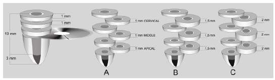
Figure 1: Schematic view of preparation of specimen preparation for push-out test

Kerem Kılıç1 Burak Sağsen2 Tuğrul Aslan2 Hasan Akbaba1 Sema Şahin2*
1Department of Prosthodontics, Faculty of Dentistry, Erciyes University, Kayseri, Turkey*Corresponding author: Sema Şahin, Department of Endodontics, Faculty of Dentistry, Erciyes University, Kayseri, Turkey, Tel: +90 352 4374937-29130; Fax: +90 352 438 06 57; E-mail: ssahin.067@gmail.com
Objectives: The aim of this in vitro study was to investigate the effects of two luting agents and various specimen thicknesses on the bond strength of fibre posts to root canal dentine.
Methods: Sixty central incisors were sectioned below the cement enamel junction. Following standardized post-space preparations, the roots were divided into two luting agent groups, and further divided into three subgroups of 10 specimens each for the push-out test of the specimen thicknesses. Three specimens with different thicknesses were taken from the cervical, middle, and apical parts of each root. Push-out test was performed. Statistical analysis was performed with three-way ANOVA followed by independent t tests (α = 0.05).
Results: Luting agent (P<0.001), specimen thicknesses (P<0.001), and the root regions (P<0.001) significantly affected bond strength values. Panavia F provided significantly higher mean bond strengths than Duo-Link (P<0.05). The highest values were obtained for 1-mm test specimens for all groups (P<0.05). The apical region of the root dentine had significantly higher bond strengths compared to the middle and cervical regions for Duo-Link (P<0.05). The region of the root did not affect the bond strengths to root dentine for Panavia F (P>0.05).
Conclusion: Bond strengthswere affected significantly by luting agents and specimen thicknesses.
Fibre post; Luting agent; Push-out; Specimen thickness
Fibre posts are commonly used to restore endodontically treated teeth when their remaining coronal tissue can no longer provide adequate support and retention for the restoration [1,2]. Important characteristics of fibre posts include a modulus of elasticity similar to that of dentine and the ability to bond to dentine using an adhesive technique [3,4]. As a result, it is suggested that loads applied to the restoration are assumed to be uniformly distributed to the supporting dentine, and the stress concentrations at the restorative interface are avoided [5,6]. As indicated by the results of both in vitro studies and the clinical trials, the fibre posts are shown to reduce the incidence of unrestorable root fractures [2-4,7].
Adhesion can be defined as a flexion force between the molecules at the interfaces of different materials. The complex organic structure and the dynamic formation and the biological activity of dentine, prevents a reliable and durable bonding [8]. Appropriate adhesive agents and luting procedures for bonding fibre posts to root dentin is challenging [9]. Fibre posts can be cemented using conventional dual cure resin-based cements in combination with total etching or self-etching adhesives, or using the recently formulated self-adhesive cements that allow simultaneous bonding between the intra radicular dentin and the post [4]. To assess the bond strength between fibre posts and root canal dentine, conventional shear and tensile tests, micro shear, micro tensile, and pull-out and pushout tests have been used [9-16]. It is suggested that the bond strength can be obtained better by the push-out test compared with the conventional shear test because the fracture occurs parallel to the dentin bonding interface in the push-out test, which makes it a true shear test [10]. Additionally, as premature failures occur during specimen preparation and because of the large data distribution observed in micro tensile tests, the push-out test has been considered more reliable [9,12,17,18]. The shear stress achieved with the push-out tests is comparable to the stress, under clinical conditions, at the interface between the dentine and the luting cement, as well as between the post and the luting cement [19].
Besides of the wide range of studies mentioning the benefits of the push-out test, the literature lacks agreement on the thickness of the push-out test specimen that should be used. In various publications, segments differing from 1 mm to 2.5 mm have been used [2,4,9,20- 22].The influence of specimen thickness on the test results has not been specifically evaluated.
In addition, the findings of different studies on the regional differences in bond strength among the three root sections are not consistent [3,18,23]. In a previous study, the most satisfactory bond strengths were reported to be present in the apical and cervical third of the root canals [23]. These findings are in contrast with the results of other studies, which show that the most reliable bond strength was usually obtained in the cervical third because of the easier access available in this portion of the root canals [24,25].
The present study evaluates whether the bond strength to dentine varies among different sections along the root, and how the bond strengths change with two different dual cure resin-based cements. Additionally, this in vitro study compares the bond strengths of the three different push-out test specimen thicknesses. The null hypotheses of the present study were as follows:
(1) Bond strength to root dentine does not vary among luting agents.
(2) Bond strength to root dentine does not vary with the thicknesses of the push-out test specimens.
(3) Bond strength to root dentine in the apical regions of the roots is higher than the other regions of the roots.
A total of 60 maxillary human central incisors of similar sizes with fully developed apices, predominantly extracted for periodontal reasons, were selected randomly. Teeth showing resorption, cracks, or caries were excluded from the study. Before the collection of teeth, the approval of local ethics committee of Erciyes University, Kayseri, Turkey was acquired (Decision number: 2015-302). External debris was removed with an ultrasonic scaler and the teeth were stored in 0.1% thymol. The teeth were cut perpendicular to the long axis at the cement enamel junction with a slow-speed diamond saw (Isomet; Buehler, Lake Bluff, IL, USA). To standardize the root canal length, the roots were cut to a uniform length of 14 mm. Working length was established 1mm short of the apex. All the roots were instrumented using Pro Taper rotary instruments (Dentsply Maillefer Ballaigues, Switzerland). The master apical file was ProTaper F3. Five mL of 2.5% NaOCl was used for irrigation between each instrument. The final rinse, with 5 mL, 17% EDTA, was used for one minute, followed by copious amounts of distilled water. Each canal was dried with paper points and obturated with cold lateral condensation using gutta-percha (Dentsply-Maillefer, Petropolis, RJ, Brazil) and resin sealer (AH Plus; Dentsply De Trey GmbH, Konstanz, Germany). The sealer was introduced into the root canals using lentulo spiral filler. After the root fillings were completed, cervical root canal openings were then filled with a provisional restorative material (CavitTM-G; 3M ESPE AG, Seefeld, Germany), and the teeth were stored at 37°C and 100% humidity for seven days to allow the sealers to set.
1.2-mm glass fibre posts (D.T. Light Post #3, Bisco Inc., Schaumburg, IL, USA) were used in this study. The post spaces were all prepared with special preparation drills (D.T #3, Bisco Inc., Schaumburg, IL, USA) to a depth of 10 mm from the cemento-enamel junction, leaving a minimum apical seal of 4 mm of gutta-percha in the canal space after the post preparation. The gutta-percha was removed with a warm plugger (Sybron Dental Specialties, Romulus, MI, USA) up to the appropriate depth. Post spaces were irrigated with saline solution and dried with paper points. One practitioner prepared all the roots in a standardized procedure. The prepared roots were randomly divided into two luting agent groups (Panavia F 2.0 and Duo-Link) with 30 specimens each for the luting procedures. The posts were cleaned thoroughly with alcohol, rinsed with distilled water, and airdried. Before the cementation procedures, no additional pretreatment procedures were applied to the post surfaces.
The ED Primer II was mixed at a ratio of 1:1, applied to the dentin walls of the post spaces using a micro brush (Micro brush X, Micro brush Corp, Grafton, WI, USA) for 30 seconds, and gently air-dried; the excess was then removed with paper points. A dual-polymerizing resin luting agent (Panavia F 2.0; Kuraray, Japan) was mixed for 20 seconds and placed in the post spaces using lentulo spiral filler. Posts were coated with cement and slowly seated by finger pressure. Excess cement was removed. Cement was polymerized for 40 seconds with a light-polymerizing unit (550 mW/cm2 , Hilux 550; Hilux, Ankara, Turkey) by placing the light tip perpendicularly through the post for 40 seconds. Before each luting procedure, the light output was measured with a light meter placed on the curing unit to ensure accurate light intensity.
Twothinlayers of ONE-STEP PLUS resin-based adhesive were applied to the dentine walls of the post holes using disposable micro-brushes. Excess bonding agent was removed carefully with paper points and the canal walls gently air-dried for 10s, before the adhesive was light cured with a light-polymerizing unit (550 mW/cm2 , Hilux 550; Hilux, Ankara, Turkey) from the canal opening for 20 seconds. The fibre posts were also coated with a thinlayer of the light cured adhesive. Duo-Link (Bisco, Inc.,Schaumburg, IL, USA) was then injected into the post spaces and the fibre posts were inserted. Excess cement was immediately removed. The tip of the light unit was placed directly on the coronalend of each fibre post and the cement light cured for 40s.
After the cementation procedures, the roots were stored at 37°C and 100% humidity for seven days before testing. Then, two groups were created according to the cementation procedures; the groups were further divided into three subgroups of 10 specimens each for the push-out test of specimen thicknesses. Perpendicular to the posts, three sections of 1-mm, 1.5-mm, and 2-mm thick were cut from the cervical, middle, and apical parts of the roots using a slow-speed diamond saw (Isomet; Buehler, Lake Bluff, IL, USA) under water cooling (Figure 1). The cervical specimens were cut from the first mm of the cervical side of the root, the middle specimens were cut from the fifth mm of the cervical side of the root, and the apical specimens were cut from the eighth mm of the cervical side of the root. The thickness of each section was carefully monitored with a digital caliper. Subsequently, all specimens were observed with a stereomicroscope (DV 4; Zeiss, Jena, Germany) to detect any artifacts caused by the slicing process; no artifacts were observed.
Figure 1: Schematic view of preparation of specimen preparation for push-out test
Each specimen was subjected to loading using a Universal Testing Machine (Instron, Canton, MA, USA) that carried a 1-mm-diameter cylindrical plunger for the cervical specimens, a 0.5-mm-diameter plunger for the middle specimens, and a 0.3-mm-diameter plunger for the apical specimens. The plunger only contacted the post during loading. The loading speed was 0.5 mm/minute-1 until the dislodgement of the post occurred (Figure 2). The values at the time of dislodgement were recorded in Newtons for each specimen.
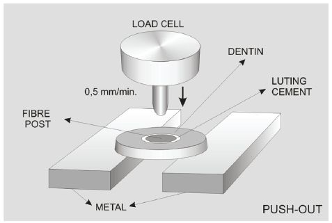
Figure 2: Push-out test method
The force needed to dislodge the fibre posts (in kN) was transformed into tension (in MPa) as similar to the study of Costa et al. [26]. The upper and lower diameters of the specimens were calculated individually, and the following formula was used (Costa et al.): Mpa=F/SL. SL was calculated using the following equation: SL=π (R + r) g; where SL=sealer adhesion area; π=3.14; R=mean radius of the cervical post, in mm; r=mean radius of the apical post, in mm; g=is the thickness of the slice in mm. Apical and cervical aspects of each slice were scanned with a digital scanner. The images were transferred to Photoshop (CS5, Adobe, USA), and the r values of the specimens were measured.
After testing the push-out bond strength, the failure mode of each de bonded specimen was analyzed by two independent operators who were blinded to the luting strategies performed. The operators used a stereomicroscope (DV 4; Zeiss, Jena, Germany) at × 40 magnification and classified the de bonded specimens according to the following criteria: (1) adhesive failure between dentine and the luting agent; (2) adhesive failure between the luting agent and the post; (3) cohesive failure within the luting agent; (4) cohesive failure within the post; and (5) mixed failure. One specimen representative of each failure mode was processed for scanning electron microscopy (SEM) evaluation to obtain SEM images of the failure patterns. The specimens were rinsed in 95% alcohol solution for one minute and air-dried. Each specimen was mounted on a metallic stub, then sputter coated with 200 Å gold-palladium in a Polaron SC7620 mini-sputter coater (Quorum Technologies Ltd., East Sussex, UK) for 5 minutes at a current of 10 mA. SEM examination was performed (Jeol JSM 6360LV, Jeol Ltd., Tokyo, Japan) at an accelerating voltage of 15 kV at a magnification of 2500× and photographed.
The data were statistically analyzed (SPSS/PC 10.0; SPSS Inc, Chicago, IL, USA) using a three-way analysis of variance (ANOVA) (luting agent type, specimen thicknesses, and root segments), making pair-wise comparisons among groups (α=0.05). Independent t-tests were used to detect differences between groups defined by the specific interacting variables.
The mean bond strength values obtained for each root region, specimen thicknesses and luting agents are shown in Table 1. The three-way ANOVA indicated that the bond strength values were significantly affected by the luting agent (F=29,929, P < 0.001), specimen thicknesses (F=228,778, P<0.001), and root regions (F=7,234, P<0.001). The statistical analysis demonstrated a significant interaction between the luting agent and the root region (F=9,589, P<0.001) and between the specimen thicknesses and the root region (F=16,971, P<0.001). The interaction between the luting agents and the specimen thicknesses was not significant (F=2,144, P=.121) (Table 2).
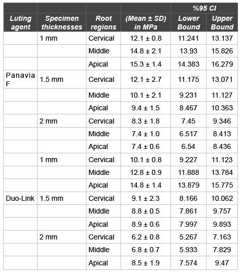
Table 1: The mean bond strength values obtained for the each root region, specimen thicknesses and luting agents
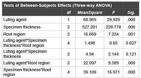
Table 2: Three-way ANOVA statistics
Df: Degree of freedom.
For Panavia F 2.0 and Duo-Link, in the cervical specimens, the mean bond strength values of the 1-mm (12.1 ± 0.8 MPa, 10.1 ± 0.8 MPa, respectively) and 1.5-mm subgroups (12.1 ± 2.7 MPa, 9.1 ± 2.3 MPa, respectively) had significantly higher values compared to the 2-mm subgroup (8.3 ± 1.8 MPa, 6.2 ± 0.8 MPa, respectively) (P<0.05), whereas there were no significant differences between the 1-mm and 1.5-mm subgroups in terms of bond strength values (P=1.000).
The mean bond strength values of the 1-mm subgroups (14.8 ± 2.1 MPa, 15.3 ± 1.4 MPa, respectively) had significantly higher values than 1.5-mm (10.1 ± 2.1 MPa, 9.4 ± 1.5 MPa, respectively) and 2-mm subgroups (7.4 ± 1 MPa, 7.4 ± 0.6, respectively) (P<0.05)in the middle and apical specimens of the Panavia F 2.0 group. The mean bond strength values of the 1.5-mm subgroups were also significantly higher than those of the 2-mm subgroups (P<0.05).
For Duo-Link, in the middle specimens, the mean bond strength value of the 1-mm subgroup (12.8 ± 0.9 MPa) was significantly higher than that of the 1.5-mm (8.8 ± 0.5 MPa) and 2-mm subgroups (6.8 ± 0.7 MPa) (P<0.05). The mean bond strength value of the 1.5-mm subgroup was also significantly higher than that of the 2-mm subgroup (P<0.05).
The mean bond strength value of 1-mm subgroup (14.8 ± 1.4 MPa) was significantly higher than that of the 1.5-mm (8.9 ± 0.6 MPa) and 2-mm subgroups (8.5 ± 1.9 MPa) (P<0.05), while there were no significant differences between 1.5-mm and 2-mm subgroups in terms of bond strength values (P=0.891) in the apical specimens of the Duo-Link group.
When the bond strengths were evaluated in terms of the root regions, for Duo-Link significantly higher bond strength values were measured in the apical (10.7 ± 0.2 MPa) compared with the middle (9.5 ± 0.2 MPa) and cervical (8.5 ± 0.2 MPa) parts (P<0.05), for Panavia F 2.0, the region of the root did not affect the bond strengths to root dentine (P>0.05).
When bond strength to root dentine was compared among luting agents, the statistical analysis revealed that Panavia F 2.0 had significantly higher bond strength value (10.8 ± 0.1 MPa) than did Duo-Link (9.5 ± 0.1 MPa) (P<0.05).
The distribution of the push-out test failure modes is shown in Table 3. For Panavia F 2.0 and Duo-Link, mixed failures at luting agent-root dentin interfaces were the most common (respectively, 71.1%, 74.4%), followed by adhesive failures between dentine and luting agent (respectively 17.7%, 18.8%), cohesive failures in the luting agent (respectively, 6.6%, 3.3%), and adhesive failures between fibre posts and luting agents (respectively, 4.4%, 3.3%). Cohesive failures in the post alone were not observed.
Table 3: Analysis of failure modes for all experimental groups A/D-L: Adhesive failure at luting agent-root dentine interface; A/P-L: Adhesive failure at luting agent-post interface; C/L: Cohesive failure of luting agent; C/P: Cohesive failure of post; M/D-L: Mix failure at luting agent-root dentine interface.
The SEM images (2500x) of representative fractured specimens of the different failure modes are presented in Figures 3 and 4.
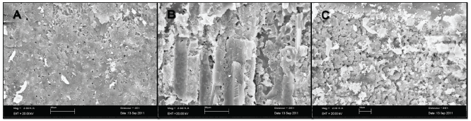
Figure 3: SEM photographs of failed specimens in the Panavia F luting agent group. A: The specimen with adhesive failure (A/D-L) between the root dentine and the Panavia F luting agent at the 1-mm thickness middle post space region. B: The specimen with mixed failure (M/D-L) between root dentine and the Panavia F luting agent at the 1-mm thickness middle post space region. C: The specimen with cohesive failure (C/L) inside the Panavia F luting agent at the 1.5-mm thickness cervical post space region. Magnification: 2500x.
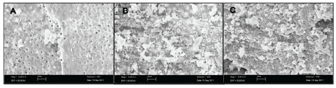
Figure 4: SEM photographs of failed specimens in Duo-Link luting agent group. A: The specimen with adhesive failure (A/D-L) between root dentine and the Duo-Link luting agent at the 1-mm thickness cervical post space region. B: The specimen with mixed failure (M/D-L) failure between root dentine and the Duo-Link luting agent at the 1-mm thickness middle post space region. C: The specimen with cohesive failure (C/L) inside the DuoLink luting agent at the 1.5-mm thickness apical post space region. Magnification: 2500x.
Bond strength provides valuable pre clinical information on the adhesive pro perties of materials, and the thin-slice push-out test method has been considered a reliable technique to measure the bond strength of fibre posts to root dentine [25].
There is a trend to reduce the number of adhesive steps. Adhesives currently in use are the etch-and-rinse with primer and adhesive applied simultaneously (two-step) and the self-etch systems, which contain a self-etch primer and an adhesive in one solution (all-in-one) [4]. Therefore, in thepresent study, theeffects of twoall-in-oneadhesives on thebondstrengths of fiber postswereevaluated.
The adhesive (ED Primer II) used with self-etching adhesive Panavia F 2.0 contains the phosphate-based functional monomer 10-methacryloyloxydecyl dihydrogen phosphate (MDP). This molecule forms chemical interactions with the hydroxyapatite remaining around the collagen within the hybrid layer, and because of the low solubility of the MDP-calcium salt in water, this bond is expected to be stable [27]. In the present study, groups bonded with Panavia F 2.0 had significantly higher push-out bond strengths than groups bonded with Duo-Link (P<0.05) that can be attributed to the MDP content of Panavia F 2.0. Similarly, Baldissara et al. [28] evaluated fatigue resistance of different resin cements of a glass-in filtrated alumina ceramic to human dentin and related the higher fatigue resistance of Panavia F toits MDP content. According to the Pereira et al. [29] study, different dual-curedres in cements show different extent of polymerization and molecula rmobility. Pereira et al. [29] used different commercially available dual-curedres in cements that present differences in their composition. Duo-Link contains lower amount of inorganic filler (61.9 wt%) compared with Panavia F 2.0 (76.9 wt%). The polymerization of dimethacrylates produces densely cross linked network and, during the polymerization period, part of the methacrylate groups involved in the formation of the cross-linked matrix remains unreacted, specially in thecase of high-molecular-weight monomers [29]. Explanations about this effect could be associated with the amount of fillers and the influence of the decrease of mobility of polymer radicals and as a consequence a decrease on the reactivity occurs. Recently, an inverse relation on polymerization shrink ageand filler loading on somedual-curedresincements has been reported [30]. The lower push-out bond strength values for Duo-Link can also be attributed to these properties. Therefore, the results obtained in the present study do not support the first research null hypothesis that bond strength to root dentine does not vary among luting agents.
In the present study, to evaluate the effects of the specimen thicknesses and different luting agents on the bond strength, push-out tests were used. There are many studies in the literature that evaluated fibre posts bond strength with the push-out test method [1,5,6,9,14,17,22,24].However, the literature lacks agreement on the thickness of the push-out test specimen that should be used. Kremeier et al. [2] evaluated the influence of the post type and luting material on the bond strength to dentin with the push-out test method. The authors used 2-mm specimen thickness for each root region [2]. Bitter et al. [5] sliced each root into six discs of 1-mm thickness representing the cervical, middle, and apical part of the root canal in their study. Silva et al. [1] obtained three 1.5-mm-thick slices perroot and identified them as cervical, medium and apical thirds. Muniz et al. [22] sectioned the specimens through their long axis into three dental slices approximately 2.5 mm each, representing the cervical, middle, and apical thirds of the root preparation. Cecchin et al. [31] sectioned bonded specimens into 1-mm-thick slabs and performed a push-out test in their study. As mentioned above, various publications have used 1.0-mm-to- 2.5-mm-thick segments [1,2,5,22,31]. In the present study, all of the 1-mm subgroups showed higher bond strength values than 1.5-mm and 2-mm subgroups. The MPa unit is used instead of the Newton unit as a bond strength value in the push-out studies. The MPa bond strength value is calculated by dividing the Newton unit to the surface area of the specimen. Therefore, the push-out bond strength values can be lower in the thicker specimens. In the present study, higher bond strength values for the 1 mm specimes could be attributed to this fact. Furthermore, Erdemir et al. [32] used thin slices (1-mm), to overcome this problem.
Regarding the fracture analysis and SEM photographs, it should be emphasized that predominant types of failure in the cervical specimens of the Panavia F 2.0 and Duo-Link groups were adhesive between the luting agent and the root dentine and the mixed type, implying the weak bond between the luting agent and the root dentine. Cervical regions of central incisor teeth are wider than circular-shaped fibre posts, and thus, resin cement thicknesses are more than the other parts, causing predominantly adhesive failures in the cervical regions. The bond strength in the middle and apical specimens of the Panavia F 2.0 and Duo-Link groups appeared to be superior because the predominant type of failure was the mixed type. This suggests that the bond strength between the luting agent and the root canal dentine in the middle and apical specimens was less affected than the cervical specimens (Table 2).
In the present study, for Duo-Link, the bond strengths were significantly higher in the apical specimens than in the cervical and middle specimens. A previous study demonstrated significantly higher bond strength values for the apical region inside the root canal similar to the present study [23]. However, no significant differences were found in regional bond strengths for Panavia F 2.0 in the present study similar to Bouillaguet et al. [17] research. Therefore, it can be concluded that bond strengths to root canal dentine seem to be related more to the area of solid dentine than to the density of dentinal tubules [5,33]. However, Ferrari et al. [24] showed that the dentine surface area available for bonding increased by 202% after etching in the cervical third, 156% in the middle third, and 113% in the apical third of the root dentine and the thickness of the hybrid layer depended on thedensity of tubules [24]. According to the results of the present study, the third null hypothesis, that bond strength to root dentine in the apical region of the root is higher than in the other regions of the root, is rejected. Although the use of size-matched drills supplied by post manufacturers permit a good fitting of posts to the canal walls, especially in the apical and middle root regions, some canals have large post space diameters in the cervical region. If the post does not fit well, especially at the cervical level, the luting agent layer would be excessively thick, and bubbles are likely to form in it, thus predisposing it to de bonding [1]. Also, when the luting agent layer is excessively thick, especially in the cervical portion of the root, polymerization shrinkage stress is higher [17]. These shrinkage stresses contribute to what has been defined as the C factor, the ratio of bonded to unbonded surface areas in root canal dentine [34]. It has been shown that a C factor in post spaces may be as high as 200 [35]. Especially with light-polymerizing materials, the high polymerization stress may cause the resin composites to detach from the dentine walls, creating interfacial gaps [20]. To attain proper polymerization in such situations, maximizing strength and adhesion of the cement, a chemically activated component of a dual-catalyst system should be effective [35].
Within the limitations of the present study, all of the null hypotheses that bond strengths to root canal dentine did not vary with the type of the resin cements, specimen thicknesses, and the regions of the root were not accepted. Although both of the luting agents are commonly used clinically, Panavia F 2.0 could be recommended as a cementation agent to prevent bond failure between fibre post and root canal dentine than self-etching luting agent Duo-Link according to the results of the present study.
In the present investigation, the push-out technique to evaluate the strength of the adhesion of fibre posts to dentine was applied more effective with 1-mm specimen thickness than 1.5-mm and 2-mm specimen thicknesses. The apical region of the root dentine was characterized by significantly higher bond strengths for Duo-Link whereas the region of the root did not affect the bond strengths to root dentine for Panavia.
The authors deny any conflicts of interest.
Download Provisional PDF Here
Article Type: Research Article
Citation: Kılıç K, Sağsen B, Aslan T, Akbaba H, Şahin S (2016) Effects of Two Luting Agents and Various Specimen Thicknesses on ThepushOut Bond Strength of Fibre Posts to Root Canal Dentine. Int J Dent Oral Health 2(3): doi http://dx.doi. org/10.16966/2378-7090.190
Copyright: © Kılıç K, et al. This is an open-access article distributed under the terms of the Creative Commons Attribution License, which permits unrestricted use, distribution, and reproduction in any medium, provided the original author and source are credited.
Publication history:
All Sci Forschen Journals are Open Access