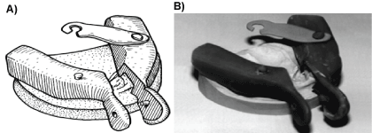
Figure 1: Design of sectional tray by Baker et al. [18]

Impression Techniques for Microstomia Patients: No longer Cumbersome - A Review
Shantanu Mulay1 Sarandha DL2 Smitha Annie Jacob3 Zina Raja2
1Senior Lecturer, Department of Prosthodontics, SMBT Dental College and Hospital, Sangamner, Ahmedabad, Maharashtra, India*Corresponding author: Smitha Annie Jacob, Reader, Department of Prosthodontics, Dr. Syamala Reddy Dental College Hospital & Research Centre, Marathahalli, Bangalore, India. E-mail: dr.smitha.jacob@gmail.com
The reduction in maximal mouth opening (microstomia) is a condition that hinders conventional prosthetic treatment of edentulous patients. In particular, the fabrication of removable prosthesis is further complicated by tongue rigidity and the constant adjustment required to accommodate the changing periphery. To rehabilitate a patient with microstomia successfully, modifications in impression making are a necessity. This article reviews the various sectional tray designs and impression procedures for microstomia patients
Impression; Microstomia; Prosthesis; Sectional Trays; Limited mouth opening
Microstomia may be the sequelae of oro-facial burns, carcinoma, cleft lip, trauma, scleroderma, Plummer Vinson’s syndrome, genetic disorders, head and neck radiation, reflex spasm, surgically treated head and neck tumours, microinvasion of muscles of mastication, connective tissue disease, fibrosis of masticatory muscles, reconstructive lip surgeries [1- 3]. The contracture of the tissue that surrounds the oral cavity may affect the patient’s ability to obtain optimal dental care and maintain good oral hygiene [4,5].
Surgical enlargement must be considered carefully because, if the rehabilitation of the surgical operation is not sufficient, a scar may result [5-7]. Without surgical operation, it is very difficult to perform prosthetic treatment for patients with microstomia, especially when the mouth circumference length is less than 160 mm2 [8]. Because the smallest diameter of a fully retentive denture may be larger than the greatest diameter of the mouth opening, sectional or collapsible dentures are indicated [9-11].
In cases, where microstomia is not manageable by surgeries or by use of dynamic opening devices [12,13], a modification of the standard impression procedures is often necessary as the overall bulk and height of impression trays makes the recording of impressions, exceptionally difficult, if not impossible, because the path of insertion and removal of impressions are compromised by lack of clearance [10,14,15].
A search in the National Library of Medicine’s Pub Med database, Google search and Science Direct was performed to include all case reports and reviews on “Impression Techniques for microstomia patients”. A total of 14 articles were included for discussion in the review out of the 19 articles.
Luebke RJ [16] used a calliper to measure the arch width of the ridge and an impression tray was selected accordingly. Plaster was poured into the selected tray to form a matrix. The impression tray was then removed and re-inserted on this matrix to ensure that the tray fits on the matrix, same way each time. The plastic tray was then cut into two sections with a disc, with the handle in the larger section. Three building blocks (LEGO Systems, Enfield, Conn) were selected to re- approximate sectional trays as one unit, which were fixed to the tray by the help of auto-polymerising unit. The authors of this study used plastic building blocks, one 32 mm × 8 mm × 3 mm and two 16 mm × 8 mm × 3 mm to re-approximate sectional tray as one unit. The two shorter blocks were fit together under the longer block.
Luebke RJ [16] also recommended that the tray should be cut into two unequal sections rather than two equal halves, so that the labial frenum is recorded accurately in the impression. The two halves of the tray are joined using LEGO building blocks [16], fins in the handle [17], metal pins [18], burs. The use of an orthodontic expansion screw, two guide pins and a screw without the screw axis to, serve as a guide or key and keyway to fabricate a split custom tray, handles, made of 1 mm generic stainless steel wire to permit placement and stabilization of each tray.
As recommended by Baker et al. [17], one half of the tray can be fabricated using light cured tray material on the duplicate cast (Figures 1a and 1b). Handle is aligned in all directions in one plane at the midline to match exactly with the second half. A knob placed inside the handle would fit into the opening on the handle in the second half tray. Horizontal locking component was made separately, the hinge of which was lubricated, and a custom impression tray can be fabricated using autopolymerizing acrylic resin.
Mirfazaelian A [3] described the use of an orthodontic expansion screw (2 guide pins and a screw) without the screw axis, to serve as a guide or key and keyway to fabricate a split custom tray (Figures 2a-2d). Preparation of a butt joint along the two pieces of a maxillary tray can enhance its stabilization during border correction and impression making. The expansion screw is placed vertically in the handle of the custom tray to accommodate the limited space. The length of the guide pins in the expansion screw can be reduced for easier insertion and removal if necessary. Maxillary and mandibular trays require different locations for the key and keyway. For the maxillary tray, the holes must be located in the overlay piece and the guide pins are placed in the other half for better access. For the mandibular tray, guide pins are placed in the overlay piece and holes are located in the other half.
Figure 1: Design of sectional tray by Baker et al. [18]
Dhanasomboon and Kiatsiriroj [2]used dental stone which was poured directly to one half of the first sectional impression made (Figures 3a and 3b). The second sectional impression, which was made, was later approximated with the first impression section and stone poured into the second half. Stock tray was modified by trimming the flange lengths.
Ohkubo et al. [10] used complete arch stainless steel trays sectioned into right and left halves that are reconnected by use of tray handle (Figures 4a and 4b). The tray handle components consisted of 4 dowel-plug-holes and screw-lock. The two halves of the sectional trays were positioned together, after screwing the dowel plug into the head screw and later screwed into the dowel hole of the handles.
Cura et al. [19] described another technique to fabricate maxillary and mandibular sectional trays. The acrylic tray design was different in that for each tray, a total of four metal pins were prepared (Figures 5a and 5b). In case of mandibular trays, two of these pins were placed near the distal end and the other two near the anterior region. For the maxillary tray, two were placed on the residual ridges and two near the midline. An acrylic resin block with a 4 × 5 mm cross section that slid tightly on the pins was prepared. The trays were cut into two pieces with a steel disc and then joined with the acrylic resin block which slid onto the parallel pins.
Yenisey et al. [20] used sectional maxillary and mandibular trays for the completely edentulous patient with microstomia, caused by scleroderma. Preliminary impressions were made with a putty silicon impression material. For each special tray, a total of four metal snaps were attached. Two female parts were attached on the canine regions and two were attached on molar regions. Another block carrying male parts of the snaps was constructed. In the mandibular tray, only one block was adequate for the stability of the right and left parts. In the maxillary tray, two blocks were constructed and they were joined together to provide stability.
Geckili et al. [21] described a modified impression procedure with a two- piece impression tray for a patient with limited oral opening as a result of the resection of a precancerous lesion on the maxillary lip. The custom tray was fabricated using auto- polymerizing resin. The tray was sectioned mesio-distally along the middle of the palate. A tungsten carbide bur was divided into three pieces of equal length. One of the bur sections was placed on top of the right alveolar crest region and another on top of the left alveolar crest region of the tray. The third bur section was placed in the palatal mid-section. All of the bur sections were fixed to the tray using auto-polymerizing resin. The acrylic resin tray and the three bur sections were lubricated with petroleum jelly and a second tray, using the same acrylic resin was fabricated to slide on the bur sections of the first tray. This two-piece custom- made tray allowed for a functional impression despite the difficulties associated with microstomia.
Jivanescu et al. [22] presented various clinical and technical steps involved in the fabrication of a flexible complete denture in case of a female patient with scleroderma induced microstomia. A standard tray was used. It was sectioned in the middle with a disc and two alginate sectional impressions were made. Afterwards, the palatine vault was marked with putty silicon. The preliminary impressions were aligned after which the first individual tray was made. The impression served to create a more adaptable individual tray of smaller sizes.
Prasad et al. [23] proposed a technique where in cross- pin slots were placed on the handle of each tray using Pindex machine, cross pins along with sleeves were placed. Tissue stops were placed on the intaglio surface to ensure stability, uniform pressure and impression material. Primary impressions were made with impression compound. The excess material crossing the midline, was trimmed to flush with the margin. The trays were reassembled extra-orally and the primary cast was poured. Maxillary and mandibular impressions were made with putty silicone impression material, placed intra-orally and moulded with finger pressure on which the dental stone casts were made.
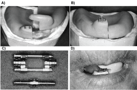
Figure 2: Split custom tray design by Mirfazaelian A [3]
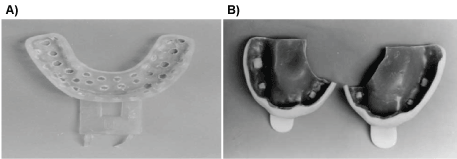
Figure 3: Sectional tray design by Dhanasomboon and Kiatsiriroj [2]
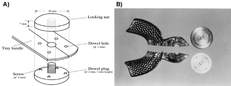
Figure 4: Sectional tray design by Ohkubo et al. [15]
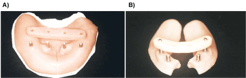
Figure 5: Maxillary and mandibular sectional trays by Cura et al. [21]
Fernanades AS, Mascarenhas K, Aras M [24] described the fabrication of a custom sectional tray with interlocking type handle for definitive impression procedures. The handle functions as an anterior lock and as two parts – the male and female unit. The male unit has an external and internal flange with an interconnecting isthmus, which is 2 mm short of the inferior portion of the internal flange. The internal flange is short of the inferior portion of the external flange by 2 mm. A horizontal plate connects the superior ends of both the flanges. The female unit has an internal recess of which the terminal ends approximate the width of isthmus. The terminal end appears as a slot in the medial wall. This slot is short of the inferior portion by 4 mm. Wax pattern of the above mentioned design are fabricated, invested, de-waxed and acrylised in auto- polymerizing acrylic resin. These patterns can also be cast in base metal alloy. This will save clinical time in fabricating the handle in future as it can be sterilized and re-used. The press button for the maxillary tray functions as a posterior lock and has a male and female part.
Limited mouth opening often complicates and compromises the prosthodontic treatment of patients. The overall bulk and the height of impression trays make the recording of impressions exceptionally difficult if not possible because the paths of insertion and removal of impressions are compromised by lack of clearance. However with careful treatment planning, prudent designing and the use of sectional impression techniques, many of the clinical difficulties encountered can be overcome.
Each of the impression tray designs mentioned in this article differ only in the complexity of the design and the successful orientation of the sectional trays together. Hence, the skill of the prosthodontist plays a pivotal role in the successful rehabilitation of these patients.
Download Provisional PDF Here
Article Type: Review article
Citation: Mulay S, Sarandha DL, Jacob SA, Raja Z (2015) Impression Techniques for Microstomia Patients: No longer Cumbersome - A Review. Int J Dent Oral health 1 (2): http://dx.doi. org/10.16966/2378-7090.109
Copyright:© 2015 Mulay S et al. This is an open-access article distributed under the terms of the Creative Commons Attribution License, which permits unrestricted use, distribution, and reproduction in any medium, provided the original author and source are credited
Publication history:
All Sci Forschen Journals are Open Access