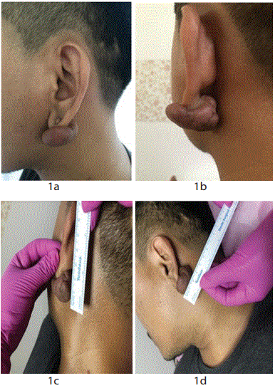
Figure 1a-d: Keloid measuring 2.5cm × 3cm in the left ear lobule with a smooth, shiny, hard, non-mobile, and telangiectasis.
Frien Refla Syarif1* Sri Lestari2 Pristia Widya Monica1 Sanfri Kefrina Syarif1
1Ermedika Clinic, South Tangerang, Banten, Indonesia*Corresponding author: Frien Refla Syarif, Ermedika Clinic, South Tangerang, Banten, Indonesia, E-mail: dr.refla.syarif@gmail.com
Introduction: Keloids are fibroproliferative lesions caused by abnormal wound healing and are characterized by excessive collagen deposits that cannot heal spontaneously. Keloid in the ear is a challenging condition to be treated by doctors, especially the large ones. This condition has a psychosocial impact on the patient and a consideration for appearance is often the main reason for patients seeking to cure keloids, although there is a high possibility of keloid recurrence. Out of various keloid therapy options, one method that can be done is surgical excision therapy with debulking technique.
Case report: A case of keloids in left ear lobule measuring 2.5 cm × 3 cm is reported after the patient does ear piercing with complaints of slight itch and aesthetical disturbance. Keloids enlarge 6 months after piercing. The case is treated by debulking excision surgery using local anesthesia Pehacain® (lidocaine 2% + adrenaline 1:80,000). At the end of the wound closure, 40 mg of triamcinolone acetone is given. It is regiven one week after the stitches are removed and repeatedly every 2 weeks for 4 times. Excision surgical results are good and the patient feels satisfied.
Conclusion: Excision surgery with debulking technique is chosen because of the large size of the lesion and to ensure there is no residual keloid tissue at the site of predilection. Corticosteroid injection is given immediately after excision to decrease fibroblast proliferation, collagen and glycosaminoglycan synthesis and suppress proinflammatory mediators in order to prevent keloid recurrence.
Excision surgery; Debulking technique; Ear keloid
Keloids and hypertrophic scars are fibrodermal proliferation abnormalities due to wound healing and are characterized by excessive collagen deposits [1,2]. These scars are associated with pain, hyperaesthesia and pruritus which can dramatically affect the quality of life of patients, especially in cases of keloids [3]. Doctors define keloids as scars that grow into normal skin tissue around the wound, while hypertrophic scars as scars that do not extend beyond the original wound [4,5]. Pathologists make a histological difference between keloids and hypertrophic scars: keloids determined by the presence of skin nodules coupled with several thick eosinophilic (hyalinising) collagen bundles called keloid collagen whereas hypertrophic scars only have skin nodules.
Keloids are observed to occur in people of all races, but are more often found in dark pigmented people, with an incidence of 6% to 16% in individuals from Africa [6,7]. Keloids appear about 15 times greater in dark-skinned people than fair-skinned people [8]. Keloids have a genetic predisposition and autosomal dominant patterns of inheritance [9,10]. Familial keloids are described as being present in two rare syndromes: Rubinstein-Taybi syndrome (OMIM 180849) [11,12] (big toes and hands, characteristic facies, mental retardation and increased keloid frequency) and Goeminne’s syndrome (OMIM 3134300) [12,13] (keloids, torticollis, renal dysplasia and cryptorchidism).
Keloid formation is partly thought to be due to an exaggerated and prolonged inflammatory phase that may result in sustained release of cytokines and growth factors, which, in turn, stimulate fibroblasts to proliferate and deposit excessive Extracellular Matrix (ECM) [14,15]. Experimental studies have demonstrated that a prolonged inflammatory period involving immune cell infiltrate increases fibroblast activities with greater and more sustained ECM deposition, thereby leading to keloid formation [14]. This finding has been supported by histological findings, which have demonstrated the presence of inflammatory cells in many keloid tissues [14-16]. The inflammatory cells found in high concentration in most keloid specimens are macrophages, neutrophils, mast cells, Langerhans cells, and lymphocytes [14-17].
Systemic factors that cause keloidogenesis include puberty and pregnancy, which is associated with an increased risk of large scar formation [18]. Pregnancy also worsens keloid conditions. This might reflect the vasodilation effect of estrogen, which can encourage the movement of immune factors and cells into the wound or wound tissue, thereby exacerbating local inflammation. Local risk factors that encourage the formation and growth of large scars including delayed wound healing, wound depth and mechanical strength can change skin tension caused by stretching [19]. This is evidenced by the fact that keloids show a high tendency to occur in areas of the body with strong and/or repeated skin tension such as the anterior chest, shoulders, deltoid, jaw and ear. Conversely, keloids rarely occur in areas with weak skin tension, such as the scalp or the anterior tibia [20].
Recent studies have shown that both the severity of inflammation and the type of immune response affect whether or not the scars will form excessively. In scars that are dense with inflammatory cells, fibrogenic factors such as Transforming Growth Factor (TGF)-β1 and β2 are released. Decreased levels of TGF- β3 and Matrix Metalloproteinases (MMP) cause accumulation of Extracellular Matrix (ECM). The growth of the Th-2 response stimulates fibrogenesis and the dominance of the Th-1 further weakens the fibrosis tissue. Keloid tissue then makes the inflammatory period longer. These things can help explain why keloids spread beyond the original wound limit [21].
Although there is a lot of information about the histomorphological structure of keloids in current literature, there are no golden treatment guidelines. There are various treatment methods that can be performed such as surgical excision, intralesional corticosteroid injection, pressure therapy, radiotherapy, laser therapy, cryosurgery, therapy with antitumor or immunosuppressive agents and a combination of these methods. However, the reported success rates vary. This is why most doctors combine existing treatment modalities.
Surgical excision causes a high recurrence rate, between 50-100% [22,23]. Therefore, this treatment is rarely used as a single therapy. Surgical removal of keloids can restore scars to their original state and subsequent post-operative scars can be reduced by additional therapy (intralesional corticosteroid injections, radiotherapy, pressure therapy, immunomodulators) [22]. Several types of steroids can be used in corticosteroid injection; the most commonly used is Triamcinolone acetonide with a concentration of 40mg/mL, given by intralesional injections at monthly intervals of up to 6 months [24]. When used together with surgical therapy, local steroid therapy is started before surgery and continued postoperatively, monthly for at least 3 month [25,26]. Rosen DJ, et al who treated ear keloids by excision method and steroid injection during intra-operative and post-operative reported keloid recurrence rate at 23% [27].
A 21-year-old male patient presents with complaints of keloids in the left ear that have enlarged 6 months after piercing. Keloids sometimes feel itchy and affect the patient's confidence so the patient wants the keloids removed. There is no previous history of treatment with keloids. There is no history of keloids on other body parts. There is no history of family members who have scars widened from the actual wound limit with a smooth surface. Physical examination results of the patient are within normal limits.
Dermatologicus status obtained keloid measuring 2.5 cm × 3 cm in the left ear lobule with a smooth, shiny, hard, mobile, and telangiectasis (Figures 1a-1d). Markers are then made for surgery (Figure 2). Laboratory tests return results as blood clotting factors, blood sugar when HBsAg normal). When patients undergoes surgical excision with debulking technique the following procedures are performed: (1) aseptic and antiseptic actions are carried out with povidone iodine and alcohol 70% (Figure 3); (2) local anesthetic is carried out at the base of the keloid using Pehacain® (lidocaine 2% + adrenaline 1:80,000) with a total injection of 2 ml; (3) incision made from the posterior part of the keloid to open the keloid skin (Figure 4); (4) keloids are excised by debulking technique and the remaining keloid tissue is cleaned from the surrounding area (Figures 5a and 5b); (5) an intra-keloidal debulking revision is carried out by means of the remaining skin of the keloid tissue then sewn over the keloid body using Prolene® size 5.0 (Figure 6); (6) antibiotic ointment is applied and the wound is covered with a bandage.
Figure 1a-d: Keloid measuring 2.5cm × 3cm in the left ear lobule with a smooth, shiny, hard, non-mobile, and telangiectasis.
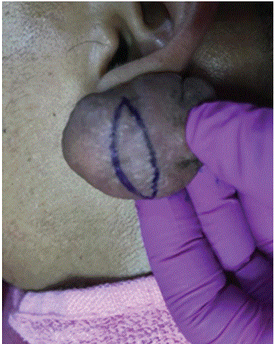
Figure 2: Markers made for surgery.
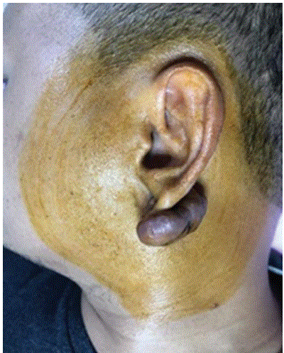
Figure 3: Aseptic and antiseptic actions with povidone iodine and alcohol 70%.
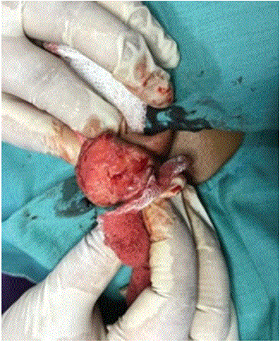
Figure 4: Incision made from posterior part of the keloid to open the keloid skin.
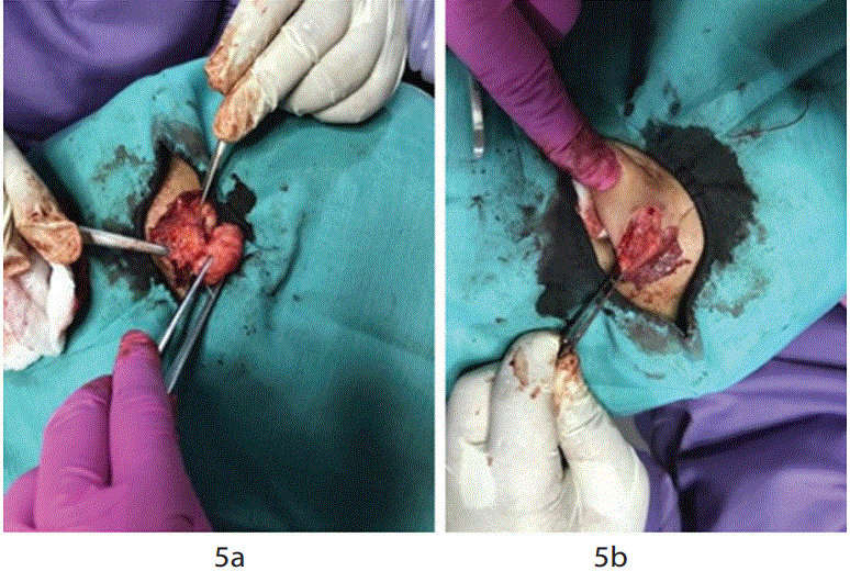
Figure 5a and 5b: Keloids excision by debulking technique and the remaining keloid tissue is cleaned from the surrounding area.
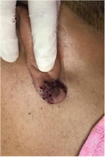
Figure 6: Intra-keloidal debulking revision.
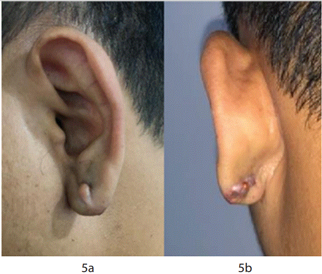
Figure 7a and 7b: The post operative image.
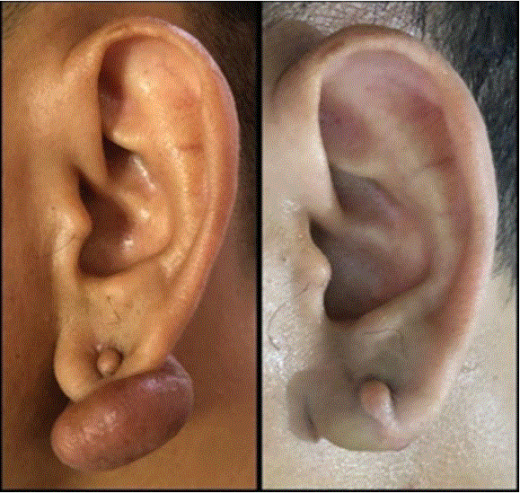
Figure 8: Pre and post operative images.
Patients were given amoxicillin systemic antibiotic + clavulanic acid 625 mg 2X a day for 5 days and mefenamic acid analgesic 500 mg 3x a day and recommended for control 10 days later. The postoperative area is recommended to not be exposed to water until the patient returns to remove the sewing. After 10 days, the wound has dried and the skin is completely fused then suture removal is taken (Figures 7a and 7b). One week after suture removal the patient returns to do the injection of triamcinolone acetonid 40 mg/ml as much as 1ml throughout the scar. Additional injection is redone for 3X at intervals of 2 weeks. Postoperative results are good and the patient is satisfied (Figure 8).
The etiology and pathogenesis of keloid disease are still unknown. Many theories have been put forward including genetic, proliferative, inflammatory, hypoxic, and mechanical stress theories [28-31]. However, none of these theories has been proven. Due to the lack of proper understanding of keloids, there is no standard disease management protocol so that various treatment centers treat this disease in different ways. The recurrence rate is therefore high, with several treatment centers reporting recurrence rates of up to 50% [28,29,32].
Predilection, size, and duration of appearance of keloids in the ear affect the choice of therapy. Excision is performed on large keloids, by debulking technique, but the recurrence rate in the selection of surgical excision therapy reaches 50%-100% [33]. Therefore, excision surgery is not used as a single therapy but is combined with adjunctive therapy such as corticosteroid injection, radiotherapy, pressure therapy, and immunomodulators [34].
Intralesional triamcinolone is the first-line therapy for the treatment of scars because it can inhibit fibroblast growth factor and increase collagen degradation. This resulted in a significant decrease in alpha-1 antitrypsin and alpha-2 macroglobulin levels, which are usually higher in keloid tissues and are natural collagenase inhibitors in human skin [35]. The International Advisory Panel on Scar Management recommends the use of intralesional steroid injections as first-line treatment for the treatment of keloids and hypertrophic scars [36]. The fractional CO2 laser and verapamil are effective and safe treatment modalities for the treatment of keloids, but that keloid treatment takes longer than TAC [35,36].
In this patient the treatment method selected are surgical excision combination therapy with corticosteroid triamcinolone acetoneid injection of 40 mg/ml to prevent the appearance of keloid recurrence. As a way of managing pathological scars, administration of Triamcinolone can suppress Vascular Endothelial Growth Factor (VEGF), inhibit fibroblast proliferation, and induce scar regression, which is perhaps the most important mechanism of action in treatment. Triamcinolone can also inhibit growth factor transformation (TGF)-β1 and induce apoptosis of fibroblasts. Depending on the size and location of the lesion and the patient's age, the dosage given varies from 10 to 40 mg/ mL and is given at intervals of 3 to 6 weeks for several months or until the scar is evenly distributed.
Keloids can have serious cosmetic effects on the ears, especially in young patients. Many treatment strategies have been proposed for the treatment of ear keloids but none are ideal. Before starting treatment, the doctor must provide education and inform the patient about the possible level of recurrence and the limitations of adequate treatment options. The use of various treatment modalities in combination is recommended because it is possible to get better results.
Excision surgery with debulking technique combined with intralesional injection of 40 mg/ml triamcinolone acetone is one of keloid management options, especially those with a large size in the ear. The technique removes the keloid tissue as complete as possible and closes the keloid body with the rest of the tissue so that it can make the ear shape look more normal aesthetically and eliminate the symptoms caused by the keloid.
Download Provisional PDF Here
Aritcle Type: CASE REPORT
Citation: Syarif FR, Lestari S, Monica PW, Syarif SK (2021) Keloid Excision with Debulking Technique on the Lobule Auricle Sinistra. J Clin Cosmet Dermatol 5(3): dx.doi.org/10.16966/2576-2826.165
Copyright: © 2021 Syarif FR, et al. This is an open-access article distributed under the terms of the Creative Commons Attribution License, which permits unrestricted use, distribution, and reproduction in any medium, provided the original author and source are credited.
Publication history:
All Sci Forschen Journals are Open Access