clinical-cosmetic-dermatology-Sci Forschen

Full Text

Research Article
Melasma-Review of Current Treatment Modalities and Efficacy Assessment of a New ResorcinolBased Topical Formulation

  Dinko Kaliterna1*       Kristina Zuzul2      Irena Kovacevic3   

1Polyclinic Poliderma, dermatovenerolog, Vukovar Street (Zagreb), Croatia
2Nada Videnic, Department of Dermatology Karlovac, Croatia
3University of Applied Health Studies, Zagreb, Croatia

*Corresponding author: Dinko Kaliterna, dermatovenerolog, Polyclinic Poliderma, Vukovar Street (Zagreb), 284, 10000, Croatia, Tel: 4828-961; E-mail: poliderma@poliderma.hr


Abstract

Melasma is one of the most common pigmentary disorders characterized by symmetrical pigmented macules on the face. It most commonly affects women of reproductive age with darker complexions, and has a considerable impact on quality of life. The pathogenesis of melasma is complex and involves various risk factors such as genetic predisposition, UV exposure, darker phototype and hormonal influence. Because of its chronic and relapsing course, as well as the fact that despite numerous treatment modalities available, no single treatment option has proven to be efficient for all patients with melasma, the treatment still remains a challenge. This paper provides a review of the current literature on melasma treatment and presents the results of a study assessing a new promising resorcinol-based topical formulation with better efficacy and safety profile than other currently available treatment options for melasma.

Keywords

Melasma; Resorcinol; Pigmentary Disorders; Melasma Treatment


Introduction

Melasma (from the Greek word “melas” meaning black) [1] is an acquired hyperpigmentation of the skin, typically affecting the sunexposed areas of the face, occasionally occurring on the neck, and rarely on the forearms. The term, “chloasma” (from the Greek word “chloazein” meaning green) is used to describe melasma developing during pregnancy (but since the pigmentation is never green, the term, “melasma” is preferred) [2].

It presents clinically as symmetrical hyperpigmented macules on the face (the color varies from light brown to bluish gray depending on the depth of melanin deposition). There are usually three patterns of involvement: centrofacial (involves the cheeks, forehead, upper lip, nose, chin (Figure 1)), malar (involves the cheeks and nose), and mandibular (involves the mandible) [3].

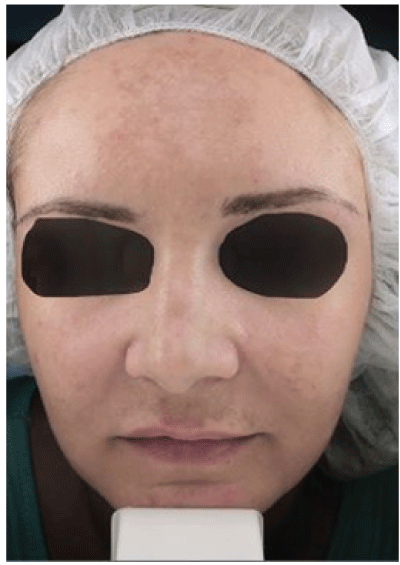
Figure 1: Image showing centrofacial pattern of melasma involvement

It is a common disorder affecting millions of people worldwide [4], with the exact prevalence varying between 1.5% and 33.3%, depending on the population. Melasma, most commonly affects women of reproductive age with darker complexions (Fitzpatrick skin type IV-VI), although it can also be seen in men. Among pregnant women, prevalence can be up to 70% [5]. Melasma of the pregnancy usually improves a year after delivery, but areas of hyper pigmentation may never completely resolve [6]. Because of its facial involvement, melasma has a considerable impact on the quality of life of the affected individuals.

The pathogenesis of melasma is complex and still not well understood. Known risk factors include genetic predisposition, ultraviolet (UV) radiation exposure, darker phototype, hormonal influence (pregnancy, oral contraceptives, increased levels of luteinizing hormone, thyroid disease), and certain medications such as phenytoin [5,7]. It is known that UV radiation induces melanocyte proliferation. Although the number of melanocytes is similar in lesional and perilesional skin, melasma may be caused by biologically more active melanocytes in the affected skin [8].

In melasma lesions, there are increased stem cell factor from fibroblast and tyrosine kinase receptor c-kit [9], as well as the expression of vascular endothelial growth factor (VEGF). Direct role of VEGF in pathogenesis of melasma is supported by the observation of increased number and size of blood vessels in melasma lesions (Figure 2), as well as by the finding that human melanocytes in vitro express VEGF receptors [10,11]. This warrants investigation of new treatment options to target the vascular component of melasma [5].

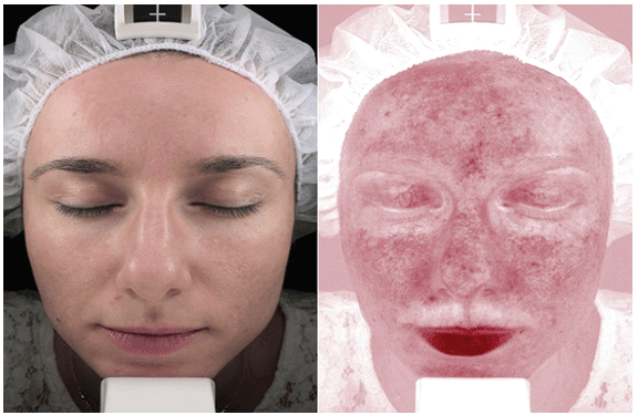
Figure 2: Image using VISIA analysis-in the areas of hyper pigmentations, there is increased number and size of blood vessels

Histopathology of melasma lesion shows an increased melanin deposition in all layers of epidermis, with elastosis and mast cells more pronounced than in normal skin [12]. The most affected biological process is lipid metabolism [8] there is down regulation of various lipid metabolism genes caused by chronic UV exposure.

Melasma is traditionally classified according to the depth of distribution of melanin pigment in three types [5]:

  1. Epidermal (light brown, enhancement of contrast with Wood’s light)-melanin is deposited in basal and suprabasal epidermis. Shows good response to treatment.
  2. Dermal (bluish gray, no enhancement of contrast with Wood’s light)-melanin laden macrophages in superficial and mid-dermis. Shows poor response to treatment.
  3. Mixed (dark brown, partial enhancement of contrast with Wood’s light)-melanin is deposited epidermis and dermis. Partial response to treatment.

Examination with a Wood’s lamp can be used to determine the location of the pigment, but is limited in detecting dermal melanin composition [4]. The severity of melasma may be assessed by using melasma area and severity index (MASI) or Melasma Severity Index (MSI) [13].

Materials and Methods

A study was conducted in a Clinic in Zagreb according to the protocol and in accordance with ethical guidelines. The study included 30 female volunteers of 25 to 45 years of age, diagnosed with melasma. The volunteers were either treatment naïve or had not used any treatment modality for melasma for at least 6 months. The exclusion criteria included any treatment for melasma within past 6 months, hypersensitivity to resorcinol or any other ingredient of the formulation, pregnancy, and any type of facial skin damage, disease, or infection.

The volunteers were given a cream (Name of the cream: My skin by Dr. Kaliterna-White skin) based on resorcinol derivative at a baseline visit, and were instructed to apply a small amount of cream once daily on a clean face at bedtime, during a period of 30 days. They were also instructed to avoid sun exposure and apply sunscreens.

The efficacy of the treatment was done using the VISIA image analysis. The images were taken before the treatment, and again after 30 days.

Results

A total of 30 volunteers were included in a study. No subjects were lost to follow-up, and all of them reported compliance to the protocol.

27 out of 30 subjects reported satisfaction with the results. Of those, 20 were extremely satisfied with results, and 7 were very satisfied. Aside from significant reduction in hyper pigmentation, they reported improved skin quality and texture, as well as reduction in skin pores. 2 out of 30 patients reported moderate satisfaction with results, while 1 patient reported that she is not satisfied.

No severe side effects were reported. Only 2 volunteers reported irritation or burning sensation during a few days after cream application, but spontaneous resolution of symptoms after a few minutes. Most of the clients reported first visible change and improvement between days 7 and 10. (Figures 3A and 3B).

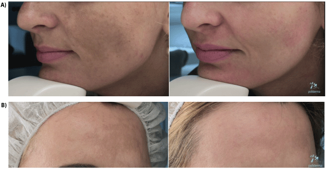
Figure 3: After 30 days before and after only with white skin 1 cream

Discussion

Because of the chronic and relapsing nature of melasma, as well as the fact that no single treatment option has proven to be efficient for all patients with melasma, the treatment may be challenging. Before choosing the treatment modality, analysis of the characteristics of melasma lesion is recommended. This can be performed by VISIA or other similar systems. The area most responsive to treatment is the forehead, while other locations and older lesions seem to be more refractory. Current treatment options include the following:

  1. Prevention of UV radiation (photo protection): No matter the chosen treatment modality, sun protection measures are a necessity (sun avoidance, wide-brimmed hat, using broad-spectrum sunscreens during and after the treatment).
  2. Hydroquinone is a gold standard of treatment of epidermal melasma, although its effects are reversible. It inhibits conversion of DOPA to melanin by inhibiting tyrosinase [4]. It is used as a topical preparation in various concentrations from 2-5% (higher concentration may be more effective, but associated with erythema, skin peeling, irritant contact dermatitis, hypopigmentation of the surrounding skin, development of milia, and exogenous ochronosis) [14]. Its long-term safety has been widely debated.
  3. Azelaic acid is derived from P. Ovale and is a weak reversible competitive inhibitor of tyrosinase. It has anti-proliferative action and is selectively cytotoxic towards hyperactive melanocytes, with minimal effects on normally pigmented skin [1]. It is available in 20% cream or 15% gel formulations. In several studies it was compared to hydroquinone, with similar efficacy but significantly more side effects such as erythema burning, pruritus and scaling [15-17].
  4. Ascorbic acid (vitamin C) is reported treatment for melasma due to its ability to chelate copper ions. It does not work well as monotherapy, it is highly unstable and rapidly oxidised. It may be a good treatment adjunct in patients who cannot tolerate hydroquinone, since it causes less irritation [4].
  5. Kojic acid is produced by A. oryzae and Penicillium spp. and inhibits tyrosinase [4]. It is used in combination with hydroquinone and glycolic acid. Kojic acid can substitute for hydroquinone if a patient is intolerant to hydroquinone [1]. Adverse effects include local irritation and contact dermatitis, as it is a known sensitizer.
  6. Topical retinoids act by stimulation of keratinocyte turnover and decreasing melanosome transfer. Treatment is contraindicated during pregnancy
  • Tretinoin has shown to be efficacious treatment for melasma [18], but the dermal pigment after the therapy remains unchanged. Its use as a monotherapy is unlikely to be as effective as hydroquinone or combination therapy, being limited by side-effects including erythema and irritation [4].
  • Adapalene is a synthetic retinoid that causes less cutaneous irritation than other retinoids, and is more appropriate for long-term use [4].
  1. Topical combination treatments
  • The Kligman-Willis formula (5% hydroquinone, 0.1% tretinoin, 0.1% dexamethasone). This combination minimizes side effects and maximizes effect in shorter period of time than when the ingredients are used individually
  • The triple therapy combination (TCC) contains 4% hydroquinone, 0.05% tretinoin and 0.01% fluocinolone acetonide. It has proven to be fairly efficacious in melasma treatment, but with often present side-effects such as erythema, irritation, burning, and xerosis, as well as a risk of post inflammatory hyper pigmentation (PIH) in patients with darker skin color [4].
  • Other combinations were also evaluated with varying success (hydroxyquinone, hyaluronic acid, glycolic acid, ascorbic acid, kojic acid)-usually show greater efficacy than individual treatments.
  1. Chemical peels are usually recommended to patients unresponsive to skin-lightening treatments, and show mixed results for melasma treatment. There are three types of chemical peels: superficial, medium, and deep (more adverse effects occur with deeper peels). They are mostly used in Caucasians, and are most useful for treating epidermal-type melasma, although recurrences are frequent. In patients with darker skin types, there is a risk of PIH. Various formulations exist [4]: Glycolic acid (GA) (concentrations from 20 to 70%), salicylic acid, trichloroacetic acid (TCA) (concentrations between 10-20%), and other such as pyruvic acid, combination SA and mandelic, phytic acid, Obagi blue and amino fruit acid, etc. Various results and little published evidence supporting their everyday use for melasma treatment.
  2. Laser therapy and intense pulsed light (IPL) numerous studies have demonstrated good efficacy of laser therapy for melasma treatment [19]. Main adverse effects are erythema, burning, dryness, scaling, edema, rebound hyper pigmentations, hypo pigmentations, de pigmentations, physical urticaria, acneiform eruption, petechiae, and herpes simplex reactivation [5]. IPL is a non-coherent broad spectrum light (500-1200 nm) effective for epidermal melasma, although recurrences are common [20]. Also higher fluences are needed, and there is a risk of PIH in darker skin types.
  3. Tranexamic acid is a lysine analog which decreases melanocyte tyrosinase activity [5]. It can be used orally (250 mg 2 × daily), topically, or by intra dermal microinjection. As an oral therapy, it is proven to be effective [21], as a topical preparation, not so effective [22], but intradermal application showed significant decrease in MASI score and minimal side effects [23].
  4. Other [5]: Oligopeptides-new class of tyrosinase inhibitors with good efficacy and safety profile. Silymarin-flavonoid with antioxidant properties, which reduces the harmful effects of UV radiation, and also inhibits melanin production in a dose-dependent manner. Orchid extracts and various botanical extracts-grape seed extract, pycnogenol, aloesin, green tea extracts, coffee berry, soy, and licorice extract.

4-n-Butylresorcinol is a resorcinol derivative that inhibits both tyrosinase and tyrosinase-related protein-1 (TRP-1) [24]. Its hypopigmenting action was first reported in 1995, and many following studies have documented its efficacy [25] and safety in melasma treatment with the 0.1% cream, but there is paucity of clinical studies that used the 0.3% cream. The biochemical assay on inhibition of human tyrosinase activity has revealed the superiority of 4-n-butylresorcinol over other hypopigmenting agents [26]. Many studies show good efficacy and safety in treating melasma [26,27].

Conclusion

Despite numerous treatment modalities available, due to its chronic and relapsing course, treating melasma still remains a challenge. Although hydroquinone and triple combination creams remain the gold standard of treatment, long term safety concerns and possible side effects warrant investigation of new, more efficacious and safer treatment modalities.

The investigated resorcinol derivative-based cream has shown the fastest onset of action (as measured by first reported improvements after 7-10 days) among all currently available topical preparations. Unlike other available topical preparations, this cream can be used throughout the year (including summer), the period of application is not limited (can be used continuously), and there are no pronounced side-effects (such as erythema or desquamation). In majority of cases, its effectiveness is better, and the results seen faster than with fractional laser. Its usage during pregnancy is not recommended, since no safety studies in this population were made.

Aside for its main purpose-the reduction in hyper pigmentation, it has shown a strong rejuvenation effect. Accordingly, other indications for using this cream include premature signs of aging and sun damage, such as excessive redness, large pores, or uneven texture.

Because of its fast onset of action, once-a-day application, and a wide range of indications, this promising product may be a revolution both in treatment of melasma and in skin rejuvenation.


References

  1. Shankar K, Godse K, Aurangabadkar S, Lahiri K, Mysore V, et al. (2014) Evidence-Based Treatment for Melasma: Expert Opinion and a Review. Dermatol Ther 4: 165-186.
  2. Achar A, Rathi SK (2011) Melasma: A Clinico-Epidemiological Study of 312 Cases. Indian J Dermatol 56: 380-382.
  3. Sanchez NP, Pathak MA, Sato S, Fitzpatrick TB, Sanchez JL, et al. (1981) Melasma: a clinical, light microscopic, ultrastructural, and immunofluorescence study. J Am Acad Dermatol 4: 698-710.
  4. Rodrigues M, Pandya AG (2015) Melasma: clinical diagnosis and management options. Australas J Dermatol 56: 151-163.
  5. Sarkar R, Arora P, Garg VK, Sonthalia S, Gokhale N (2014) Melasma update. Indian Dermatol Online J 5: 426-435.
  6. Elling SV, Powell FC (1997) Physiological changes in the skin during pregnancy. Clin Dermatol 15: 35-43.
  7. Lee AY (2015) Recent progress in melasma pathogenesis. Pigment Cell Melanoma Res 28: 648-660.
  8. Kang HY, Suzuki I, Lee DJ, Ha J, Reiniche P, et al. (2011) Transcriptional profiling shows altered expression of wnt pathwayand lipid metabolism-related genes as well as melanogenesis-related genes in melasma. J Invest Dermatol 131: 1692-1700.
  9. Kang HY, Hwang JS, Lee JY, Ahn JH, Kim JY, et al. (2006) The dermal stem cell factor and c-kit are overexpressed in melasma. Br J Dermatol 154: 1094-1099.
  10. Kim EH, Kim YC, Lee ES, Kang HY (2007) The vascular characteristics of melasma. J Dermatol Sci 46: 111-116.
  11. Kim EJ, Park HY, Yaar M, Gilchrest BA (2005) Modulation of vascular endothelial growth factor receptors in melanocytes. Exp Dermatol 14: 625-633.
  12. Hernández-Barrera R, Torres-Alvarez B, Castanedo-Cazares JP, Oros-Ovalle C, Moncada B (2008) Solar elastosis and presence of mast cells as key features in the pathogenesis of melasma. Clin Exp Dermatol 33: 305-308.
  13. Majid I, Haq I, Imran S, Keen A, Aziz K, et al. (2016) Proposing Melasma Severity Index: A New, More Practical, Office-based Scoring System for Assessing the Severity of Melasma. Indian J Dermatol 61: 39-44.
  14. Martins VM, Sousa AR, Portela Nde C, Tigre CA, Gonçalves LM, et al. (2012) Exogenous ochronosis: case report and literature review. An Bras Dermatol 87: 633-636.
  15. Lowe NJ, Rizk D, Grimes P, Billips M, Pincus S (1998) Azelaic acid 20% cream in the treatment of facial hyperpigmentation in darkerskinned patients. Clin Ther 20: 945-959.
  16. Verallo-Rowell VM, Verallo V, Graupe K, Lopez-Villafuerte L, GarciaLopez M (1989) Double-blind comparison of azelaic acid and hydroquinone in the treatment of melasma. Acta Derm Venereol Suppl (Stockh) 143: 58-61.
  17. Baliña LM, Graupe K (1991) The treatment of melasma. 20% azelaic acid versus 4% hydroquinone cream. Int J Dermatol 30: 893-895.
  18. Griffiths CE, Finkel LJ, Ditre CM, Hamilton TA, Ellis CN, et al. (1993) Topical tretinoin (retinoic acid) improves melasma. A vehiclecontrolled, clinical trial. Br J Dermatol 129: 415-421.
  19. Kwon SH, Hwang YJ, Lee SK, Park KC (2016) Heterogeneous Pathology of Melasma and Its Clinical Implications. Int J Mol Sci 26: 17.
  20. Wang CC, Hui CY, Sue YM, Wong WR, Hong HS (2004) Intense pulsed light for the treatment of refractory melasma in Asian persons. Dermatol Surg 30: 1196-1200.
  21. Wu S, Shi H, Wu H, Yan S, Guo J, et al. (2012) Treatment of melasma with oral administration of tranexamic acid. Aesthetic Plast Surg 36: 964-970.
  22. Kanechorn Na Ayuthaya P, Niumphradit N, Manosroi A, Nakakes A (2012) Topical 5% tranexamic acid for the treatment of melasma in Asians: A double-blind randomized controlled clinical trial. J Cosmet Laser Ther 14: 150-154.
  23. Lee JH, Park JG, Lim SH, Kim JY, Ahn KY, et al. (2006) Localized intradermal microinjection of tranexamic acid for treatment of melasma in Asian patients: A preliminary clinical trial. Dermatol Surg 32: 626-631.
  24. Kim DS, Kim SY, Park SH, Choi YG, Kwon SB, et al. (2005) Inhibitory effects of 4-n-butylresorcinol on tyrosinase activity and melanin synthesis. Biol Pharm Bull 28: 2216-2219.
  25. Madan Mohan NT, Gowda A, Jaiswal AK, Sharath Kumar BC, Shilpashree P, et al. (2016) Assessment of efficacy, safety, and tolerability of 4-n-butylresorcinol 0.3% cream: an Indian multicentric study on melasma. Clin Cosmet Investig Dermatol 9: 21-27.
  26. Huh SY, Shin JW, Na JI, Huh CH, Youn SW, et al. (2010) The efficacy and safety of 4-n-butylresorcinol 0.1% cream for the treatment of melasma: a randomized controlled split-face trial. Ann Dermatol 22: 21-25.
  27. Khemis A, Kaiafa A, Queille-Roussel C, Duteil L, Ortonne JP (2007) Evaluation of efficacy and safety of rucinol serum in patients with melasma: A randomized controlled trial. Br J Dermatol 156: 997-1004.

Download Provisional PDF Here


Article Information

Aritcle Type: Research Article

Citation: Kaliterna D, Kristina Zuzul, Kovacevic I (2017) Melasma-Review of Current Treatment Modalities and Efficacy Assessment of a New Resorcinol-Based Topical Formulation. J Clin Cosmet Dermatol 1(3): doi http://dx.doi.org/10.16966/2576-2826.116

Copyright: © 2017 Kaliterna D, et al. This is an open-access article distributed under the terms of the Creative Commons Attribution License, which permits unrestricted use, distribution, and reproduction in any medium, provided the original author and source are credited.

Publication history: 

  • Received date: 04 Feb 2017

  • Accepted date: 19 Jul 2017

  • Published date: 25 Jul 2017

  •