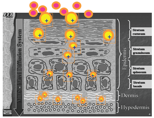
Figure 1: Releasing of the active ingredients at the designed sites


Pierfrancesco Morganti1* Pietro Febo2 Maria Cardillo3 Giovanna Donnarumma4 Adone Baroni5
1Dermatology Department, 2nd University of Naples, Italy, R&D Director, Nanoscience Centre MAVI, Aprilia (LT), Italy*Corresponding author: Pierfrancesco Morganti, Dermatology Department, 2nd University of Naples, Italy, R&D Director, Nanoscience Centre MAVI, Aprilia (LT), Italy, Tel: + 39 06 92 86 261; E-mail: pierfrancesco.morganti@mavicosmetics.it
Chitin and lignin are two building materials, obtained from waste, giving strength to the exoskeleton of crustaceans and plant cells respectively. Both the polymers seem to be a dynamic structural source of molecules that trigger immune responses to humans as well. Moreover, due to their different electrical charges covering the surface of these natural ingredients, they may be bound to form micro/nanoparticles and innovative nanocomposites to be embedded into non-woven tissues for producing advanced medications, more effective when used in their nano dimension. By the use of these polymers, it is possible to produce porous scaffolds that, mimicking the natural Extra Cellular Matrix can facilitate the appropriate cell infiltration, proliferation, and differentiation. Both chitin and lignin have, in fact, an interesting antioxidant, anti-inflammatory and cicatrizing effectiveness, being easily metabolized from the environmental and human enzymes without producing toxic secondary ingredients. Some data are reported in this paper to support these activities.
Chitin; Lignin; Chitin nanofibril; Chitinase; Biocomposite; Nanofiller
Degradable and natural polymers represent the class of biomaterials more often used for cosmetic and biomedical applications as nanocarriers or tissue engineering scaffolds [1]. For their interesting properties, scientists have been encouraged to use them also as drug delivery systems, increasing their efficiency and improving their functionality and bioavailability to achieve maximum clinical upshot. Example of the most focused nanocarriers, which can load active ingredients to intracellular sites, are polymeric compounds such as liposomes, chitosan, chitin, and chitin nanofibrils reported by different scientific papers [2-7]. These natural fibers are used to make biocomposite materials as the most advanced and adaptable engineered polymers for producing safe matrices and innovative carriers. The right combination of polymeric matrices and reinforcing natural fibers may produce composites possessing the finest properties of each component. The fiber-reinforced composites, in fact, have the scope to improve or adjust the altered or variable properties (mechanical, thermal, optical, or electrical) of the matrices into which they are incorporated at the concentration rate from 1% to10% [8]. However, the ideal material for biomedical use should be biocompatible, biomimetic, non-toxic, and nonimmunogenic [9], having also the capacity to facilitate the cell adhesion, growth, migration and being responsible at molecular and physical level of the in vivo milieu [10,11]. Moreover, the biodegradability of all the materials used should be another important characteristic function of the tissue engineered design, remembering that a slow biodegradation is to be preferred for a long-term human implant, while a rapid biodegradation results fundamental for the wright remodeling of the tissue to be repaired [10-12]. Additionally, cell proliferation and differentiation can be enhanced by the use of polymeric tissues having the same structure of the skin Extra Cellular Matrix (ECM) [13]. Also, the nanoparticle technology may be of help to maximize the therapeutic efficacy and minimize the undesirable side effects of the polymer system-of-choice by the control of its drug/active ingredient’s bioavailability and release [14,15]. This kind of release system, for example, can transport a wide variety of active ingredients through the epidermis, keeping them at the action site, in the right time and dosage [16,17]. Therefore, the nanotechnology may aim to (a) facilitate the ingredient transport, increasing its efficacy and reducing the possible toxic side effects; (b) maximize contact time with the skin, minimizing transdermal absorption; and (c) release the actives in the designed sites (Figure 1). Additionally, these polymeric nanostructures favour more contact with the skin stratum corneum, increasing the quantity of incorporated active ingredients to reach the action site [17]. Thus, the necessity to design the right protocol, (a) characterizing size and morphology of nanoparticles and fibers, (b) achieving both the proper nano encapsulation of the active ingredients and their inclusion in the polymer fiber/tissue, taking into account the stability of the entire system and its loading efficiency, release pattern and activity

Figure 1: Releasing of the active ingredients at the designed sites
Chitin (Figure 2) represents the second most abundant natural polysaccharide after cellulose [18]. On the other hand lignin (Figure 3), responsible for the strength and rigid structure of the cell walls in plants, is an available by-product of the paper manufacture and ethanol production from lignocellulosic biomass. It represents the second major component of wood and annual plants [19,20].
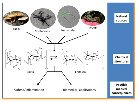
Figure 2: Chitin as building material of crustacean ‘exoskeleton and fungi’ wall
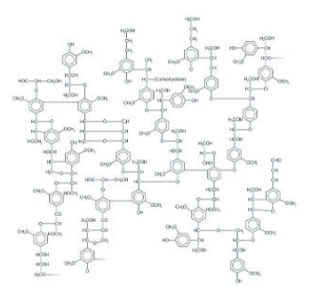
Figure 3: Lignin proposed chemical structure
Unfortunately both chitin and lignin as natural polymers are principally used to produce fuel, being underutilized for making added value products, also if they represent a waste material of about 300 billion tons /year [21].
It is interesting to underline that the renewable polysaccharide chitin, consisting of both crystalline and amorphous domains, has shown to be useful organic nanofiller [22]. According to a patented technology [23], the amorphous part can be removed and isolated from the nanocrystals which, used as supporting material for living bodies, are characterized for their high modulus and bioavailability. Chitin nanocrystals, in fact, known also as chitin nano fibrils (CN) form a microfibril arrangement in living plants as well as in animals, with size increasing from simple molecule and highly nano crystalline fibril, the composite matrix at micrometer level upward (Figure 4) [18,24] In the same way lignin (LG), solubilized in alkalized water and covered by a thin corona of PEG, can be easily treated by the spray drier to obtain a brown powder of micro/ nanoparticles with a mean dimension of about 163nm (Figure 5). Furthermore, combining the electropositive CN with the electronegative LG, it is possible to obtain micro/nanoparticles which, entrapping a wide variety of active ingredients (hydrophilic and/or lipophilic), can be used in medicine for their specific and effective characteristics and properties. Particularly, the simple CN-LG nanoparticles (Figure 6) have shown to possess interesting antibacterial and anti-inflammatory properties [25], useful to make innovative cosmetics for aged [6,26], problematic and sensitive skin [22,27] or non-woven tissues and perforated films for feminine or baby pads [28].

Figure 4: Microfibrils arrangement of chitin in crustaceans (Source: Raabe et al modified)
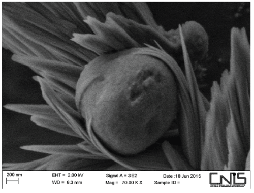
Figure 5: Micro/nanoparticles of lignin covered by a thin corona of PEG at SEM
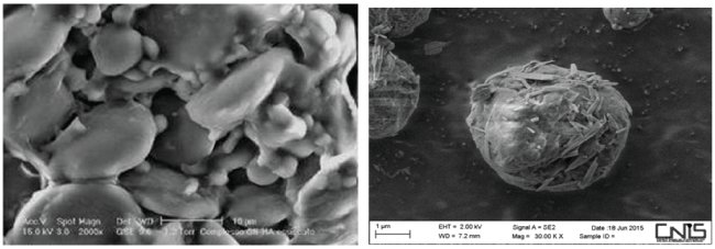
Figure 6: Micro/nanoparticles of the complex chitin nanoparticlenanolignin
Coming back to the chemical and biological characteristics of chitin, it has been hypothesized that, this natural and ubiquitous polymer is a size-dependent regulator of innate immunity [29]. It is used, in fact, by fungi, crabs, and insects not only to protect their structures from the environmental harsh conditions, but also as host anti-parasite/pathogen immune response. The balance between biosynthesis and degradation of chitin, which regulates its accumulation on the relative settings, is mediated by chitinases (i.e., endo-Beta-1,4-N-acetylglucosamidases), enzymes produced as an immune response to chitin containing pathogens [30,31]. While the role of chitin and chitinases have been clearly established as signaling sensor of microbial and parasitic invasion in the field of plant and microbial immunity [32], their ability to regulate local inflammatory cell function in humans, is not enough clear [29-31]. In any way, this sugar-like polymer is the major component of many allergy-triggering environmental components, such as house-dust mites, crustaceans food, and fungal spores [33-36] (Figure 7), being appreciated as an under diagnosed disease entity. In fact, different studies clearly demonstrate that chitin and chitin derivatives can stimulate innate immune cells, such as macrophages, basophils and eosinophils, and modulate adaptive Type I or Type 2 responses, via a variety of cell surface receptors, including macrophage mannose receptors, toll-like receptors 2, and dectin-1, mediating their cellular and tissue effects in a size-dependent pathway (Figure 8) [37-39]. In addition recently, our group has shown that chitin nanofibrils have the capacity to complex different active ingredients for obtaining micro/nanoparticles and polymers capable to increase, for example, the whitening activity and effectiveness of a specifically designed cosmetic emulsions and non-woven tissues (Figure 9). Moreover, CN complexed with nanolignin has evidenced an interesting anti inflammatory and immunomodulatory activity, accelerating the repairing activity of burned skin (Figure 10) [25,40]. At this purpose, it is interesting to remember that the chitin-degrading enzymes, produced by humans as part of the 18-glycosyl-hydrolase and mainly expressed and secreted by neutrophils and macrophages, are induced at sites of inflammation and infection for remodeling the tissue structure [41]. This suggests that such proteins could play an active role in anti-infective defense and resource responses [31,42-44], as shown from our recent studies also [25-27,40]. This the reason why CN has evidenced an increased release of skin defensins with a contemporary modulation of metalloproteinases, when used in culture of human keratinocytes and fibroblasts (Figure 11 and 12) [25-27,40]. It is to remember, in fact, that chitin has a repeating molecular pattern analogous to other Toll-like receptor 2, i.e., TLR-2, encoded in humans by the TLR-2 gene [45]. These TLR receptors have shown to function as sensors of microbial and parasitic invasion by the inflammation cascade, as immune response to the pathogens [46,47]. Recently [48], it has been also evidenced that chitin seems to be a pathogen-associated molecular pattern (PAMP), capable to mediate some cellular and tissue effects in a size-dependent and specific-manner pathway. It seems to serve as a PAMP that, according to the chitin size, activates macrophages via TLR-2, regulating also in vitro and in vivo both their function and the acute inflammation phenomena, by a stimulated release of pro- and antiinflammatory cytokines. Chitin fragment, in fact, has shown to exert a size-dependent effect on macrophage activity, demonstrating a potential use as immune adjuvant (Figure 7) [48,49]. The mean size chitin has evidenced a pro-inflammatory activity, while its small size fragment (<40 µm) has shown an anti-inflammatory function, activating in macrophages both TNF and IL-10 (Figure 8). Probably, the interesting effectiveness shown by CN is due not only to its very small size of 240 × 7 × 5 nm, but also to show the same backbone of hyaluronic acid (Figure 13), which could serve as an alarm signal by its degradation molecules [32,50]. At this purpose it has been suggested that chitin, recognized by some receptors, triggers the induction of chitinases, leading to the generation of smallsized chitin particles that are taken up by the host cell [35]. However, the ability of polysaccharides to induce inflammation, according to their size, could be a general principle of glycol biology [35,39,51], where chitin fragments or nanochitin could modulate the intensity and chronicity of local inflammation and the consequential cell apoptosis [25,52].
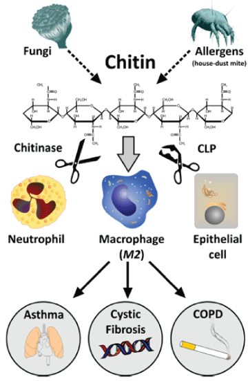
Figure 7: The proposed role of chitin, chitinases and chitinase-like proteins as allergy/triggering environmental components. [35].

Figure 8: The proposed role of chitin size in anti-pathogen responses to produce different pro inflammatory cytokines. [49].
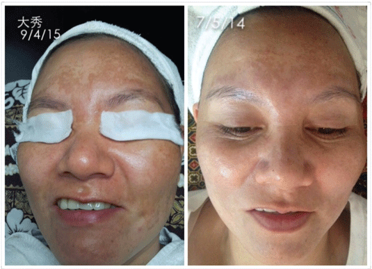
Figure 9: Whitening activity of a cosmetic emulsion based on the use of CN-LG nanoparticles entrapping different active ingredients
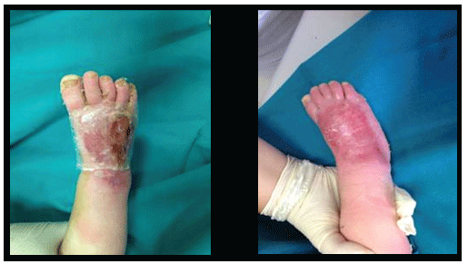
Figure 10: Repairing activity of a burned skin by a non-woven tissue based on CN-LG nanoparticles entrapping nano structured Ag
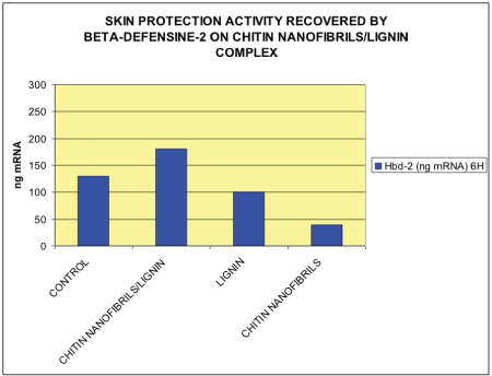
Figure 11: mRNA expression of Defensine-2 in HaCat cells after 24 hours of treatment with non-woven tissue made of CN-LG fibres
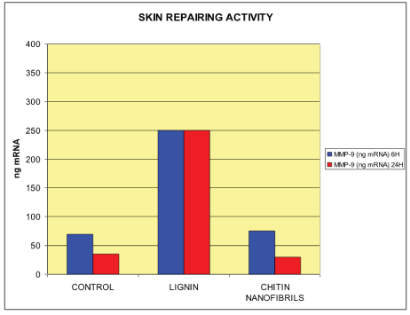
Figure 12: mRNA expression of Metalloproteinases-9 in HaCat cells after 6 and 24 hours of treatment with by non-woven tissue Made of CN-LG fibres
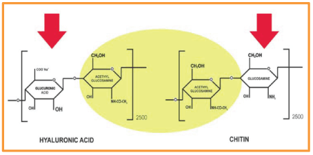
Figure 13: Chitin has the same backbone of hyaluronic acid
Lignin is an industrial by-product, which available in large amount from the plant biomass, is mainly used for energy production [20]. However, this interesting natural polymer contains in its branched molecule many phenolic, hydroxyl, carboxylic carbonyl and methoxyl groups, the structure of which represents an excellent source for the production of valuable molecules and functional products with antioxidant, antimicrobial and anti mutagenic properties [53]. One of the most studied characteristic of lignin is its antioxidant property connected with the hydroxyl and methoxyl groups which, functioning as proton donors could stabilize the radical in the quinone resonance structure [54]. Naturally, different types of lignin possess different antioxidant, antimicrobial and UV absorption properties, depending on the source and processing method used, as well as on the post-treatment and the different geographical origin of the plants. Any polymer, however, is recognizable for its own molecular weight, physicochemical characteristics and polydispersity [55,56]. This is the reason why a fine-tuning of the process condition is necessary to balance the recovery and the quality of lignin with those of other wood components. It contains, in fact, various phenolic groups, mainly based on the structure of benzoic acid and cinnamic acid (i.e., p-coumaric and ferulic acids). The phenolic groups confer to this natural macromolecule the interesting antimicrobial and antioxidant properties [57,58], centered specifically on the nature and side structure of the phenolic functional group [59], especially strengthened by its nano dimension. For this reason, lignin, hindering its phenolic groups, can stabilize the reactions induced by oxygen, reactive oxygen and nitrogen species (ROS and RNS), slowing down the aging processes of polymer composites realized with this natural macromolecule, as well as of the biological systems, when used as active ingredient in cosmetic and medical formulations. It is considered, in fact, as safe antioxidant and antimicrobial compound, being also a biodegradable ingredient with a low toxicity when used as filler for polymeric matrices or biological active ingredient for medical purpose [60].
Additionally, for its antioxidant, UV protectant, and antibacterial property, lignin represents also a promising green and natural ingredient useful to balance the general microbiota, allowing reducing the environmental problems related to petrol-derived chemicals [61]. For all these reasons we prepared, by the gelatin method, CN-LG nanoparticles which, blended with different active ingredients, have been used to make a non-woven tissue by the electro spinning technology. In fact, the CN is a cationic polymer while nanolignin is an anionic ion, so that the polyelectrolyte complex CN-LG intersects via ionic linkages. These innovative tissues have been applied on burned skin with the purpose to obtain a quick, antioxidant, anti inflammatory, antibacterial and reepithelialization activity, as previously reported [25,40,62].
Due to the interesting specific characteristics of both Chitin nanofibrils and Lignin, it has been designed to use the CN-LG nanoparticles as functional delivery carrier of active ingredients for the human tissues regeneration. At this purpose, major efforts have been taken to develop non-woven tissues mimicking the behavior of the skin. By different studies [22,46], it has been shown that a non-woven tissue, made by the electro spinning of a CN-LG blend, incorporated into PEO and Chitosan, seems to play an important role in providing a platform capable to influence the perception and response of human cells to the skin tissue. This natural matrix, applied on wounded and/or burned skin, has evidenced to possess a chemo attractive property. It seems capable not only to activate both macrophages and neutrophils for initiating the healing process, but also to attack the microbial bio film (Figure 14) [22]. Moreover, it has been shown that CN promotes the tissue granulation and re-epithelialization, limiting the formation of hyper trophic and keloid scars (Figure 15) [63]. Chitin nanoparticles, in fact, activate keratinocytes and fibroblasts proliferation, regulating also the collagen synthesis and the cytokine and macrophage secretion [64]. According to a recent study [65], the wound healing capacity of this innovative non-woven tissue seems to be also based on the activation of the immune competent cells. These specialized cells, acting on chitin by the enzymatic activity of chitinases, cause the release of glucosamine and acetyl-glucosamine, which could modulate the ECM synthesis. In conclusion, the introduction of CN into the chitosan/PEO matrix, make it possible to form bio-reasorbable composite fibers and non-woven tissues, skin-friendly and with good adhesion characteristics. Likewise, the CN electro spun nanofibers, exhibiting an ECM-like architecture (Figure 16) together with interesting antibacterial and anti inflammatory effectiveness, seem to open new perspectives to make future innovative and biodegradable babies and feminine pads [28].
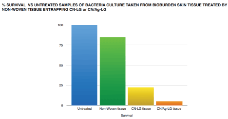
Figure 14: Bactericidal activity of Chitin Nanofibrils on the microbial biofilm

Figure 15: The activity of CN-Chitosan as re-epithelialized agent that limiting the formation of hypertrophic scars and keloids
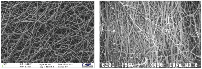
Figure 16: The CN scaffold (left) has the same structure of the skin ECM (right)
In conclusion, the defense from biological and environmental stressors could be centered on the endocrine regulation, synthesis and production of chitin by the immune system, in both invertebrate and vertebrate [66]. Thus, on one hand chitin and chitin-like proteins seem to play an important role in normal processes, such as cell growth, turnover and remodeling, while the skin antimicrobial peptides seem to be tightly trigged by the chitinases activities in mammals and humans also [67].
On the other hand lignin, as carbohydrate-based polymer could be considered not only a mechanical and passive defensive barrier against the pathogen attack, but also a dynamic structural source of signaling molecules that trigger immune responses to humans [66]. Moreover, as previously reported, it is a versatile biopolymer that seems to possess several other useful characteristics, such as UV-absorption, antifungal, antibiotic and anti-carcinogenic properties [68]. Thus, according to the obtained findings, it is possible to explicate the antimicrobial and skin repairing response obtained by the use of the non-woven tissue incorporating both CN and LG nanoparticles. This innovative matrix has shown to re-epithelialize in a shorter time the burned skin, improving the release of the incorporated active ingredients, in the right dose and time [6,25,40]. Moreover, it was also obtained a re-equilibrium of the skin superficial microbiota, while the cicatrization process was concluded without the formation of anomalous scars. In conclusion, a better knowledge of all the physicochemical and biological implications involving the chitin-chitinase and CN-lignin activity seems to be of great interest for understanding their possible interactions with the different layers of the human skin. From these studies and by the use of nanochitin, nanolignin and other natural by-products human and environmentallyfriendly, it will be possible to find innovative cosmetic and biomedical applications, as well as to identify new bio markers and/or therapies for fungal and other pathological conditions. This the goal of our future research projects necessary, in our opinion, to preserve the biodiversity of our planet, maintaining the natural raw materials for the future generations.
Download Provisional PDF Here
Aritcle Type: Review Article
Citation: Morganti P, Febo P, Cardillo M, Donnarumma G, Baroni A (2017) Chitin Nanofibril and Nanolignin: Natural Polymers of Biomedical Interest. J Clin Cosmet Dermatol 1(2): doi http://dx.doi.org/10.16966/2576-2826.113
Copyright: © 2017 Morganti P, et al. This is an open-access article distributed under the terms of the Creative Commons Attribution License, which permits unrestricted use, distribution, and reproduction in any medium, provided the original author and source are credited.
Publication history:
All Sci Forschen Journals are Open Access