Abstract
An unusual case of superior vena cava syndrome is presented where the diagnostic path was riddled with initial oversights. Demanding surgical treatment provided symptomatic relief to the patient while adjuvant oncological treatment aimed at achieving prolonged remission.
Keywords:
Endometrial adenocarcinoma; Metastasis
Case Report
A 55-year old female patient was admitted in February 2014 because of suspected myocardial infarction. She experienced nausea, dizziness, had high blood pressure, but reported no chest pain and her main complaint was facial swelling accompanied by heavy sensations in her head. In the emergency room a marginally positive troponin I (0.511 µmol/l) and T wave inversion in the infero-lateral leads in the ECG were noted. A few weeks before the patient had been evaluated in another hospital because of swelling of the face and neck. Cushing syndrome was excluded and a CT scan of the thorax without intravascular contrast (because of presumed anaphylactic reaction to contrast agent in the past) reported no pathological masses. The presumed diagnosis was angioedema ascribed to ACE inhibitors and the patient’s antihypertensive treatment was switched to losartan 100 mg per day.
The patient’s past medical history included a total abdominal hysterectomy with ovariectomy and pelvic lymphadenectomy in June 2012 because of endometrial adenocarcinoma (endometrioid type, FIGO IB, grade II). The operation was followed by vaginal brachytherapy. A few months later local metastases were found in the vagina. The lesions were electrocuted and the patient was treated by additional vaginal brachytherapy and external beam radiotherapy. Treatment was completed in March 2013 and no morphological signs of relapse were found later, although a small increase in the tumor marker CA-125 was detected.
Upon admission, treatment with dual antiplatelet therapy, fondaparinux and bisoprolol was started. Transthoracic echocardiography showed no abnormalities other than diastolic dysfunction grade 1. Since the patient reported a history of anaphylactic reaction to radiographic contrast agents more than 35 years ago, cardiac MRI with gadolinium contrast agent was performed according to a myocardial perfusion protocol instead of conventional X-ray coronary angiography. The MRI did not show myocardial ischemia or signs of myocarditis, but detected a large mass in the apical portion of the right atrium measuring 60 × 55 × 50 mm which nearly obstructed the superior vena cava inflow (Figure 1). The diagnosis of superior vena cava syndrome due to obstruction at the level of the right atrium was confirmed, explaining the facial swelling and heavy sensations in the patient’s head. A repeated transthoracic echocardiography confirmed that the right atrial mass was heterogeneously echoic and could be detected only by the absence of color Doppler flow in the affected region (Figure 2).
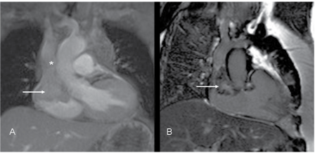
Figure 1: A: Post-gadolinium T1-weighted cine gradient echo image, coronal plane. Large mass in the upper half of the right atrium (arrow), obstructing the inflow of vena cava superior (asterisk) B: Late gadolinium enhancement phase sensitive inversion recovery image, semi-coronal plane. Heterogeneous enhancement of the mass (arrow)
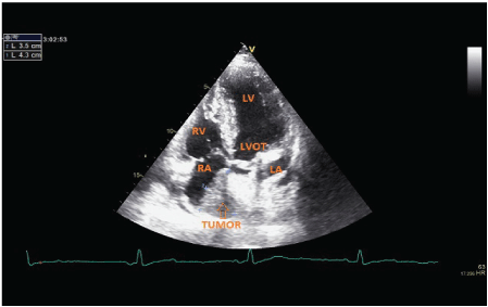
Figure 2: Transthoracic echocardiography (5 chamber view) of the patient showing a heterogeneously echoic mass filling out the vast majority of the right atrium in the field of view. LA – left atrium, RA – right atrium, LV – left ventricle, RV – right ventricle, LVOT – left ventricle outflow tract
In preparation for surgical treatment, skin testing excluded allergy to commonly used radiological contrast agents. Therefore coronary angiography was performed, revealing a significant stenosis of the right coronary artery which was successfully stented, while a CT scan of the head and an ultrasound examination of the abdomen and pelvis showed no signs of a metastatic tumor.
Surgery was performed with the use of extracorporeal circulation. A solid formation covered most of the upper part of the right atrium, particularly the right atrial appendage and opening of the superior vena cava (Figure 3). The tumor was resected, and the atrium and superior vena cava were reconstructed using CorMatrix™ ECM for cardiac tissue repair as an acellular xenograft.
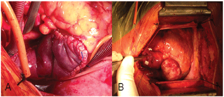
Figure 3: A: Tumor of the right atrium filling out the right auricule. B: Surgical reconstruction of the right atrium and the superior vena cava by CorMatrix™
Histological analysis of the tumor showed endometrioid adenocarcinoma confirming the diagnosis of cardiac metastasis.
The patient recovered well from the operation, clinical signs of face congestion regressed and she was referred to the Institute of Oncology Ljubljana. Chemotherapy was proposed as an additional treatment to surgery for microscopic residual tumor in metastatic disease. The patient received six cycles of paclitaxel with carboplatin. Two months after the last cycle of chemotherapy she was readmitted to the hospital with the symptoms of headache and vomiting due to high intracranial pressure. CT scan revealed brain metastases. She received central nervous system radiation treatment which led to improvement of her symptoms so that she was able to return to her home environment.
Discussion
The described case is unusual because of the initial diagnostic difficulties in identifying the rare cause of the superior vena cava syndrome and because of the difficult surgical treatment.
Our patient’s facial swelling was first evaluated in a hospital specialized for pulmonary diseases and allergy. Superior vena cava syndrome was considered, but was ruled out because a CT scan of the thorax performed without a contrast agent due to the patient’s presumed contrast allergy, did not show any pathological masses. Although the patient’s complaints were essentially unchanged, the reason for the admission to the University Medical Centre of Ljubljana was a suspected myocardial infarction in view of the positive troponin, which was in retrospect likely due to the cardiac involvement by the metastatic tumor, while the ECG changes were non-specific. Fortunately, MRI of the heart was chosen as the imaging modality, because the patient reported an allergy to radiographic contrast agent, which was later dismissed. MRI clearly showed the tumor in the right atrium, while conventional X-ray coronary angiography would not have led to the main diagnosis. The patient’s concomitant stenosis of the right coronary artery would likely have been interpreted as the cause of the elevated troponin and stenting would have concluded the patient’s in-hospital treatment. Interestingly, initial transthoracic echocardiography failed to show the mass in the right atrium, although it is the recommended initial imaging modality for detecting cardiac tumors [1,2]. Repeated transthoracic echocardiography confirmed the presence of a heterogeneously echoic mass in the right atrium (Figure 2) which was missed on the first examination, probably because the tumor mass was nearly filling out the entire right atrium and failing to provide enough contrast to the flowing blood to be detected by a less experienced ultrasonographer.
Superior vena cava (SVC) syndrome is caused by any condition that obstructs blood flow through SVC either from the outside or intraluminally. The former is usually caused by a mediastinal mass, generally to the right of the midline, which may consist of lymph nodes, lymphoma, thymoma, an inflammatory process (fibrosing mediastinitis), aortic aneurysm or a pathological process involving the right lung [3]. Thrombosis of the superior vena cava without extrinsic compression is also a well recognized cause of the superior vena cava syndrome [3]. The most common causes of SVC thrombosis are intra-thoracic malignancies, e.g., lung cancer (non-small cell and small cell) or non-Hodgkin lymphoma [4,5] or indwelling intravascular devices, such as central venous catheters or cardiac pacemaker leads [6]. Intracavitary tumors of the right atrium are not generally listed among the causes of the superior vena cava syndrome. Treatment of SVC syndrome should preferably address its cause, most commonly by radiation treatment, chemotherapy, venous stenting, and/ or supportive medical treatment [7].
Primary tumors of the heart are found in 0.0017% to 0.19% of unselected patients at autopsy [8,9].Three quarters of primary heart tumors are benign; most of those are myxomas, which are usually located on the left side of the interatrial septum [10], with the remainder being papillary fibroelastomas [11], fibromas [12] and others. Primary malignant tumors of the heart are even rarer, the majority being different types of sarcomas [13]. Metastatic heart tumors are 20 to 100-times more frequent than primary cardiac tumors, but intracavitary growth of secondary heart tumors is very rare [1,14,15]. Cardiac metastases are usually small and multiple and only rarely gain clinical attention, mostly by causing pericardial effusions [14,15]. The cancers which most commonly metastasize to the heart are malignant melanoma, carcinomas of the lung, breast and esophagus, lymphoma, leukemia and soft tissue sarcomas [14-16]. There are only 8 described cases of metastatic colon cancer involving the right atrium in the literature, 2 of which caused the superior vena cava syndrome [17] while descriptions of endometrial cancer with cardiac metastases are even more scarce with only 6 cases involving the right atrium reported [18-20].
Right-sided cardiac tumors present surgeons with a technical challenge because of the difficult placement of the cannula for cardiopulmonary bypass [21]. In addition, there is the problem of reconstructing the large veins and the heart wall when the tumor is invasive. In our case, reconstruction of the superior vena cava and the right atrium was solved by using CorMatrix™, acellular porcine small intestinal submucosa, serving as an acellular xenograft. In spite of the technical challenges, surgical removal of the metastatic tumor prolongs survival and is considered the treatment of choice. Radiation therapy is reserved only for patients who are not medically fit to receive the operation. There are only data from case reports on radiation of cardiac metastases and there is no consensus on the total radiation dose required for cardiac metastases. Radiationinduced heart disease is the dose limiting factor in treatment of cardiac metastases. Doses of 45 Gy with a possible additional boost to 60 Gy have been described in the literature [22].
Even with adjuvant chemotherapy the prognosis of patients with malignant SVC syndrome remains grave and reflects the underlying malignant disease [3].
Conclusion
In conclusion, metastatic endometrial adenocarcinoma of the right atrium may present as an unusual cause of the superior vena cava syndrome, posing diagnostic and therapeutic challenges. Therefore, a multidisciplinary approach such as in this particular case is mandatory for optimal management.
Article Information
Article Type: Case Report
Citation: Perme RL, Logar HB, Berden P, Juvan KA, Kneževič I, et al. (2016) Superior Vena Cava Syndrome Due to Metastasis of Endometrial Adenocarcinoma in the Right Atrium – A Case Report. J Clin Case Stu 1(4): doi http://dx.doi. org/10.16966/2471-4925.128
Copyright: © 2016 Perme RL, et al. This is an open-access article distributed under the terms of the Creative Commons Attribution License, which permits unrestricted use, distribution, and reproduction in any medium, provided the original author and source are credited.
Publication history:
Received date: 01 Jun 2016
Accepted date: 25 Jul 2016
Published date: 29 Jul 2016
