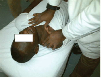
Figure 1: Positioning for sternoclavicular inferioanterior glide

Ganiyu Sokunbi*
Department of Physiotherapy, Bayero University Kano, Nigeria*Corresponding author: Ganiyu Sokunbi, Department of Physiotherapy, Bayero University Kano, Nigeria, Tel: 0023481 3846 8412; E-mail: ganiyusokunbi@gmail.com
Background: Thoracic outlet syndrome (TOS) is a condition that is somewhat controversial and there are no clear-cut clinical guidelines as regards the best way to manage this condition. This case study was designed to evaluate the effect of manual therapy, postural correction and acupuncture like TENS (ACUTENS) in the management of TOS.
Methods: The subject was a 33 years old male patient with a four month history of sudden onset of severe pain in the neck with radiation into right upper limb. Patient complained of lower neck pain, muscle weakness in the right upper limb and numbness and tingling sensation in his right hand on his first visit. Following a careful subjective assessment and objective assessment including physical examination, a rehabilitation program comprising of postural correction, manual therapy, pectoralis muscle stretching exercises and ACUTENS were carried out three times weekly for six weeks. Neck pain and disability scale (NPADS) was used to assess pain intensity and the impact of the pain on function, Activity of daily living (ADL) and emotion of the patient.
Results: After 6 weeks of treatment, both pain intensity and the impact of pain on emotion was reduced by 80% and 50% respectively. The impact of pain on function and ADL were reduced by 60% and 70% respectively. Patient also reported remarkable improvement with reduction in numbness and tingling sensation.
Conclusion: Findings from this case study showed that manual therapy, postural correction, pectoralis muscle stretching exercises and ACUTENS were effective in managing TOS. However, the long term and short term efficacy of using these treatment modalities either singly or in combination and comparison with a non- intervention control group is warranted.
ACUTENS; Manual therapy; Thoracic outlet syndrome; Physiotherapy
Thoracic Outlet Syndrome (TOS) is a broad term which is non-specific in the sense that it consists of a group of distinct disorders that affect the nerves in the brachial plexus and various nerves and blood vessels in the anatomical area above the first rib and behind the clavicle. The disorders are complex, somewhat confusing, and poorly defined, with various signs and symptoms of the upper limb [1]. The signs and symptoms encountered in the upper extremity may include paraesthesia, numbness, pain, oedema, muscle weakness, claudication, discoloration, temperature and tropic changes, ulceration, gangrene, and, in some cases, Reynaud’s phenomenon [2]. There are various causes of TOS. The thoracic outlet is an area in the neck above the uppermost rib on both sides of the body [1]. Thoracic outlet syndrome (TOS) occurs when the narrowing of this space causes pressure on nerves and/or blood vessels. It can result from injury, disease, or a congenital abnormality. Poor posture and obesity can aggravate the condition, which is more common in women than in men [1].
Based on the neurovascular structure that may be affected, TOS can be classified as neurologic (NTOS), arterial (ATOS) or venous (VTOS) [2]. The most common type NTOS constitute 90%-95%, while VTOS constitute approximately 3% and the least common ATOS, and constitute 1% of cases [3]. NTOS is rare and is caused by congenital anomalies but it generally occurs in middle-aged women and almost always on one side of the body [2]. Symptoms include weakness and wasting of hand muscles, and numbness in the hand [2]. ATOS occurs on one side of the body. It affects patients of both genders and at any age but often occurs in young people [3]. Like NTOS, ATOS is rare and is caused by a congenital anomaly [3]. Symptoms can include sensitivity to cold in the hands and fingers, numbness or pain in the fingers, and finger ulcers or severe limb ischemia. VTOS is also a rare disorder that affects men and women equally. The exact cause of this type of TOS is unknown. It often develops suddenly, frequently following unusual, prolonged limb exertion. Psychological changes are often seen in patients with thoracic outlet syndrome. It is not clear whether these precede the onset of the syndrome or are the result of dealing with the pain and frustration of diagnosing and treating this condition [4].
Treatment for individuals with TOS varies depending on the type. NTOS is generally effectively treated with surgery. Most other forms need only symptomatic treatment [4]. Most cases of TOS require conservative treatment which may include drugs such as analgesics, and physiotherapy to; increase range of motion of the neck and shoulders, strengthen muscles, and induce better posture [1]. Physiotherapy treatment of TOS is usually preceded by a thorough history taken followed by a physical examination. Many patients will have a history of neck or shoulder trauma or a repetitive physical stress at work. Physical examination will necessarily include the following tests such as pulse examination, presence or absence of oedema/cyanosis/collateral veins, tenderness over scalene muscles (trigger points) or pectorals minor, reduced sensation to very light touch in fingers and provocative maneuvers etc. [3]. Diagnosis of TOS remains controversial. Other testing besides physical testing procedure that may be useful in making diagnosis includes radiography, neuroradiology, Magnetic Resonance Imaging (MRI) angiography, ultrasonography and electrophysiology. Gillard (2001) reported that of the physical testing procedures, best predictive results appear to be elicited by combining several tests. Gillard (2001) found best values for the Adson test, hyperabduction test and Wright test [4]. Sander (2007) found 98% of patients positive to modified version of Elvey’s upper limb tension test (ULTT). There is limited evidence in the form of randomised controlled trial as regards the efficacy of the reported therapeutic intervention for TOS and varying opinions exist as to the best way to manage this condition in the light of current evidence. The aim of the present study was to present via a case study, the effect of manual therapy, exercises for postural correction and Acupuncture like Transcutaneous Electrical Nerve Stimulation (ACUTENS) in the management of TOS.
A 33 years old male referred to the Physiotherapy department, University of Maiduguri Teaching Hospital with a four month history of sudden onset of severe pain in the neck with radiation into right upper limb. Patient complained of lower neck pain and muscle weakness in the right upper limb on his first visit. Lying on the right upper limb while sleeping at night and carrying his briefcase to work aggravate his pain. Resting in the form of avoidance of lying on the right side and lifting with the right upper extremity were the only alleviating factors reported. Patient derived occasional pain relief from vigorous shaking of the right upper extremity. Pain severity was reportedly constant throughout the day.
Patient reported recurrent numbness and paraesthesia in the right hand which were usually made worse by lying on the right side. However, his main concern about pain, inability to lift with the right hand and having had to stop driving due to difficulty in selecting the gear and turning the steering with the right hand. The patient had no history of other medical and/or surgical conditions that could be linked to his present complaint. Previous treatment with analgesics relieved his pain slightly on a temporary basis and physiotherapy treatment in the form of short wave diathermy to the right shoulder for 2 weeks at 2 sessions per week produced little benefit. Red flag factors such as cord signs and symptoms, signs of adherent dura, drop attack, basilar artery insufficiency and rheumatoid arthritis were not reported by the patient.
General observation: Patient has a mesomorphic body somatotype. The patient weighed 65 kg and was 1.63 m in height (body mass index=24.5 kg/m2 ). He walked into the examination room with normal gait.
Local observation: Patient was presented with cervical spine postural asymmetry (neck deviated towards the left side of the body) the right shoulder held in a brought forward position. Patient also presented with protracted scapulae, flat upper back and poking chin posture. Patient did not appear pale and there were no signs of skin trophic changes and cyanosis.
Palpation: The skin felt normal temperature to touch. Digital palpation on the cervical spine in the posteroanterior direction elicited pain centrally and right paramedially at C4-C5, C5-C6, and C6-C7 spinal segmental levels. Pain was elicited on palpation of the right pectoralis muscles but no pain was reported on palpation of the scalene muscles. Pectoralis minor muscle was palpated with the patient in supine lying and with the right shoulder joint passively abducted to 90 degrees. The therapist right fingers were slide below the clavicle where its s-shape is most concave to find the coracoid process of the scapula and then medially and interiorly along the anterior ribs and onto the fibers of pectoralis minor. Patient was requested to resist scapular depression to assure proper location of pectoralis minor muscles.
Movements: Assessments of cervical spine for joint signs of capsular pattern of movement restriction and non-capsular pattern of movement restrictions caused by internal derangements as described by Youdas et al. [5] was carried out. It shows full active range of movement (ROM) of the cervical spine in extension which measured 70°, right and left side flexion measured 20° each and right and left side rotation measured 45°. Cervical spine AROM in flexion measured 80°. Limitation in Side flexion and Side rotation were accompanied with pain. Similar pattern of pain and movement restriction were elicited with passive ROM. Right and left shoulder, elbow and wrist joints AROM were within normal limit.
Oxford muscle grading of all the muscles in the right shoulder, elbow and wrist muscles measured 3/5, while all the muscles in the left shoulder, elbow and wrist muscles were within normal limit (5/5). Biceps, brachioradialis and triceps jerk reflexes were normal and intact on the left but were reduced on the right side. The pulse rate which was determined by taking radial pulse measured 72 beat per minute. The blood pressure assessment carried out with mercury sphygnomanometer (Dengiglian, Germany) measured 120/79 in both upper extremities. The SemmesWeinstein monofilaments test as described by Muller et al. [6] was performed to assess superficial and deep skin sensation at the following dermatomal levels; C4 (Top of the acromioclavicular joint), C5 (Lateral side of antecubital fossa), C6 (Thumb), C7 (Middle finger), C8 (Little finger), T1 (Medial side of antecubital fossa) and T2 (Apex of axilla) were carried out. It showed impairment of superficial and deep skin sensation at all the dermatomal levels tested in the right upper extremity but normal and intact sensation in the left upper extremity.
Adson test was carried out with the patient seated on a stool and arms held close to the sides. The examiner palpates the patient’s radial pulse and also listens for bruits above the clavicle. The right arm was then elevated and the patient was instructed to turn the chin away from the right side. A positive test results was obtained with diminished radial pulse, bruit, and numbness and tingling [3]. Wright test was carried out with the examiner taking the radial pulse of the patient in same position as described above. Patient’s right arm was then hyperabducted and the radial pulse was measured again. The result was positive with a decrease in the radial pulse [3].
Upper limb nerve tension test was carried out with the patient in a seated position. The examiner asked the patient to abduct both arm up to the shoulder level with elbow joint held in extension. Patient was asked to maintain this position for 20 seconds. The result was positive with increasing pain and tingling sensation reported in the right upper extremity.
Cervical spine X-ray report dated 15/5/2014 revealed straightening of the cervical curvature due to muscular spasm from pain. It also showed cervical rib which was noticed at the top end of left of the ribcage. Cervical MRI was requested and carried out to ascertain the involvement of other tissues that could not be revealed by X-ray. The Cervical MRI report dated 22/10/2014 indicated multilevel C4-C5 C5-C6 C6-C7 moderate disc protrusion.
Neck and right upper extremity pain possibly caused by neurovascular compression by disc cervical disc protrusion and cervical rib at the top end of right thoracic cage. Asymmetrical neck and upper back posture and poor body mechanics that may be causing or contributing to neurovascular compression and irritation at the thoracic outlet. The forward head and poking chin posture could have also induced excessive tension in the scalene muscles. Limitation of right side flexion and rotation at the cervical spinal segment levels due to pain. Weakness of the muscles in the right upper extremity, impaired sensation at C4, C5, C6, C7, C8, T1 and T2 dermatomal levels possibly due to impaired conduction of sensorimotor nerve roots due to neurovascular compression at the right thoracic outlet. Impaired functional use of the right hand (lifting and driving) possibly due to pain and muscle weakness. Patient also rated constant tingling and numbness in the right palm and fingers as a major problem.
Clinical impression: Impaired functional use of the right upper extremity associated with cervical spine movement limitation and neck pain radiating to the right upper extremity secondary to right thoracic outlet syndrome.
Outcome measures: The main outcome measure used in this study was a multidimensional Neck Pain and Disability Scale, NPAD [7]. It was used to measure pain intensity and the impact of the neck pain on function, interference with activity of daily living and emotions. The NPAD was chosen for this study as it was specifically developed to evaluate the multidimensional effects of neck pain disorders. It has been demonstrated to have a good temporal stability (test-retest reliability) and construct validity [8]. The NPAD consists of 20 questions that examine pain intensity and the impacts of neck pain disorders on the patient neck function, interference with activity of daily living and emotions. Patient responds to each question by marking a point along a 10-cm line Visual analogue scale (VAS), which is marked at 2-cm intervals so that the minimum score for each question is 0 and the maximum is 5. Following completion of the NPAD, the score for each question was calculated by measuring the distance of the patient’s mark from the zero (0) end. The questions scores were added to give a total of score out of 100 which was then converted to a percentage. A low score represent a lower level of pain and disability and vice versa. Subsequently scores for each of the four key domains were calculated using the method describe by Wheeler et al. [9]. For each domain, a higher score (closer to 10) represents a higher impact. Patient was also asked to subjectively rate the severity of tingling and numbness in the right hand in his own word. Assessments were carried out prior to intervention, 3 weeks after the initial treatment and 6 weeks after the initial treatment.
Treatment procedure: Treatment procedure including postural correction, manual therapy, pectoralis muscle stretching exercise and acupuncture-like TENS (ACUTENS) was carried out as follows. All treatment was carried out thrice weekly for six weeks.
Postural corrections: Patient was instructed on postural awareness and how to identify the proper posture by using postural overcorrection exercise [10]. Patient was in a seated position before a mirror. He was initially asked to hyperflex ‘slouch’ the upper back. While in this position, patient was instructed to think about and feel the tension in the neck, upper back and shoulders and to maintain this position for10 seconds. This was followed by hyperextension ‘chest-out’ posture. Patient was instructed to think about and feel the tension in the neck, upper-back and shoulders and to maintain this position for10 seconds for another 10 seconds. From the chest out position patient was instructed to slowly reverse to adopting the initial slouch posture but to stop at the point he noticed a drop in the tension (relaxation) in his neck, upper-back and shoulders muscles. Patient was asked to think about and feel the tension at different positions in order to facilitate his awareness of muscular tension and relaxation at both extreme postures He was asked to repeat this exercise 10 times and to take note of this position which he was also encouraged to adopt at all-time whether at home, work, rest or play, in the office, when standing, sitting or lifting.
Manual therapy: Manual therapy was carried out to open up the thoracic outlet, mobilise the sternoclavicular joint and the scapular. Also, to stretch and facilitate relaxation of the scalene muscles. Manual therapy techniques used in this study consists of sternoclavicular glide, stretching of the scalene muscles, soft tissue mobilisation (STM) to the scalene muscles and scapular mobilisation. Sternoclavicular inferior anterior glide was carried out with the patient in supine lying position and the therapist faces the patient from the right side (Figure 1). One hands of the therapist supported the head of humerus and the acromion while the other hand griped the subcutaneous lateral boarder of the clavicle. The clavicle was then glided in upward and posterior directions for 3 sets of 10 repetitions. This was followed with stretching of the scalene muscles and a gentle kneading massage to the scalene muscles as described by Cyriax and Cyriax (2000) [10]. Scapular mobilisation was carried out with the patient in side lying position (Figure 2). Five repetitions of 3 sets of scapular distraction, upward and downward rotation were carried out. To achieve scapular distraction, the therapist one hand is placed on the spine of the scapula. The other hand is brought under the humerus and the fingers are placed on the inferior medial boarder of the scapular. The patient trunk is brought closer to the abdomen of the therapist to stabilize it. The therapist then retracts the scapular by an anteriorly directed stabilizing force and using the fingers on the medial boarder of the scapular to gently distract the scapular from the thoracic cage. Scapular upward rotation was achieved by the therapist applying a distracting force directed in a superior direction on the inferior medial boarder of the scapular to rotate the scapula upward and outward. For downward rotation of the scapular, the thenar eminence of the therapist hand positioned on the spine of the scapular impart mobilization force directed in an inferior direction on the lateral boarder of the scapular to rotate the scapular downward.
Figure 1: Positioning for sternoclavicular inferioanterior glide
Corner stretching exercise: This exercise was carried out to stretch tight pectoral muscles and fascia. The patient stood and faces the corner of the treatment room with each palm on the walls of the room that form the corner. He was then instructed to leans his body toward the corner without moving his hands or forearms on the wall and then reverses the movement. 3 sets of 10 repetitions of this exercise was carried out in a session.
Dual-channel TENS (Chinese, model 7000) was used to provide TENS treatment. All the cables from TENS unit to the electrode were intact and properly connected. The examiner tested the machine by switching ‘on’ the machine to lighten up the indicator light. Patient was in a forward lean sitting position with hands and forearms resting on a pillow on the plinth in front of the patient for comfort. Patient was well draped to expose only the part of the body where TENS were positioned. Patients were properly instructed on what to expect during the ACUTENS treatment. The TENS electrodes were positioned at selected acupuncture points (acupoints) widely accepted for treating cervical spine disorders [11], namely LI4 (highest point of the adductor pollicis muscle), GB 20 (point of intersection between Sternocleidomastoid muscle and Trapezius muscle behind the neck) and GB 21 (middle of the line between C7 spinous process and coracoid process). At each acupoint, the skin was wiped with alcohol. The following TENS variables were used: Lowest rate/frequency possible (2 Hz), the highest intensity that will be tolerable for the subject for 40 minutes. Tolerance was defined as the level at which the participants ask the investigator to stop increasing the stimulation [12].
The patient was seen and treated between September 3, 2014 and October 15, 2014, a period of six weeks. After 3 weeks of treatment pain intensity was reduced by more than 50%, function and Activity of daily living (ADL) improve by less than 50% and with no changes on the impact of pain on the emotion of the patient (Figure 2). After 6 weeks of treatment, both pain intensity and the impact of pain on emotion was reduced to 10% while the impact of pain on function and ADL were reduced to 30% and 20% respectively (Figure 2). Also patient reported that he was able to lift his suitcase to and fro the work place and drive his car without any noticeable neck and/or right upper extremity pain. He also regarded the earlier constant irritating tingling and numbness sensations as negligible and almost totally abolished after 6 weeks of treatment.
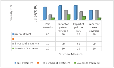
Figure 2: Effects of treatment on pain, function, ADL and emotion
The outcome of this case study has shown the effectiveness of manual therapy coupled with postural correction, pectoralis muscle stretching and acupuncture-like TENS in the management of thoracic outlet syndrome. Patient did not give any history of road traffic accident and /or previous trauma to the neck and the shoulder joints. However the presence of cervical rib, positive upper limb tension and other TOS provocative tests coupled with MRI report of multiple levels cervical disc prolapse are quite suggestive of a diagnosis of thoracic outlet syndrome. There appear to be limited reports on the efficacy of these treatment modalities in patients with TOS either in the form of case studies or randomised controlled trials, thus limiting comparisons of the findings of this study with others. However the treatment techniques used in the present case study were selected based on the findings from subjective assessment and physical examination of the patients in this study. Postural correction and re-education was carried out to reverse and/or reduce the impact of faulty posture on the symptoms and its aggravation. The poking chin posture and flat upper back posture have been reported to be capable of causing prolonged and excessive flexion of the cervical and upper thoracic spine [4]. This posture may further aggravate compression on the neurovascular structure due to the displaced intervertebral disc [4]. It has been demonstrated that intervertebral pressure is greatest when the spine is in flexion. Moreover when the spine is in flexion the disk is pushed posteriorly by the tilt on the surface of the vertebrae [10]. Thus, it is important for this patient to develop a greater awareness of his corrected posture. Initially, a new postural attitude might be uncomfortable, constant practising with visuobiofeedback, reminders and encouragement by members of the family and therapist might be essential to reinforce correct postural attitude.
A positive Adson test could imply compression of the vascular component of the neurovascular bundle at the thoracic outlet caused by stiffness of the thoracic spine, hypomobility of the cervical facet, cervical ribs and/or a tight scalene anterior muscle. This provided the rationale for the use of manual therapy consisting of mobilisation of the sternoclavicular joints coupled with stretching and soft tissue mobilisation to the anterior scalene muscles in this study. Sternoclavicular glide and scapular mobilisation could be used to increase the mobility of the shoulder girdle and the 1st and 2nd ribs on the affected side [10]. The shoulder girdle and the 1st and 2nd ribs on the unaffected side were also treated in order to make postural adjustments easier from the kinesthetic standpoint. . Excessive tension in the scalene muscles can result in elevation of the 1st rib, thus reducing the size of the costoclavicular space [1]. Thus, stretching of the scalene muscles is an important preliminary step to increasing the mobility of the 1st and 2nd ribs. Also, if nerve compression is occurring in the inter scalene triangle, the patient’s symptoms can be diminished at times by restoring or improving the elasticity of the scalene muscles, especially if they have been in a guarding position for an extended period of time. However, caution is to be demonstrated in performing this procedure. The muscle stretching should always be done in a slow, gradual manner and stretching of the scalene muscles should not be performed if there is an irritation of the brachial plexus in which traction on the plexus causes pain (Cyriax and Cyriax, 2000) [10].Similarly, a positive wright test is suggestive of compression of the axillary artery caused by stiffness of the thoracic spine, a tight pectoralis muscle or a deformed or hypertrophied coracoid process. Thus, necessitating the use of pectoralis muscle stretching exercise in this study.
It is also possible that neck pain radiating to the right upper extremity was not caused by the mechanical effects of pressure on the neurovascular structure alone. It could also be as a result of inflammatory changes at the site of the compression. There has been report that acupuncture stimulate the body’s self-regulation of anti inflammation regulation of the immune system, improve the vertebral blood circulation, alleviate disc degeneration and their respective organizations [13]. Studies have reported the success of acupuncture in the treatment of spinal disorders [11,14,15]. Possible therapeutic effects of acupuncture could be linked to enhancing activation of A- δ and C afferent fibres in muscle during needle stimulation of acupuncture points thus; signals are transmitted to the spinal cord, and via afferent pathways to the midbrain [11]. The resulting flow and integration of this information among specific brain areas will lead to a change in the perception of pain via descending pain modulatory system. Acupuncture analgesia improved the noxious descending inhibitory controls and pain gate mechanism and therefore helped to reduce the patients’ pain levels [13].
Findings from this study could influence the choice of assessment techniques and treatment modalities in the management of TOS. Also, it could stimulate further research in a large randomised control trial to establish the short and long term benefits of using either one or a combination of the treatment approaches in this case study in the management of TOS.
Discussion on comparison of the findings from this study with other studies with regards to the choice of the modalities used in this case study and their effects were not exhaustive. This could be partly due to the facts studies investigating the effects of ACUTENS, postural correction exercise and stretching exercise on TOS appeared not to have been widely reported. Also, the present study is a single case study, and thus its findings cannot be generalised to a wide range of population with TOS. Also, more comprehensive neurological and laboratory based tests such as nerve conduction test, electromyography test etc could have been used to further strengthen the diagnosis of TOS. Other limitation to this case report is primarily due to the fact that cervical spine active range of motion was not objectively measured with a goniometer. However, Cyriax and Cyriax (2000) approach to diagnosing cervical spine internal derangements with passive and active cervical ROM to determine the suitability for the application of manual therapy was carried out.
Download Provisional PDF Here
Article Type: Case Report
Citation: Sokunbi G (2016) The Effect of Manual Therapy, Postural Correction and ACUTENS in the Management of Thoracic Outlet Syndrome - A Case Study. J Clin Case Stu 1(6): doi http://dx.doi. org/10.16966/2471-4925.113
Copyright: © 2016 Sokunbi G. This is an openaccess article distributed under the terms of the Creative Commons Attribution License, which permits unrestricted use, distribution, and reproduction in any medium, provided the original author and source are credited.
Publication history:
All Sci Forschen Journals are Open Access