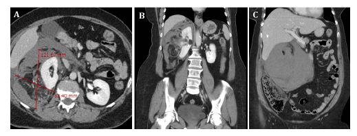
Figure 1: Appearance of ruptured AML mass (A); The coronal image of mass (B); Huge retroperitoneal hematoma and pushing organs (C).

Eyup Burak Sancak*1 Murat Tolga Gulpinar1 Alpaslan Akbas1 Mustafa Resorlu2 Gurhan Adam2 Gökhan Basturk1 Ali Riza Erdogan3
1Department of Urology, Faculty of Medicine, Canakkale Onsekiz Mart University, Canakkale, Turkey*Corresponding author: Eyup Burak Sancak, MD, Department of Urology, Canakkale Onsekiz Mart University, Faculty of Medicine, Canakkale, Turkey, Tel: 90 5323782204; Fax: 90 286 2180393; E-mail: eyupburaksancak@comu.edu.tr
Renal angiomyolipoma (AML) is the most common mesenchymal tumor of the kidney composed of fat, smooth muscle and blood vessels. Although AML is usually asymptomatic, it may sometimes be symptomatic because of retroperitoneal haematoma. The treatment modalities are the stabilization of the patient pursued by either selective transarterial embolization as a first line treatment or the second option of surgery. Dilated and tortuous renal artery anomalies can be observed in AML and may complicate selective embolization. In our study we present a case with large AML developing spontaneous rupture. Selective angioembolization for pseudoaneurysms was unsuccessful due to aberrant artery and torsioned vessel structures, finally the patient underwent surgical treatment.
Aberrant renal artery; Retroperitoneal hematoma; Ruptured angiomyolipoma; Super selective embolization
Renal angiomyolipoma (AML) is the most common mesenchymal tumor of the kidney. AML is four times more frequent in women than in men, and has an incidence of 0.3-3% in the general population. AML is characteristically present as benign lesion without local invasion, consisted of varying proportions of mature adipose tissue, smooth muscle and abnormal thick-walled blood vessels. Arteries within the structure of AML typically are numerous, do not include internal elastic membrane and have an irregular smooth muscle pattern [1-3].
Renal artery anomalies are very common and are estimated to have an incidence of 35%. It is stated in numerous studies that AML can be found together with anomalies of the renal artery. In the presence of renal artery anomalies, embolization and even surgery may be difficult [4-6]. Here we present planned treatment of a case with spontaneous rupture of large angiomyolipoma accompanied by aberrant artery.
A 63 year old female patient, weighing 85 kilograms and 165 cm tall, presented to our hospital from a state hospital with a diagnosis of retroperitoneal haemorrhagia and/or bleeding. The patient had presented at the abovementioned hospital with a spontaneous and sudden dull ache in the right flank region of 2 hours duration. A contrast enhanced computed tomography (CECT) scan revealed a probable retroperitoneal haematoma that required angiography. Therefore, the patient was urgently transferred to our hospital.
General condition of the patient was good-mediocre. There was no additional complaint except for flank pain. The personal and family history of TS was negative. There was a pain with deep palpation on the right flank and right inguinal area. Costovertebral angle tenderness was positive in the right flank area. Serial laboratory tests were performed which showed a decrease in haemoglobin level from 10.3 g/dl (haematocrit 30.4%), to 7.9 (haematocrit 24.1%) in twelve hours; blood pressure decrease 110/70 mmHg to 90/60 mmHg, with a mean heart rate of about 95 bpm.
The CECT scan was evaluated again. It was understood that it showed the presence of an abundant retroperitoneal haematoma, mainly located in the posterior and lateral peri-renal space and along the right parietal colic sulcus, with diameter 121.67 mm × 83.40 mm, extending to the pelvis (Figures 1a-1c). The haematoma also was pushing the vena cava, second duodenal portion, lower liver margin and same kidney encompassing the entire proximal-inferior tract of the same side (Figure 1c). Since the retroperitoneal haematoma was predominantly composed of fat, it was thought to be caused by rupture of an exophytic renal AML. No active bleeding was documented during CT imaging.
The patient had angiography planned. Right renal artery was entered, via the right transfemoral arterial approach. Following a selective arteriography, irregular torsioned arterial branches supplying the mass and two pseudoaneurysms localised in the middle pole were determined (Figures 2 and 3). During the process, due to technical difficulties related to tortuous and irregular arteries, superselective embolization of the segmental renal artery of the middle-pole was unsuccessful (Figure 3). During following hours, upon the deterioration of the patient’s hemodynamics, the patient underwent surgery. In the course of the operation, owing to bleeding from irregular arterial branches and clinical condition of the patient, nephrectomy was performed. Material sent for pathology after surgery confirmed AML. In addition to these, the renal angiography showed atypical aberrant renal arterial branch originating directly from the renal artery, ascending parallel to vena cava, giving branches to surrenal gland and the upper pole of the kidney, ending in a mass posterio-lateral of the renal area (Figure 4). It was thought that it may be a renal capsular artery which gained dominance; however, it followed a different way from the classical course of the renal capsular artery.
Figure 1: Appearance of ruptured AML mass (A); The coronal image of mass (B); Huge retroperitoneal hematoma and pushing organs (C).
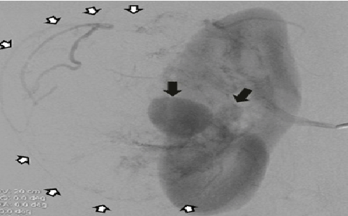
Figure 2: The blood supply of AML mass (white arrow) and middle pole localized two pseudoaneurysm (black arrow).
Renal angiomyolipoma (AML) is a benign mesenchymal tumor of the kidney composed of fat, smooth muscle, and blood vessels. Although AML is usually asymptomatic, may be symptomatic because of retroperitoneal haematoma. The choice of treatment varies according to the patient’s clinical status. The patients may consult as sporadic cases or with regard to tuberous sclerosis (TS). TS is identified in 20% of all renal AML cases, with 80% of them applying as multiple or bilateral lesions [1-3]. Although AML is known as usually benign, renal perivascular epithelioid angiomyolipoma (PEComa), a subtype of AML, is defined as a potentially malignant mesenchymal tumor which was recently added to the 2004 WHO classification of renal tumors [7].
AML is usually an asymptomatic. In a minority of the patients it is symptomatic and presents with complaints known as ‘Lenk’s triad’, consisting of haematuria (23%), palpable mass (47%) and flank pain (53%) [3,8]. In addition to this, AML is regarded as the most common cause of spontaneous renal bleeding [9]. The risk of haemorrhage is related to the angiogenetic component of AML, due to the existence of aneurysmatic vessels with an aberrational course, the size of the tumor and association with TS [10]. The patients with larger AML are at greater risk of spontaneous or traumatic rupture ending with haemorrhagic complications [11]. If there is a retroperitoneal haematoma secondary to ruptured AML, the treatment modalities are stabilization of the patient pursued by either selective transarterial embolization as a first line treatment or surgery. There are indications for surgery, which is radical or partial nephrectomy, if there is failed embolization, suspect malignant or persistent bleeding [3,9]. In this case, because of failed embolization, the patient was taken for operation. Due to tortuous and irregular arteries, the superselective embolization might be unsuccessful. It is stated that in AML’s arteries may originate from renal, adrenal, ureteral, gonadal, phrenic and lumbar arteries [2]. In our case, due to atypical vascularized tumor mass, bleeding might be persistent. Because of atypical vascularization of the huge mass and clinical features it was decided to perform nephrectomy.
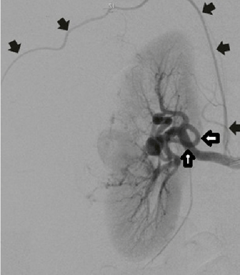
Figure 3: Tortuous and irregular arteries (white arrow) and unidentified aberrant artery (black arrow)
Each kidney is supplied from the single and wide renal artery which is a lateral branch of aorta abdominalis. These vessels are usually rooted in the aorta between L1-2 vertebrae and just below of the start of arteria mesenterica superior. Left artery usually emerges slightly above the right artery and is usually shorter. Each of the renal arteries enters the renal hilum and is divided into segmental branches feeding the renal parenchyma [12].
Renal arterial anomalies including their number, source, and course are common entities. Variations of the renal vessels are found in about 35% of cases studied. The renal artery may originate from the aorta or from the common iliac, internal iliac, inferior mesenteric, middle sacral, or from a common intercostal-renal trunk. The right artery may cross in front of, instead of behind, the vena cava. The renal artery may give off the inferior phrenic, hepatic, right renal, middle and inferior adrenal, gonadal, pancreatic, some of the colic arteries, and one or more of the lumbar arteries [2,13,14].
In our case, the right renal artery started at the level of the second lumbar vertebra. Atypical vessel originating from renal artery and feeding the AML followed an unusual course. Although this artery followed a track similar to renal capsular artery, it was still different because of the fact that it ascended parallel to vena cava, gave branches to surrenal gland and the upper pole of the kidney, and disappeared in a mass at the posterio-lateral of the renal area [15]. With this characteristic structure, the artery which did not have features of accessory renal artery was considered an aberrant artery. Such an artery was not encountered in the literature. The AML originating from both the middle pole segmental and the aberrant artery was not been documented in the literature.
The first treatment choice for ruptured angiomyolipoma is selective embolization. While performing embolization, it should be kept in mind that the kidney may have numerous aberrant blood supply. After unsuccessful embolization, surgery is an alternative treatment choice.
The authors report no conflicts of interest.
Download Provisional PDF Here
Article Type: Case Report
Citation: Sancak EB, Gulpinar MT, Akbas A, Resorlu M, Adam G, et al. (2015) Angiomyolipoma accompanying with renal aberrant artery. J Blood Disord Med 1 (1): doi http://dx.doi.org/10.16966/ 2471-5026.102
Copyright: © 2015 Sancak EB, et al. This is an open-access article distributed under the terms of the Creative Commons Attribution License, which permits unrestricted use, distribution, and reproduction in any medium, provided the original author and source are credited.
Publication history:
All Sci Forschen Journals are Open Access