
Figure 1: A pedunculated and twisted leiomyoma located in the fundus of the uterus.

Gaye YFO1* Niang MM1,2 Gassama O1 Thiam O3 Thiam M4 Cisse CT1
1Department of Gynecology and Obstetrics, Cheikh Anta Diop University, Dakar, Senegal*Corresponding author: Gaye YFO, Department of Gynecology and Obstetrics, Cheikh Anta Diop University, Senegal, Tel: 00221775235277; E-mail: yifigaye@gmail.com
Background: The torsion of pedunculated uterine leiomyoma is a rare complication of uterine leiomyoma.
Case: A 34-years-old woman was admitted at gynecological emergency for acute pelvic pain. The emergency trans-abdominal ultrasound was in favor of a right ovarian twisted cyst associated with leiomyomas. Laparotomy was performed and showed torsion of pedunculated uterine leiomyoma inserted on the fundus which was removed.
Conclusion: Torsion of a pedunculated uterine leiomyoma is a rare surgical emergency. Imaging should not delay emergency surgery that confirms or sometimes rectifies the diagnosis.
Pedunculated uterine leiomyoma; Torsion ultrasound laparotomy; Myomectomy
Uterine leiomyoma, a benign tumor of the uterine muscle, is a ubiquitous pathology affecting 20 to 25% of women in reproductive period [1]. Its incidence is 70% at the age of 50 years [2]. It is only symptomatic in 20 to 50% of cases and may have several and various characteristics depending on the number, size and the topography of the leiomyomas [2,3]. Among the multiple complications of the uterine leiomyoma, one is an exceptionnel, the torsion of pedunculated uterine leiomyoma. Its incidence is not well known, only few cases have been described in the literature. It is a surgical emergency whose clinical expression can simulate an adnexial torsion. Imaging can guide the diagnosis. However, laparoscopy or even laparotomy performed in an emergency ensures the management. We report a case of twisted pedunculated uterine leiomyoma discovered during the laparotomy.
A 34 years-old woman with no medical history (gravida 0, para 0) was admitted at gynecological emergency for acute pelvic pain appeared 5 days ago. She also complained of three-month bleeding and a lack of improvement of the pelvic pain despite analgesics. The general condition was normal. Physical examination revealed a rounded right painful mass measuring about 10 cm.
The emergency trans-abdominal ultrasound was in favor of a right ovarian twisted cyst which measures 110 mm associated with leiomyomas. Laparotomy rectified the diagnosis by highlighting a necrotic pedunculated uterine leiomyoma that had undergone torsion. The twisted fibroma was 15 cm in diameter (Figure 1).
Figure 1: A pedunculated and twisted leiomyoma located in the fundus of the uterus.
The annexes were healthy. An uncomplicated myomectomy was performed; the twisted fibroma weighed 3700 grams (Figure 2). The surgical follow-up was uneventful and the discharge was authorized 4 days after the surgery.
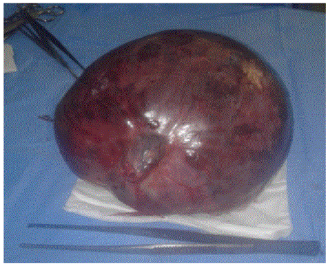
Figure 2: Surgical specimen (length: 15 cm, weight: 3700 grams).
The torsion of pedunculated uterine leiomyoma is one of the most uncommon surgical emergency [3]. Its symptoms are the same with surgical acute abdomen. This disease imitates different pathologies as the ectopic pregnancy and the adnex twist [4,5].
This torsion occurs only on pedunculated uterine leiomyoma especially if the pedicle is long and thin. The size of the leiomyoma is a key factor in the onset of an irreversible twist. Tsai, in 2006 [6], had reported a case of sub-serosal fibroma measuring 8 cm in diameter. Our patient had a leiomyoma bigger than Tsai’s with 15 cm of diameter and 3700 grams of weight. The pregnancy with the increase of the uterine size and the postpartum with the uterine involution are periods at risk of torsion of pedunculated leiomyoma [7]. Gaym [8] described a case of twisted leiomyoma in a patient in the first trimester of pregnancy. However, this complication can occur outside the gravido-puerperium period. Our patient had no history of gravidity and was in her gynecological period.
Clinical examination and imaging (CT/ultrasound) help to make the diagnosis by highlighting the pedunculated fibroma and checking the integrity of the ovaries. However, the preoperative diagnosis of twisted pedunculated fibroma is difficult to make. In Tsai’s observation [6], ultrasound had uncovered the pedunculated leiomyoma but had not detected the twist. For our patient, ultrasound found a twist of adnexs. Therefore, this confirms the lack of sensibility of the ultrasound in the diagnosis of twisted pedunculated leiomyoma [9]. Thus, in any case, imaging (CT/ultrasound) should not delay the surgery which confirms or sometimes rectifies the diagnosis. The treatment depends on the precocity of the diagnosis, the size of the leiomyoma, the medical equipment and the surgeons experience. In 2005, Katsumori [10] had described the embolization of the uterine arteries for the treatment of pedunculated leiomyoma. Cuillier [4], who reported a twisted pedunculated leiomyoma with 12 cm of diameter, performed a laparoscopy associated with suprapubic mini-laparotomy in order to extract the leiomyoma. However, abdominal myomectomy (laparotomy) is the most common technic in the management of twisted pedunculated fibroma. For our patient, as Tsai [6] and Foissac [1], we performed a median laparotomy. The surgical procedure depends on the patient’s age and whether or not she wants to become pregnant. Due to the young age of our patient and her gravidity status, we realized a myomectomy. In the opposite, Foissac [1] had performed a total hysterectomy associated with bilateral adnexectomy in an old multiparous.
Torsion of pedunculated leiomyoma is a rare surgical emergency. The clinical expression can simulate a twisted adnex. We must mention it in case of acute pelvic pain especially if the patient is treated for uterine leiomyoma. Abdominal myomectomy by laparotomy remains the treatment of reference.
Download Provisional PDF Here
Aritcle Type: Case Report
Citation: Gaye YFO, Niang MM, Gassama O, Thiam O, Thiam M, et al. (2017) The Torsion of a Pedunculated Uterine Leiomyoma, a Rare Etiology of Acute Pelvic Pain: about an Observation and a Review of The Literature. Gynecol Women’s Health Res 1(1): http://dx.doi.org/10.16966/2689-3096.104
Copyright: © 2017 Gaye YFO, et al. This is an open-access article distributed under the terms of the Creative Commons Attribution License, which permits unrestricted use, distribution, and reproduction in any medium, provided the original author and source are credited.
Publication history:
All Sci Forschen Journals are Open Access