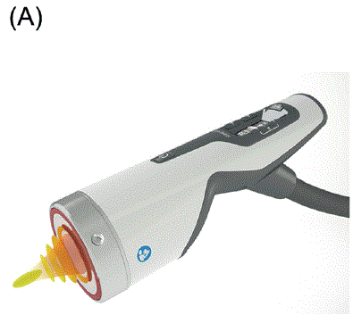
Figure 1A: Forced AWT.

Kuriko Kimura*
Health & Beauty Clinic Le Coquelicot, 5F KOTY Bldg, 5-9-1, Ginza, Chuo-ku, Tokyo 104-0061 Japan*Corresponding author: Kuriko Kimura, Health & Beauty Clinic Le Coquelicot, 5F KOTY Bldg, 5-9-1, Ginza, Chuo-ku, Tokyo 104-0061 Japan, Tel: 03-3572-5965; E-mail: kuriko_k@coquelicot.co.jp
The author executed a series of self-biopsies following focused acoustic, and focused and radial acoustic wave therapy to examine the effectiveness of the two types of acoustic wave therapy and to identify and investigate mechanotransduction; the mechanism of action produced by acoustic wave therapy. A case of a 28-year-old patient’s acoustic wave therapy that improved a vascular malformation is reported as a successful example. The series of histopathologic examinations following acoustic wave therapy strongly suggest that mechanotransduction causes neither fatal damage nor critical degeneration of tissue, and rather create moderate changes; indicating that the treatment is safely effective. There are distinctive differences in histopathologic changes between the two types of acoustic waves; the combination of focused and radial acoustic wave therapy leads to a faster normalizing change with less infiltration of cells than that following only focused-acoustic wave therapy. Further research on the mechanism of acoustic wave therapy is awaited, and a clear elucidation of this mechanism will bring about widespread recognition of this modality as a promising treatment for skin tightening and rejuvenation.
Acoustic wave therapy; Focused acoustic wave; Focused and radial acoustic wave; Skin tightening; Histopathology
Acoustic Wave Therapy (AWT) has been applied for a variety of medical purposes and is currently receiving increasing attention [1,2]. AWT is also known as Extracorporeal Shock Wave Therapy (ESWT) and was initially used to disintegrate kidney stones. AWT is also able to treat many orthopedic disorders, such as plantar fasciitis and calcific tendonitis, and is available to improve refractory skin ulcers as well as rejuvenate skin. Moreover, AWT is able to mitigate capsule contracture caused by breast augmentations and reduce dysfunctional fat tissue, including cellulite; therefore, it is used for body slimming and contouring.
The objective of this study was to examine the effectiveness of AWT and to investigate mechanotransduction [3-8]; however, the concept of mechanotransduction is abstract and thus difficult to understand intuitively, as substantial visible evidence is not present. The author was curious about what was occurring inside and wanted to visualize mechanotransduction, even if to a limited degree.
The treatment method and technique the author adopted for acoustic wave therapy was slow in motion, repeatedly dragging saggy skin pulling it upward to where the former skin was supposed to be [1].
The author has experience in more than 18000 cases of facial tightening treatment by acoustic wave therapy from 2016 to 2023, has executed a series of self-biopsies following AWT and has reported the histopathologic findings as well as the diagnostic surgical case of a 28-year-old patient. To the author’s knowledge, there is no internationally published paper that has reported chronological histopathological changes following AWT.
It was proven several decades ago that ESWT can be applied in urologic and orthopedic disorders. Recently, in cosmetic and aesthetic medicine, AWT has introduced as an effective treatment method for body slimming and for reducing cellulite and excessive malfunctional fat tissue, as well as for skin tightening and rejuvenation [9,10]. AWT is a noninvasive, nonthermal and nonfreezing technique based on the propagation of mechanical stimulation in tissue. Acoustic waves are special pressure waves that are generated when atmospheric conditions suddenly change, such as when a jet plane accelerates beyond supersonic speeds or when there is thunder [1]. Acoustic waves are characterized by single, mostly positive pressure pulses followed by a small tensile wave component. Acoustic waves induce a variety of biological reactions through the shear and pressure forces that they produce, which are collectively called mechanotransduction [1,2]. The stimulation creates signals inducing growth factors and cytokines, reducing redundant fat and activating blood and lymphatic flow in tissues [1,2,11].
Unlike the continuous periodic wave form of ultrasound devices such as High-intensity Focused Ultrasound (HIFU), acoustic waves do not generate heat, even if the energy output is very high, and this characteristic feature prevents thermal damage to the skin and the body. Acoustic waves have single amplitude with high pressure force, whereas ultrasound exhibits continuous amplitude. Another notable characteristic of AWT is its versatility in accordance with its intensity: The emission intensity used in kidney stone lithotripsy is approximately 4.0 mJ/mm2, whereas regeneration and pain reduction require less than one-tenth that of lithotripsy.
This histopathologic tracing study started when the author was checking the directions for how to use the biopsy kit, using the author’s own abdominal subcutaneous fat tissue prior to applying the biopsy to a patient. The biopsy trepan was the Kai Medical Disposable BIOPSY PUNCH (4 mm) with PLUNGER made in Gifu, Japan.
The acoustic wave device used in this study was DUOLITH® SD1 TOWER »ultra« AWT® (STORZ MEDICAL ITALIA S.R.L. Roma Italy), which loads a pair of acoustic waves: the planar hand piece generates deep focused acoustic pulses electromechanically, having as light stimulative breaking effect. The emission intensity was 1.24-0.45 mJ/mm2 with a pulse frequency of 3-5 Hz (Figure 1 (A,B)). The linear hand piece generates radial acoustic pulses pneumatically, removing dysfunctional fat and accelerating blood and lymphatic flow in tissues The emission intensity was set at 4 bars with a frequency of 21 Hz (Figure 2 (A,B)).
Figure 1A: Forced AWT.
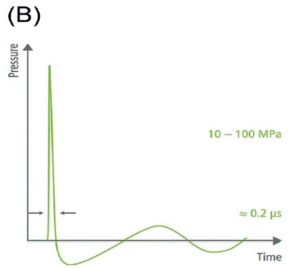
Figure 1B: Forced waveform.
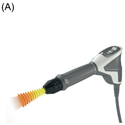
Figure 2A: Radial AWT.
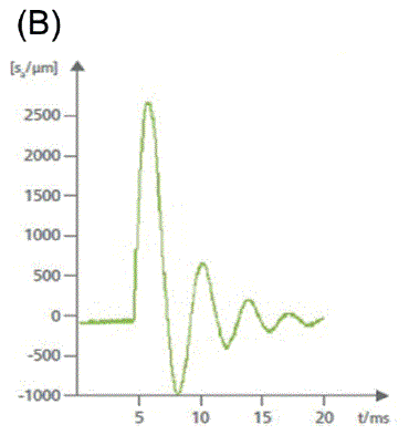
Figure 2B: Radual wave form.
For the objective evaluations of AWT, three-dimensional digital photographs from a Canfield Scientific Vectra Handy camera and software (Figure 3) and skin-analyzing digital photographs from VISIA were used (Figure 4) (Canfield Scientific Inc, Fairfield, NJ, USA).
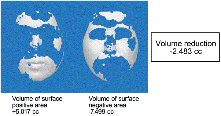
Figure 3: Vectra.
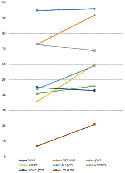
Figure 4: Visia.
Each collected biopsy specimen was immediately soaked in fresh 20% formalin in the specified container, brought to a major Japanese pathological laboratory and processed for Hematoxylin-eosin (HE) staining by professionals. The histopathologic findings were assessed, and diagnostic consultations were conducted by a proficient emeritus professor of pathology and dermatology.
The author was prudent regarding the treatment and care of the patient, considering medical ethics throughout the research. Written informed consent, including that for publication of this report, was obtained from the patient.
The first study the author conducted was the pre-and post-onemonth change in subcutaneous fat tissue following AWT; however, every cellular change seemed to be complete within at least one month after the author proceeded to the next trial (Figure 5a) (Day 1). The second study was the comparison of the shorter time axis after AWT. The “granular transformation” finding was particularly noteworthy. It occurred 3 days after the focused and radial-AWT, producing slight membranous degeneration in the subcutaneous fat tissue. The HE staining was slightly faded, and some of the fat cells were fragmented and nearly obliterated (Figure 5b) (Day 3).
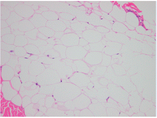
Figure 5A: The very first time histoptho.
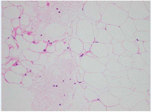
Figure 5B: Granular transformation.
Another suggestive finding was observed after 7 days of AWT; the cells treated with focused AWT had a little infiltration of neutrophils and histiocytes (Figure 5c) (Day 7) whereas the cells treated with focused and radial AWT had no cell infiltration (Figure 5d) (Day 7).
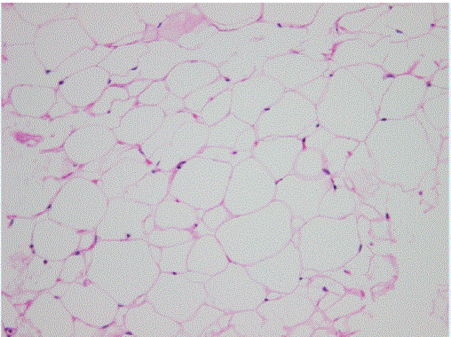
Figure 5C: Some cell infiltration focused only.
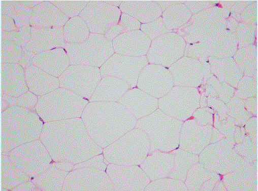
Figure 5D: No cell infiltration focused and radial.
The next study the author carried out was a chronological review of the biopsy, which was full of new findings: for the 24 to 48 hours after the AWT, there was no change observed in the epidermis, dermis and subcutaneous fat tissue, and the cell bodies of the fat tissue were characteristically stained pink. There was no disappearance or degeneration of the nucleus or destruction of the cell membrane.
After 72 hours of focused and radial-AWT, there was another unique finding of “a small air cavity in part of the subcutaneous fat tissue surrounded by collagen fibers” (Figure 6a) (Day 3). The color tone of the HE staining was slightly different; in the cells treated with focused-AWT, the stain became reddish pink, and in the cells treated with focused and radial-AWT, the stain became pale pink.
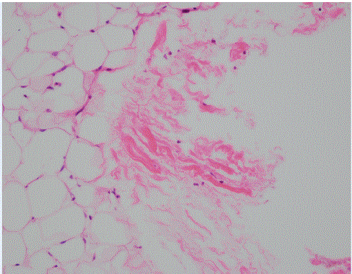
Figure 6A: Small air cavity.
After 96 hours, conspicuous cell infiltrations with lymphocytes were caught in the very deep layer of fat tissue (Figure 6b) focused only, and (Figure 6c) focused and radial-AWT (Day 8) there was no marked change observed in the subcutaneous fat tissue other than the cell bodies of the fat tissue, which were stained pale pink in both the focused-acoustic and focused and radial AWT. No fatal destructive degeneration was observed in the subcutaneous fat tissue throughout the study.
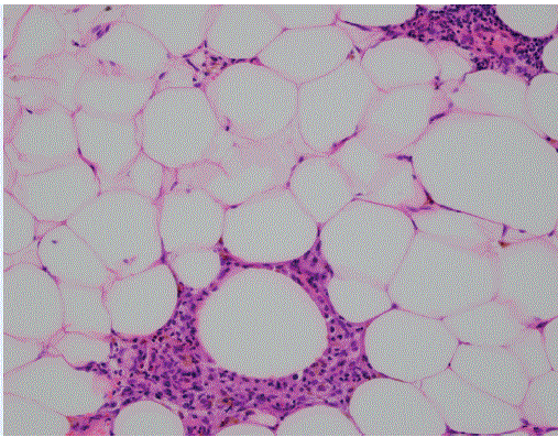
Figure 6B: After 96 hours (focused only).
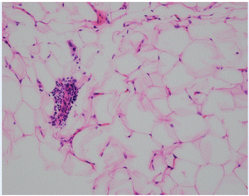
Figure 6C: After 96 hours (focused and radial).
For the final histopathological examination, the author tried to identify when changes in the subcutaneous fat tissue would stop after one treatment session of AWT; the changes stopped after approximately 336 hours (Day 14), and very slight infiltration of lymphocytes was observed.
The present patient was born with a nonhereditary hemangioma (left head, left face and left neck). Hermain concern was that her left ear was asymmetrically larger than her right ear, so she wanted to remove some of the hemangioma lesions. She also wanted to undergo facial tightening treatment by AWT to reduce unwanted facial volume. The patient had previously been diagnosed with hemangioma simplex at two university hospitals; however, after all the detailed examinations, including CT scanning and angiography, the correct diagnosis was a high-flow type of Arteriovenous Malformation (AVM).
From the age of 5 to 8 years, the patient had undergone several laser treatments but dropped out due to pain. At approximately age 17, the patient developed hyperhidrosis with a heat sensation on the left side of the face and neck. At the age of 23, the patient tried another laser treatment at a nearby hospital, which lightened the redness of her left cheek, but the heat sensation remained the same.
With the patient’s consent, facial tightening treatment by AWT was performed by the author as AWT has a beneficial effect on reducing volume and rejuvenating skin [1,2] her left thigh was treated with focused-AWT, and her right thigh was treated with focused and radial-AWT. Three days later, AVM reduction surgery was performed on the left ear of the patient, and biopsy specimens were collected from the left ear and thighs by a skilled plastic surgeon.
The author performed one session of facial tightening treatment on the patient following focused and radial-AWT. The vascular malformation area on the left side of the patient’s face and neck was reduced, and the redness of the left cheek decreased. The patient’s facial volume calculated by VECTRA was-2.483 cc after 5 months of AWT and the surgery (VECTRA).
The skin analysis (VISIA) showed improved data for pores, porphyrins, UV spots, skin texture, wrinkles, and red areas. The data for brown spots and other spots became slightly worse, affected by the seasonal disadvantage of August (i.e., summer time) (VISIA) (Figure 7).
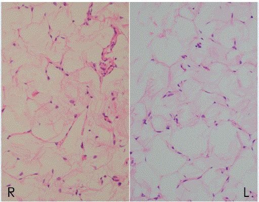
Figure 7: 27 Y old patient. Focused only and focused and radial 14.
Regarding the changes resulting from AWT and left ear surgery, the patient’s pre- and post treatment photographs showed that the redness and thickness of her upper, middle and lower cheeks were reduced and that the red-colored area of her neck looked thinner. The patient was pleased with the compact, balanced left ear after the hemangioma reduction surgery, and moreover, she mentioned that the annoying heat sensation had diminished by more than half in intensity after AWT (Figure 8).
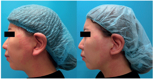
Figure 8: Pre-treatment/ Post treatment.
By means of a histopathologic approach, the author investigated and tried to disclose the mechanisms of AWT and mechanotransduction. The unexpected histopathological finding was that there was a period of no change for the first 24 to 48 hours after AWT, with no formative alterations visible from the outside. The author speculated that the cessation period could have been caused by the relatively strong intermittent mechanical stimulations by AWT, and on reflection, and possibly, the first 48 hours might have been an indispensable period during which the mechanical stimulations were converted to biological signals.
The very first positive histopathological finding the author encountered was “granular transformation” 3 days after (72 hours focused and radial-AWT, which was almost the same timing or phase of “the small air cavity surrounded by collagen fibers” following focused and radial-AWT. Although strict scientific reproducibility was not attained, the author suspected these unique and random findings could have been a part of metabolic change induced by AWT, and those types of changes may have been nothing but mechanotransduction.
On the 7th day of AWT, the focused-AWT and the focused and radial-AWT differed regarding cell infiltration. This may suggest that each AWT has respective and specific features, possessing synergetic effects. It was clear that the combination of focused and radial AWT brings faster and smoother changes with less infiltration of cells and is more effective.
The reason for the differences in color tone of the HE staining is not definite but could be related to the accompanying dynamic changes in the permeability of the cell membrane.
Regarding the 28-year-oldpatient’s biopsy results, the Right thigh (R) was treated with focused AWT, and the cell bodies of the right thigh were stained reddish pink. The Left thigh (L) was treated with focused and radial-AWT, and the cell bodies were stained pale pink. The author reconfirmed that focused and radial-AWT is a more beneficial and all-inclusive treatment than only focused-AWT.
Lastly, the author unexpectedly encountered the conspicuous cell infiltration with lymphocytes after 96 hours: (Figure 6b) (Day 8) (Figure 6c) (Day 8). The cell infiltrations were caught in the deep layer of fat tissue, not subcutaneous but visceral fat tissue. Subcutaneous fat and visceral fat are considered different in nature but the visceral fat may be related and affected by the acoustic wave therapy.
The author thinks that the mechanotransduction signals appear naturally from time to time.
The author was aware of the limitations of the self-biopsy method assessed with a small number of people, but this study and idea came in the midst of the COVID-19 pandemic. There may be differences in age, race, lifestyle, and the methods and timing of biopsy
Mechanotransduction changes naturally take place and they do not cause fatal damage or critical degeneration in tissue. Acoustic wave therapy can be safely and effectively applied for a variety of medical uses, and the author believes it will gain wide acceptance from medical professionals as well as patients.
Download Provisional PDF Here
Aritcle Type: CASE REPORT
Citation: Kimura K (2024) Histopathologic Examination Changes Observed as Mechanotransduction Following Focused and Radial Acoustic Wave Therapy. J Clin Cosmet Dermatol 8(1): dx.doi.org/10.16966/2576-2826.181
Copyright: ©2024 Kimura K. This is an open-access article distributed under the terms of the Creative Commons Attribution License, which permits unrestricted use, distribution, and reproduction in any medium, provided the original author and source are credited.
Publication history:
All Sci Forschen Journals are Open Access