Introduction
Facial volume loss management is one of the commonest esthetic facial treatments particularly for the area of the temporal fossa. This area is one of the areas of the face that tends to be significantly affected by volume loss (facial skeleton action) as a result of aging.
Hyaluronic Acid (HA) is an extracellular matrix key molecule that is important for the maintenance of tissue structure and vascularity [3,4]. In addition to its hydrophilic properties, HA plays an important role in fibroblast metabolism, nutrient transport and inter-cell signalling [5-8].
HA bio-implants are commonly used through the body for tissue volume loss correction. We have previously published on our experience with regards to the use of Stylage® XL and XXL (LABORATOIRES VIVACY, France), which are non-animal in origin sterile dermal fillers manufactured using “Inter-Penetrating NetworkLike” (IPN-Like) cross-linking technology. We previously reported on clinical outcomes, and gel tissue stability following injection and up to 24 months in 4 Caucasian patients treated in our clinic [1,2]. These patients had HA gel injections to their temporal fossae for volumizing and reshaping purposes.
However, in view of the observation that the residual gels and their clinical effects were still present 24 months after injection, we obtained patients’ consents to continue collecting photographic, imaging and clinical outcome data 42 months after the single injection session of their temporal hollows. Therefore, the aim of this study was to describe the behavior and volume resorption of these HA volumizing gels at the long term.
Methods
Participants
Our study population comprised 4 Caucasian patients who were enrolled into our two previous studies that compared Stylage XL versus Stylage XL-Lido (incorporating lidocaine added at manufacturing) [1] and Stylage XL versus Stylage XXL [2].
When enrolled into the initial studies, the two participants injected with the same HA volumizing gel with or without added lidocaine were aged 58 and 71 and the other two participants who participated in the Stylage® XL versus XXL, were 67 and 75 years old. Participants demographics, the HA product type, HA volume injected, site of injection, skin type according to the Fitzpatrick classification [9] and aging grade according to the Glogau classification [10] are presented in table 1.
| Participant |
Age |
Right Temple |
HA types |
Left Temple |
Fitzpatrick’s
Classification [5] |
Glogau’s
Classification [6] |
| Participant 1 |
71 |
XL
1.00 ml |
XL
versus
XL-Lido |
XL-Lido
1.00 ml |
III |
III |
| Participant 2 |
58 |
XL-Lido
0.55 ml |
XL
0.50 ml |
II |
III |
| Participant 3 |
75 |
XL
1.00 ml |
XL
versus
XXL |
XXL
0.95 ml |
III |
III |
| Participant 4 |
67 |
XXL
1.00 ml |
XL
1.00 ml |
III |
III |
Table 1: Patients’ demographic data, skin type according to the Fitzpatrick classification [9] and aging grade according to the Glogau classification [10], the product chosen and the side injected.
Prior to enrollment into the original studies, patients were provided with written and oral information about the procedure and study and were only included after obtaining a written valid informed consent from them. Participants also consented separately to the follow-up extension of the study. All the study procedures were in full compliance with the Helsinki declaration and participants had full ownership of their images and were made aware that these would only be used for any purpose with their full agreement. The HA products were used within the recommended manufacturer and licensing protocol.
Procedure
All injected HA gels were produced using the registered and patented ‘Inter Penetration Network Like®” (IPN-Like® cross-linking technology) characterized by a double, interpenetrating matrix, with added mannitol for its anti-free radical effect, particularly against hydroxyl radicals. The dual nature of the matrix influences the gel’s viscoelastic properties and hence facilitating injection and making the gel suitable for wrinkle filling wrinkles and correction of facial shape and volume. Gels of low elasticity and low viscosity are used to fill wrinkles where their physical characteristics allow them to penetrate between extracellular matrix fibers. In contrast, a highly viscous and elastic gel is more suitable for deeper injection into subcutaneous fatty tissue (hypodermis and muscle, even close to bone. The three HA-based gels used in this study were cross-linked with the wellknown and most widely used cross-linking agent in the aesthetic field: BDDE (1,4-ButaneDiol Diglycidyl Ether). This step creates covalent bonds between HA fibers. The network is thus modified, increasing the rheology of the gels and indirectly reducing water absorption. The degree of cross-linking ranges between 5 to 6%.
The HA gels used in this study have the same therapeutic indications, namely, the correction of facial volume. Therefore, they are of high viscosity and elasticity. The following gels were used:
• Stylage® XL without lidocaine (XL), 26 mg/ml HA. Lots: -EXI18075F-Exp 2020 - 08 - EXH18031F - Exp 2020-07.
• Stylage® XL with lidocaine (XL-L), 26 mg/ml HA. Lot: -LXD16362F -Exp: 2019-05.
LXD16362F -Exp: 2019-05. • Stylage® XXL (XXL), 21 mg/ml HA. Lot: -EDF18012H-Exp: 2020-06.
All the patients, the 2 expert assessors and the radiologist were blinded to the type of injected HA. The 4 patients were each injected in both temporal fossae using the 2 different HA gels being compared (one on either side). In the study where Stylage® XL with and without lidocaine was compared, the gel without lidocaine was injected first to mitigate the risk of distorting participants' pain perception due to the lidocaine effect. In the study where Stylage® XL was compared to XXL, the Stylage® XL was injected first. The needle was gently advanced until it was felt to come in contact with bone (Figures 1 and 2). If necessary, a second or even third injection point was used, depending on what was deemed necessary to achieve optimal outcome. However, the volume injected never exceeded 1 ml per temporal fossa. The technique of injection was previously reported in detail [1,2].
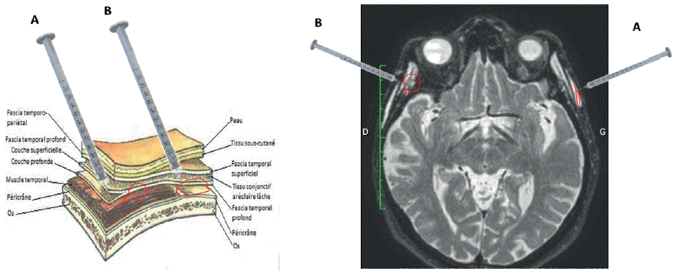
Figure 1: Diagram of injection into the temporal fossa. Examples of needle placement in the temporal fossa during a bolus injection, with simulation of the behavior of the injected hyaluronic acid gel.
A: in the temporal muscle or between the fascias; diffusion between superficial and deep fascia.
B: against the bone; diffusion upwards, downwards and possibly between the fascia.
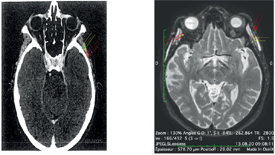
Figure 2: Examples of needle placement in the temporal fossa during a bolus injection.
Left image: Red needle is against the bone whereas yellow needle has no bone contact.
Right image: Yellow needle has no bone contact, and the bevel is pointing upwards. Red needle has no bone contact, bevel is pointing downwards (right of the image) and upwards (left of the image).
Photographs
As previously described, participants were photographed before and just after the injection, then every six months for 2 years [1,2]. Following the same protocol and for the purpose of this study, images of the face, profile, right and left three-quarter views were taken in the principal investigator’s medical room at month 42 (M42). One patient was followed up at month 44, the delay being due to an illness. She was photographed at the MedImage radiology institute in Geneva, for practical reasons. The camera used was a Nikon® DX digital camera, lens AF-S DX Nikkor, ED 18-55 mm 1 : 3.5-5.6 GII (Figures 3 and 4).
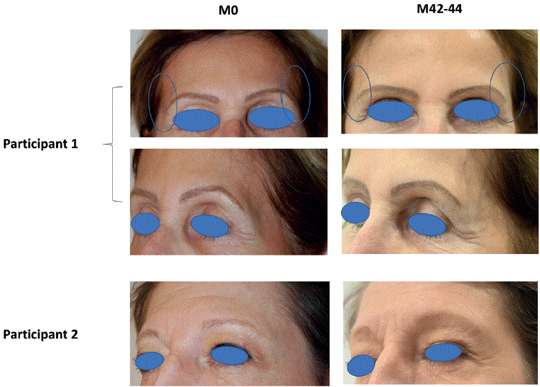
Figure 3: Photographic images of patients 1 and 2 (Stylage® XL versus XL-lido) at baseline and M42-44.
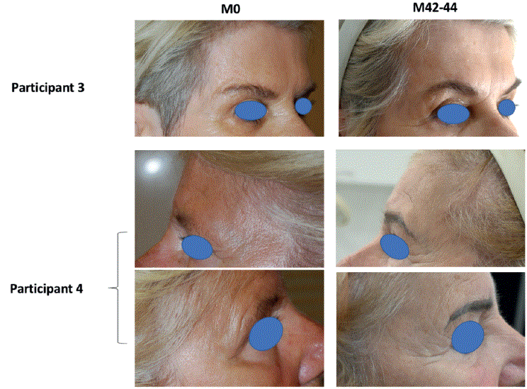
Figure 4: Photographic images of patients 3 and 4 (Stylage® XL versus XXL) at baseline and M42-44.
MRI
MRIs were performed at MedImage, medical imaging institute Geneva, Switzerland-using an Achievia 1.5 Tesla MRI (nuclear magnetic resonance imaging) unit (Philips SA Health Systems, GlandSwitzerland). On MRI images, the richer the tissue water content the whiter the image is, while bone appeared black, and other structures (muscles, fascia, etc.) showed as gradients of grey. Assessments were carried out blindly by the radiologist. The sequences used were 3DFLAIR, 3DT1, 3DT2.
MRIs were performed at baseline, post-injection and the same follow-up time points as the photographs. For the purpose of this study, an additional MRI was performed at M42 (M44 for one patient) month (Figure 5).
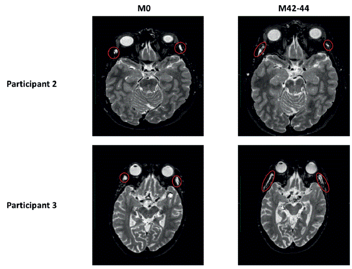
Figure 5: MRI images of participants 2 and 3. Participant 2. Right temple-0.55 ml Stylage® XL-Lido; Left temple: 0.50 ml Stylage® XL. Participant 3: Right temple-1.00 ml Stylage® XL; Left temple:-0.95 ml Stylage® XXL.
Outcomes
The treating physician and 3 independent experts assessed aesthetic outcomes using M42-44 photographs compared to those taken at baseline using the Global Aesthetic Improvement Scale (GAIS) [11] and the Merz-Pharma MAS© scale [12].
MRI scans of the subjects’ faces were used to monitor the behavior and evaluate the residual volume of HA gels over time. Assessments were undertaken using the Philips’ IntelliSpacePortal 7.0 image post-processing software which includes a tool for gel visualization and volume calculation from images acquired in 2D. This “tissue segmentation” tool defines an area of interest on a 2D image based on pixel intensity, tissue homogeneity, variance and regularity. Following zone selection, the software identifies all pixels with similar characteristics. An extrapolation from the 2D cross-sections is then performed providing a volume measurement expressed in mm3.
Results
Photographs at M42-44
Participant 3 had their photograph taken on month 44 in the radiology institute prior to the MRI examination. The lighting in the imaging institute was different from that in the investigator’s office, where the rest of the photographs were taken, hence the noticeable color differences. The 3 experts (injector+2 blinded evaluators) evaluated the potential persistence of a clinical improvement, solely on photographs, 42-44 months after their initial treatment of the temporal fossae of the 4 participants. Table 2 shows experts’ assessments using the Merz-Analogic Scale® (MAS®), in comparison to previous assessments undertaken at baseline before treatment, M12 and M24. Except for participant 2 (Stylage® XL vs. Stylage® XL-Lido) on the Stylage® XLLido side, all the other participants were considered to have a degree of persisting improvement compared to baseline. However, there was a gradual deterioration in the level of improvement compared to the other assessment time-points.
| |
Expert |
BT |
|
M12 |
ΔR |
M12 |
ΔL |
M24 |
ΔR |
M24 |
ΔL |
M42-44 |
ΔR |
M42-44 |
ΔL |
|
Stylage® XL vs. Stylage® XL-Lido |
Participant 1 |
| |
R |
L |
R XL |
|
L XL-
lido |
|
R XL |
|
L XL-lido |
|
R XL |
|
L XL-lido |
|
| 1 |
3 |
4 |
3 |
0 |
2 |
2 |
3 |
1 |
2 |
3 |
4 |
-1 |
2 |
2 |
| 2 |
2 |
3 |
0 |
2 |
2 |
1 |
1 |
1 |
1 |
2 |
1 |
1 |
1 |
2 |
| 3* |
4 |
4 |
3 |
1 |
2 |
2 |
4 |
0 |
2 |
2 |
2 |
2 |
2 |
2 |
| Mean |
3.0 |
3.7 |
2.0 |
1.0 |
2.0 |
1.7 |
2.3 |
0.7 |
1.3 |
2.3 |
2.3 |
0.7 |
1.7 |
2.0 |
| |
Participant 2 |
| |
|
R |
L |
R X L
-lido |
|
L XL |
|
R X
L-lido |
|
L XL |
|
R X L
-lido |
|
L XL |
|
| |
1 |
2 |
0 |
1 |
1 |
0 |
1 |
1 |
0 |
0 |
-1 |
1 |
0 |
1 |
0 |
| |
2 |
2 |
2 |
1 |
1 |
1 |
1 |
2 |
0 |
2 |
0 |
1 |
0 |
1 |
0 |
| |
3* |
2 |
0 |
1 |
2 |
0 |
0 |
1 |
1 |
0 |
0 |
1 |
1 |
1 |
-1 |
| |
Mean |
1.7 |
1.0 |
0.7 |
1.3 |
0.3 |
0.7 |
1.3 |
0.3 |
1.3 |
-0.3 |
1.0 |
0.3 |
1.0 |
0.3 |
| |
Participant 3 |
| |
|
R |
L |
R XL |
|
L XXL |
|
R XL |
|
L XXL |
|
R XL |
|
L XXL |
|
| |
1 |
3 |
3 |
1 |
1 |
0 |
1 |
1 |
2 |
0 |
1 |
2 |
2 |
1 |
3 |
| |
2 |
2 |
3 |
2 |
0 |
2 |
1 |
2 |
0 |
2 |
1 |
1 |
1 |
1 |
2 |
| |
3* |
3 |
3 |
1 |
2 |
0 |
3 |
1 |
2 |
0 |
3 |
2 |
1 |
1 |
2 |
| |
Mean |
3.0 |
2.3 |
2.0 |
1.0 |
1.7 |
1.7 |
1.7 |
1.3 |
1.7 |
1.7 |
1.7 |
1.3 |
1.0 |
2.3 |
|
Stylage® XL vs. Stylage® XXL |
Participant 4 |
| |
R |
L |
R XXL |
|
L XL |
|
R XXL |
|
L XL |
|
R XXL |
|
L XL |
|
| 1 |
4 |
4 |
3 |
2 |
2 |
2 |
1 |
3 |
1 |
3 |
2 |
2 |
2 |
2 |
| 2 |
3 |
2 |
2 |
1 |
1 |
1 |
2 |
1 |
2 |
0 |
1 |
2 |
2 |
1 |
| 3* |
2 |
2 |
0 |
2 |
0 |
2 |
1 |
1 |
1 |
1 |
1 |
1 |
1 |
1 |
| Mean |
3.0 |
2.0 |
1.3 |
1.7 |
1.0 |
1.7 |
1.3 |
1.7 |
1.3 |
1.3 |
1.3 |
1.7 |
1.7 |
1.3 |
Table 2: Evaluation of the efficacy temporal fossa treatment with «IPN-Like ®» HA volumizer gels according to the MAS® scale by experts.
*Physician who performed the injections. XL: Stylage® XL without lidocain, XL-L: Stylage® XL with lidocaine, XXL: Stylage® XXL without lidocaine; R: Right, L: Left; BT: before treatment; M12, M24, M42-44=12,24 and 42-44 months follow-up time points.
|
Stylage® XL vs. Stylage® XL-Lido |
EXPERT |
AT |
AT |
M12 |
ΔR |
M12 |
ΔL |
M24 |
ΔR |
M24 |
ΔL |
M42-44 |
ΔR |
M42-44 |
ΔL |
| Participant 1 |
| |
R XL |
L XL-li |
R XL |
|
L XL-li |
|
R XL |
|
L XL-li |
|
R XL |
|
L XL-li |
|
| 1 |
3 |
4 |
2 |
-1 |
4 |
0 |
2 |
-1 |
4 |
0 |
2 |
-1 |
4 |
0 |
| 2 |
2 |
2 |
1 |
-1 |
1 |
-1 |
1 |
-1 |
2 |
0 |
3 |
1 |
4 |
2 |
| 3* |
3 |
4 |
2 |
-1 |
4 |
0 |
2 |
-1 |
4 |
0 |
3 |
0 |
3 |
-1 |
| Mean |
2.7 |
3.3 |
1.7 |
-1 |
3.0 |
0.3 |
1.7 |
-1.0 |
3.3 |
0.0 |
2.7 |
0 |
3.7 |
0.3 |
| Participant 2 |
| |
R XL-li |
L XL |
R X L -li |
|
L XL |
|
R XL -li |
|
L XL |
|
R X L -li |
|
L XL |
|
| 1 |
4 |
4 |
4 |
0 |
4 |
0 |
3 |
-1 |
4 |
0 |
2 |
-2 |
2 |
-2 |
| 2 |
1 |
1 |
1 |
0 |
1 |
0 |
1 |
0 |
1 |
0 |
1 |
0 |
4 |
3 |
| 3* |
4 |
4 |
4 |
0 |
4 |
0 |
4 |
0 |
4 |
0 |
3 |
-1 |
4 |
0 |
| Mean |
3.0 |
3.0 |
3.0 |
0 |
3.0 |
0 |
2.7 |
-0.3 |
3.0 |
0 |
2.0 |
-1 |
3.3 |
0.3 |
| Participant 3 |
|
Stylage® XL vs. Stylage®XXL |
|
R XL |
L XXL |
R XL |
|
L XXL |
|
R XL |
|
L XXL |
|
R XL |
|
L XXL |
|
| 1 |
4 |
4 |
3 |
-1 |
4 |
0 |
3 |
-1 |
3 |
-1 |
2 |
-2 |
3 |
-1 |
| 2 |
2 |
3 |
1 |
-1 |
2 |
-1 |
2 |
0 |
2 |
-1 |
4 |
2 |
4 |
1 |
| 3* |
4 |
4 |
3 |
-1 |
4 |
0 |
3 |
-1 |
4 |
0 |
3 |
-1 |
3 |
-1 |
| Mean |
3.3 |
3.7 |
2.3 |
-1 |
3.3 |
-0.3 |
2.7 |
-0.7 |
3.0 |
-0.7 |
3.0 |
-0.3 |
3.3 |
-0.3 |
| Participant 4 |
| |
R XXL |
L XL |
R XXL |
|
L XL |
|
R XXL |
|
L XL |
|
R XXL |
|
L XL |
|
| 1 |
4 |
4 |
4 |
0 |
4 |
0 |
3 |
-1 |
3 |
-1 |
1 |
-2 |
1 |
-3 |
| 2 |
3 |
3 |
1 |
-2 |
1 |
-2 |
4 |
1 |
4 |
1 |
1 |
-2 |
1 |
-2 |
| 3* |
4 |
4 |
4 |
0 |
4 |
0 |
3 |
-1 |
3 |
-1 |
1 |
-3 |
2 |
-2 |
| Mean |
3.7 |
3.7 |
3.0 |
-0.7 |
3.0 |
0.7 |
3.3 |
0.7 |
3.3 |
0.7 |
1.0 |
2.7 |
1.3 |
2.3 |
Table 3: Evaluation of the efficacy of temporal fossa treatment with «IPN-Like®» HA volumizer gels according to GAIS scale by experts.
*Physician who performed the injections. XL: Stylage® XL without lidocain, XL-li: Stylage® XL with lidocaine, XXL: Stylage® XXL without lidocaine; R: Right, L: Left; AT: after treatment,; M12, M24, M42-44= 12, 24 and 42- 44 months follow-up time points.
MRI at 42 and 44 months
One of the participants had some movement during the examination, causing artifacts that could potentially interfere with image interpretation and even the machine's calculation of HA volume.
Residual volume
In our previously reported 24-month follow-up of the 4 participants, we noted that the calculated residual volume after injection increased progressively until it stabilized at around 18 months (M18). Thereafter, MRI images did not appear to change between M18 and M24 [1,2].
Table 4 shows the volumes injected at baseline for the 4 patients and the MRI calculated residual volumes 30-45 minutes post injection and then at M 12, 18, 24 and 42-44. For all participants, the calculated residual volume on the post-injection MRI was substantially higher compared to the injected volume. The calculated residual volume reached its maximum calculated volume by M12 and M18 in 1 and 3 patients respectively, followed by a gradual decrease in volume on MRI. At the M42-44 follow-up, the calculated residual volume remains equivalent to 2.25-3 times the volume injected at baseline.
|
Time |
R |
L |
R |
L |
R |
L |
R |
L |
R |
L |
R |
L |
|
|
Volume injected |
After treatment |
M12 |
M18 |
M24 |
M42-44 |
|
Participant 1 |
HA gel |
XL |
XL-Li |
XL |
XL-Li |
XL |
XL-Li |
XL |
XL-Li |
XL |
XL-Li |
XL |
XL-Li |
| Amount
(mm3) |
1,000 |
1,000 |
770 |
1,129 |
1,571 |
1,458 |
2,160 |
1,495 |
1,577 |
1,458 |
3,548 |
3,847 |
|
Participant 2 |
HA gel |
XL-Li |
XL |
XL-Li |
XL |
XL-Li |
XL |
XL-Li |
XL |
XL-Li |
XL |
XL-Li |
XL |
| Amount
(mm3) |
550 |
500 |
1,929 |
2,096 |
3,221 |
3,868 |
3,647 |
4,369 |
4,266 |
3,953 |
1,468 |
1,423 |
|
Participant 3 |
HA gel |
XL |
XXL |
XL |
XXL |
XL |
XXL |
XL |
XXL |
XL |
XXL |
XL |
XXL |
| Amount
(mm3) |
1,000 |
950 |
1.732 |
1.909 |
3.187 |
4.023 |
4.023 |
3.308 |
3.308 |
3.069 |
2,272 |
3,178 |
|
Participant 4 |
HA gel |
XXL |
XL |
XXL |
XL |
XXL |
XL |
XL |
XXL |
XXL |
XL |
XXL |
XL |
Amount
(mm3) |
500 |
500 |
1.154 |
0.751 |
1.571 |
1.458 |
1.384 |
1.180 |
1.757 |
1,484 |
1,275 |
1,302 |
Table 4: HA IPN-L® resorption over 42-44 months.
M12, 18, 24, 42-44: month 12, 18, 24, 42-44 after injection; R=right temple, L=left temple. XL=Stylage® XL without lidocaïn, XL-Li=Stylage® XL with lidocain, XXL=Stylage® XXL
Table 5 shows the volumes injected at D0 compared with residual volumes calculated only at M42-44. In view of the small sample size, we did not perform any sensitivity analysis comparing these results by HA gel type.
| Time |
R |
D0 T0 |
L |
R M42-44 |
RΔ |
L M42-44 |
LΔ |
| Participant 1 |
|
|
|
|
|
|
|
| HA gel |
XL |
|
XL-Li |
XL |
|
XL-Li |
|
| Amount (mm3) |
1,000 |
|
1,000 |
3,548 |
+2,548 |
3,847 |
+2,847 |
| Participant 2 |
|
|
|
|
|
|
|
| HA gel |
XL-Li |
|
XL |
XL-Li |
|
XL |
|
| Amount (mm3) |
550 |
|
500 |
1,468 |
+918 |
1,423 |
+923 |
| Participant 3 |
|
|
|
|
|
|
|
| HA gel |
XL |
|
XXL |
XL |
|
XXL |
|
| Amount (mm3) |
1,000 |
|
950 |
2,272 |
+1,272 |
3,178 |
+2,228 |
| Participant 4 |
|
|
|
|
|
|
|
| HA gel |
XXL |
|
XL |
XXL |
|
XL |
|
| Amount (mm3) |
500 |
|
500 |
1,275 |
+775 |
1,302 |
+802 |
Table 5: Comparison between the amounts inject at D0 and the calculated residual volume at M 42-44.
D0 T0=day of injection, M 42-44=month 42-44 after injection, R=right temple, L=left temple. XL=Stylage® XL without lidocaïn, XL-Li=Stylage® XL with lidocain, XXL=Stylage® XXL. Δ=variation between D0-injected amount, and M 42-44-calculated residual volume.
MRI images
Tables 6 and 7 give descriptions of the MRI images from baseline to M42-44. There was a progressive change in shape observed immediately post-injection where the gels seemed to infiltrate along the anatomical planes and muscle fibers, and to slowly spread caudally.
|
|
Right temple |
Left Temple |
|
Participant 1 |
|
Stylage®XL, without lidocaine(1.0 ml) |
Stylage®XL-Lido (1.0 ml) |
|
AT |
Gel in the form of a papule with a linear appearance, a dense uniform structure and convex edges located mainly under the aponeurosis, not touching the bone, (770 mm3) |
Gel infiltrated underneath the aponeurosis, not touching the bone, no papule but with a dense uniform structure and concave edges (1.129 mm3) |
|
M6 |
Gel extended antero-posteriorly, flattening out and infiltrating caudally (1,950 mm3). |
Gel became narrower and extended caudally (1.226 mm3). |
| M12 |
The gel had a stable appearance (1.571 mm3). |
The gel had a stable appearance (1.458 mm3). |
|
M18 |
Appearance similar to that observed at 6 and 12 months (2.160 mm3). |
Gel infiltrated significantly distally in a fan-shaped manner (1.495 mm3). |
|
M24 |
The shape of the gel remained stable (1.577 mm3) |
The gel almost completely infiltrated distally, still in a fan- shaped manner (1.458 mm3). |
| M42 |
The shape of the gel remained stable (1468 mm3). |
The shape of the gel remained stable (1423 mm3). |
|
|
Stylage®XL-Lido,with lidocaine(0.55 ml) |
Stylage®XL, without lidocaine (0.50 ml) |
|
AT |
Gel in the form of a papule with smooth convex edges and uniform structure located very deep in pre- and particularly post-aponeurotic spaces and caudally in the intra-muscular region infiltrating the muscle fibers (1.929 mm3). |
Gel was cigar-shape with a uniform structure and smooth convex edges located deep in pre- and post-aponeurotic spaces, but less deeply than on the right, extending in a fan- shaped manner distally infiltrating the muscle fibers to a significant extent (2.096 mm3). |
|
Participant 2 |
M6 |
Gel still had convex edges and uniform structure extending caudally with an equal spread in pre- and post-aponeurotic spaces infiltrating the muscle fibers. It extended along the tendons and fibers, which connect the coronoid apophysis (2,936 mm3). |
Gel still located deeply but moved further in the post- than pre-aponeurotic space. It lost its convex appearance and appeared less uniform in structure (3.314 mm3). |
| M12 |
The gel had a stable appearance (3.221 mm3). |
The gel had a stable appearance (3.868 mm3). |
| M18 |
The gel had a stable appearance (3.647 mm3). |
The gel had a stable appearance (4.369 mm3). |
|
M24 |
The gel had a stable appearance and shape but linear edges (4.266 mm3). |
The gel had a stable appearance (3.953 mm3). |
| M42 |
Structure unchanged, shape is refined (3.548 mm3). |
Appearance unchanged (3847 mm3). |
|
|
Right temple |
Left Temple |
Table 6: MRI characterization of gel behavior and estimated volume in the temporal fossae at different time-points.
AT: After treatment; M6, M12, M18 and M24: 6, 12, 18 and 24-month follow-up time points
|
|
Stylage®XL, (1.0 ml) |
Stylage®XXL (0.95 ml) |
|
AT |
Gel not touching the bone, located in the pre-aponeurotic plane as a linear uniform dense papule with convex, smooth edges and little infiltration of the fibres (1.732 mm3). |
Gel located in the pre & post aponeurotic planes in the shape of a small and very linear uniform papule with smooth convex edges (1.909 mm3). |
|
Participant 3 |
M6 |
Gel has spread into the pre>post-aponeurotic plane, flattening out significantly, linear appearance with smooth edges (1.477 mm3). |
Gel has a linear appearance located equidistant between the pre-and post-aponeurotic regions. The structure remained uniform with smooth edges (2.098 mm3).* |
| M12 |
Appearance remained stable (3.187 mm3). |
Appearance remained stable (4.023 mm3). |
| M18 |
Appearance remained stable (2.236 mm3). |
Appearance remained stable (3.308 mm3). |
| M24 |
Appearance remained stable (2.467 mm3). |
Appearance remained stable (3.069 mm3). |
| M42 |
Appearance remained stable (2.272 mm3). |
Appearance remained stable (2.504 mm3). |
|
|
Stylage®XXL (0.50 ml) |
Stylage®XL, (0.50 ml) |
|
AT |
Gel not touching the bone located more markedly sub- aponeurotic than super- aponeurotic as a linear uniform dense papule with convex edges (1.154 mm3). |
Gel infiltrated underneath the aponeurosis not touching the bone with a dense uniform structure, concave edges and no papule (0.751 mm3). |
|
Participant 4 |
M6 |
Gel extended antero-posteriorly flattening out and infiltrating caudally (1.664 mm3).* |
Gel became narrower and extended caudally (1.191 mm3).* |
| M12 |
Appearance remained stable (1.571 mm3). |
Images show no more papules but remained visible extending distally (1.458 mm3). |
| M18 |
Appearance remained stable (1.384 mm3). |
Papule not visible in the temporal fossa (1.180 mm3). |
| M24 |
Appearance remained stable (1.757 mm3). |
Volume is 1.484 mm3, or a little less than 3 times the injected volume. |
| M 42 |
Appearance remained stable, spread caudally (1275 mm3) |
Appearance remained stable, spread caudally (809 mm3) |
|
AT |
Gel not touching the bone located more markedly sub- aponeurotic than super- aponeurotic as a linear uniform dense papule with convex edges (1.154 mm3). |
Gel infiltrated underneath the aponeurosis not touching the bone with a dense uniform structure, concave edges and no papule (0.751 mm3). |
Table 7: MRI characterization of gel behavior and estimated volume in the temporal fossae at different time points.
*The images were not optimal because of movement but still able to be interpreted by the radiologist and by the machine.
Once the maximum volume was reached, the intensity of the MRI signal (white appearance) did not appear to change until M42-M44. However, the shape continued to change together with a reduction in the calculated residual volumes. We observed no signs of inflammatory reaction in tissues adjacent to the gel throughout the follow-up period.
Discussion
Summary of results
In this report we focused on long-term outcomes of 4 patients treated with different Stylage® HA gel fillers for their temporal fossae. This enabled us to monitor the gel behavior and any residual effect 42- 44 months after injection.
On T2-weighted MRI sequences, water appears as a white hypersignal and the more hydrated the tissue, the more intense the white color. On this sequence, bones appear black, while soft tissues appear as different shades of grey. Considering the volume increase between its injection and M24, it seems that IPN-Like® HA gels, similar to other cross-linked HA gels; attract water from the body as soon as they are injected. As described in our two previous publications on this subject, the gels seem to capture water from surrounding tissues within 30-45 post-injection as the MRI calculated residual volume was substantially greater than the injected volume [1,2]. The calculated residual volume continued to increase till M12-18, following which it started decline in. Nevertheless, still 2.25 to 3 times the volume injected. Interestingly, this MRI-detected sustained volume increase was not associated with an equivalent sustained clinical improvement.
Interpretation of the results in light of what is known
Native HA (unsulfated linear and negatively charged glycosaminoglycan) acts as a ‘sponge’ imbibing water. Indeed, 1 gram of HA is capable of absorbing 1,000 times its weight in water [13-16]. Injected cross-linked gels are already hydrated but still increase in volume by a factor 2 to 3. Some gels absorb up to 6 time their weight in water [17,18]. Nonetheless, it must be noted that these results were obtained in vitro. Indeed, in those conditions, there are no external constraints to the gel compared to in vivo. In vivo, HA gels are degraded by hyaluronidases, oxidative and mechanical stress. The pathways include HA monomers circulating in the blood and metabolism by the liver and kidneys [13-17].
Furthermore, in addition to water entering the cross-linked HA inter-mesh spaces; it is possible for water to adhere to the cross-linked HA fibers, due to electrostatic forces such as van der Waels forces. On MRI generated images, it is currently not possible to distinguish HA from bound water [19]. It is also possible that, a physiological, non-inflammatory encapsulation of the gel fibers could occur. Over time, this would lead to its isolation from endogenous hyaluronidases and free radicals reducing the rate of HA gel degradation. Although speculative, this could be a plausible explanation for the MRI findings.
It has been reported that some gels show little displacement, either cohesive or non-cohesive, when injected into subcutaneous fatty tissue, according to the Sundaram-Gavard-Molliard scale, even after 24 months of follow-up [20-24]. In this study, injection of the different HA gels was intended to be close to the bone, into the participants’ temporal fossae. It is possible that, if a more superficial tissue plane was touch or indurated, may be as result of the aging process, the HA gel might have been sub-optimally placed. Moreover, depending on needle’s bevel orientation the gel may have been directed away from the bone into the overlying soft tissues rather than towards bone (Figures 1-3). However, we do not have evidence that this has happened to any of our patients. If the injection was more superficial than in close contact with bone, it is possible that the gel, through gravity and natural muscular movements, migrated caudally along these structures. The shape of the gel bolus, as visualized after injection was spherical. This changed over time to a more elongated, stretched shape, spreading caudally along the fascia and/or muscle fibers (Figures 1-3) [25].
However, to understand what actually happens in vivo would require not only a larger-scale study, but also tissue biopsies spread out over time, or an alternative more powerful diagnostic modality rather than MRI which is beyond our current state of art technology.
Implications of our finding as on clinical practice and future research
During the time-course of our studies, we have observed that if Stylage® XL was not injected at the bone level contact, it integrates itself into adjacent anatomical structures as soon as it is injected, producing an intense white streak on the MRI image. The same pattern can be seen at M42. Caudally, a few hyper-dense deposits were seen on MRI following injection. At M42-44, there was a decrease in the white signal on MRI, suggesting that the gel was resorbing. If injected as a bolus in contact with the bone, the gel had the appearance of a sphere. Over time, however, it migrated caudally. This observation could be the consequence of the lighter viscosity of Stylage® XL, compared with Stylage® XXL. Stylage® XL with lidocaine showed exactly the same mechanical behavior as its lidocaine free variant following injection and at M42-44.
In contrast, Stylage® XXL, probably because of its higher viscosity, although following injection the bolus had a spherical shape, at M42, there was a slight caudal extension of the gel, partially infiltrating the soft tissues. There was evidence that some gel particles were present in more superficial structures. This could be the consequence of the needle removal itself or secondary to a retrograde migration of gel along the needle track. Caudally, we observe a lower intensity of the signal on MRI. We could therefore hypothesize that the caudally infiltrated part was resorbing due to its smaller volume compared to the initial supra-osseous bolus.
We assume that the apparent slow resorption rate observed in this study could be explained by what was described a long time ago on NASHA™ cross-linking technology-based gels as isovolumic resorption [26,27]. Progressively, gel deposits contain more water and less HA, and hence, less concentrated. The space occupying effect of HA filler is due to its hygroscopic (hydrophilic) property that allows imbibition of local tissue water. This volumizing effect, however, is not maintained as the HA filler becomes fragmented and absorbed [28].
This would explain why, during this long-term follow-up study, MRI images showed only few changes once stabilization has been achieved. However, there are several in answered questions; How can the stability of appearance be explained; is this stability the consequence of HA gels isovolumic resorption. Why the MRI images no longer change once this stability has been obtained. Pünchera J, et al. [29] reported that on MRI imaging, injections of a monophasic cross-linked Cohesive Polydensified Matrix© (CPM©) HA Volumizer persisted for 3.6 to 10.16 years. Moreover, yet contrary to our results, they reported that clinical effects, based on GAIS evaluation, persisted in 3 out of 4 patients. To our knowledge, apart from these data, there are no other long-term clinical follow-up studies following HA gel subcutaneous injections.
In view of the discrepancy between MRI findings and clinical outcomes demonstrated in our study, it is prudent that MRI white hyper-signals following HA injections should be interpreted with caution, particularly when clinical effects tend to disappear with time. Indeed a ‘hydrated area’ on MRI is not necessarily representative of intact HA gel in the tissues. Furthermore, all CE marked products have the legal obligation to demonstrate the total degradation of their product in vivo. Those histological data exist for STYLAGE gels and other manufacturers but are not usually published.
Finally, HA gels seem to tend to remain for longer duration than suggested by manufacturers. Therefore, it is prudent that aesthetic injectors are aware of the type, volume and exact duration of any previous HA injections prior to attempting any aesthetic treatments to mitigate the risk of side-effects.
Conclusion
We report on a 42-month follow-up of 4 patients, treated by temporal injections with two different cross-linked hyaluronic acid gels, manufactured with the patented and registered cross-linking technology “Inter-Penetrating Network-Like™”. Both gels are officially registered for volumetric correction of the face.
The subjects, the radiologists and the independent clinical evaluators were blinded for product type. Two subjects were injected in their temporal fossae with a bolus of two variants of the same gel, with versus without lidocaine. The remaining two patients were treated for the same indication with two volumizing gels with differing hyaluronic acid concentrations and rheological properties.
We observed that the different gels tended to keep their spherical shape post injection if the products were deposited in close contact with bone. However, if injections were performed in the soft tissues, gels immediately infiltrate caudally between these structures. This diffusion seemed to also occur over time irrespective of the site of injection.
On MRI follow-up, IPN-Like® HA gels seem to absorb a certain volume of water upon injection. This volume continues to increase for 12-18 months; this is then followed by volume stabilization till approximately 24 months then a gradual decline. However, this observation did not correspond to clinical outcomes.
We did not observe any radiological or clinical evidence of a tissue inflammatory reaction to the gel. It would be interesting to carry out the same type of study on more subjects, and in a multi-centric setting. This would allow us to confirm or refute what we observed and concluded after this preliminary observation.
Acknowledgement
The authors would like to thank Superviseme LTD-medical writing services-(http://www.superviseme.eu) for their support with the medical writing and editing of the manuscript, which was funded by LABORATOIRES VIVACY.
Declarations of Interest
Hyaluronic acid gel syringes were kindly suppied by the Vivacy laboratory, Archamps, France.
Doctor St. Besse received an honorarium for her work.
Doctors P. Micheels, B. Elias, J. Vandeputte declare no conflict of interest, having received no compensation for work performed.
Dr Micheels is a consultant and/or trainer for Allergan, CROMA, IBSA, Galderma Q-Med Suisse, KioMed, Kylane, Kylis, Merz, Sinclair, Teoxane and Vivacy.
Dr Vandeputte is a consultant and trainer for Merz and Advanced Aesthetic Technologies.