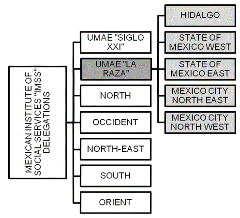
Figure 1: Above, “IMSS” delegations and reference center organigram with the regions of Mexico involved.

Héctor Bizueto-Rosas1* Samuel Gutiérrez-Vogel2 Ramiro Hernández-Salgado1† Juan López-Silva2 María Elizabeth Enríquez-Vega2 Alfonso Cossío-Zazueta2 Hugo Alonso Pérez-González3 Hilario Gómez-Valdez1 Carlos Martínez-López2 Roberto López-Rodríguez1† Gonzalo Maldonado-Ibarguen1 Juan Ernesto Cruz-Castillo1 Oscar Andrés Rodríguez-Jiménez1 Ernesto Pacheco-Pittaluga1 Ely Guadalupe Moran-Reyes1 Gabriela Jimena Muñoz-Paredes1 Roberto Carlos Serrato-Auld1 Perla Elin Leyva-Rivera1 Armando Guzmán-Caballero4 Carlos Daniel Gómez-Calvo5
1Department of Vascular Surgery and Angiology, “UMAE Hospital de Especialidades”, Centro Médico Nacional “La Raza”, Mexican Institute of Social Security, Mexico*Corresponding author: Héctor Bizueto-Rosas, Department of Vascular Surgery and Angiology, “UMAE Hospital de Especialidades”, Centro Médico Nacional “La Raza”, Mexican Institute of Social Security, Mexico, E-mail: dr_bizueto_h@yahoo.com
Objective: Present our experience as a tertiary referral center in the surgical management of the carotid paraganglioma.
Introduction: Carotid paraganglioma is a slow-growing tumor, originated at the chemoreceptor cells of the carotid bulb and due to factors as chronic hypoxia, familiar history or female gender it can present hyperplasia. Most of these tumors are benign and asymptomatic which delays its diagnosis and treatment. Surgical excision is the gold standard treatment.
Methods: We performed an ambispective study from 1987 to 2019 of the patients with the diagnosis of carotid paraganglioma that underwent surgical excision in our center, using the Shamblin classification system to describe them.
Results: A total of 964 tumors were resected from patients of 21 to 82 years old, in 32 years, with an average of 30 procedures each year. 92% of the patients were female, Shamblin type I 7%, type II 47% and type III 46%; bleeding went from 50 to 10,000 ml, with previous embolization in 8 cases, usage of stent-graft in 10 patients, 6 with external carotid artery ligation and only 1 patient with bypass using PTFE (Polytetrafluoroethylene) graft. Morbidity and mortality: cerebral vascular event in 3%, bleeding >2000 ml in 11%, nerve injury in 23%, vascular injury in 25% and overall mortality 2%.
Conclusion: In our center experience, surgical removal is the first treatment of choice; there is no benefit from using other therapies such as embolization, usage of stent-graft in the external carotid artery and only certain chosen patients could benefit from radiotherapy. Malignancy is less than 1%, severe neurological complications in about 3%. Genetic factor in our population, associated with the area height can be determinant.
Carotid paraganglioma; Surgical excision
Carotid body tumors are mostly benign, of slow-growth and originated at the chemoreceptor cells of the carotid bulb; they correspond to nearly half of the total paragangliomas [1,2]. According Glenner GG, et al. [3], the term paraganglioma is the most accurate to denominate these tumors [4].
Paragangliomas of the head and neck and the Carotid Body Tumor (CBT) are neuroendocrine tumors, they represent 0.6% of the head and neck total tumors and originate from the paraganglia of the autonomous nerve system [5]. They are associated with the parasympathetic nervous system so can be found from the skull base to the aortic arch [6]. Associated sudden death has been described [7] and can be locally invasive causing bone destruction and cranial nerve dysfunction [8]. About 10% are bilateral, although most are unilateral and sporadic [4]. Incidence of malignant tumors is below 10% but they can cause metastasis. Familiar presentation is usually around 10%, in which cases the multi-locate presentation is of greater risk [9].
The incidence of these tumors is proportional to height due to a chronic hypoxia that stimulates carotid bulb hyperplasia [2] or due to chronic obstructive pulmonary disease. Due that most of the CBT are asymptomatic and of slow-growing rates (between 1 to 2 mm per year), diagnosis is belated [10,11].
They are transmitted in an autosomal dominant manner through the gene and locus 11q23 [12] and the bilateral incidence is about 3 to 8% in sporadic cases and up to 33% in familiar cases [12]. In 79% of the head and neck paragangliomas, a mutation in the sub-unit D of the SDH gene is present, and it suggests the cause for multiple paragangliomas [13]. Female gender and age surrounding 60 years old are predominant [10].
The gene mutation SDHD is present in about 6-36% of the patients with head and neck tumors and up to 80% in familiar cases. The mutation P81L is used as a screening method for familiar paragangliomas in the USA [14].
According Shamblin WR, et al. [15], CBT can be classified in type I, II and III. Type I is the smallest tumor with very low adherence to the carotid arteries and with usually an easier surgical excision; type II is a tumor partially surrounding the carotid arteries and with firm adherence to the adventitia layer; and type III with an intimate, adherent relationship to the circumference of the carotid bifurcation [15]. Type III tumors are present in up to 25% of the cases, and in its surgical management vessel replacement is considered [15].
They are other suggested classifications in which the type III is divided in a) or b), according to Luna-Ortiz K, et al. [16], considering a) as originally described or b) if vessel wall infiltration is present. Mayo clinic considers as type IIIa those that do not reach skull base and as type IIIb those reaching skull base and have no distal artery for considering suitable anastomosis (Zanaret and cols, 2000).
Another suggested classification states a difference regarding hemodynamic repercussion, for which a tumor between two and 5 centimeters without hemodynamic changes is considered type II, and those greater than 5 centimeters, skull base extension, wall infiltration, atheroma plaque or significant hemodynamic change is a type III (Maldonado-Diaz HO and cols, 2017).
In a 32-year period, from 1987 to 2019 a total of 964 carotid body tumors were resected, from 24 to 65 cases per year with an average of 30 yearly. In this period a continuous record was kept, which was reviewed. Age went from 21 to 82 years old (mostly older patients whom specifically asked for surgical procedure). Diagnostic tools included clinical findings, Doppler ultrasound, CT scan and angiography; all discerned by the Shamblin classification.
Of the total 964 carotid body tumors resected, one corresponded to a Castleman disease (giant lymph node hyperplasia) and other to an unusual neurofibromatous type of carotid body tumor. Histopathological report of the 964 tumors concluded as a “carotid paraganglioma, lymph hyperplasia”, with malignancy confirmed in only one case; two other patients were associated with other malignant tumors. Mutation p81L of the SDHD gene (11q23) was found in 16% of the patients in heterozygous form [14].
Up to 92% of our patients were female with an average age of 55 years old. Regarding frequency as Shamblin type classification, type II were 47%, type III were 46% (Figures 1 and 2) (Table 1).
Figure 1: Above, “IMSS” delegations and reference center organigram with the regions of Mexico involved.

Figure 2: Patients with carotid body tumors distribution by size in millimeters and Shamblin classification, in the last five years.
| Mexican States | State Population | Insurance Holders | Population Allowed For Medical Service | Asigned to referal center u mae h cmn “la raza” |
| Hidalgo | 294,206 | 223,084 | 847,719 | 847,719 |
| State of Mexico | 17,363,387 | 1,559,744 | 5,771,053 | 5,771,053 |
| Mexico City | 8,811,266 | 3,340,440 | 11,357,496 | 5,678,748 |
| Total | 12,297,520 |
Table 1: Below, the total population of those regions being 12,297,520 potential patients. All stated regions are above the 2000 meters of sea level. UMAE H CMN “High specialty medical unit, hospital, national medical center.”
Our average surgical time was 180 minutes (90 to 240 minutes) in those patients with Shamblin type III. We found no significant difference regarding bleeding using Cell Saver technology or bipolar electrocautery.
Bleeding was from 50 to 10,000 ml, with an incidence of major bleeding (considered as greater than 2000 ml) of 11% (Figure 3).
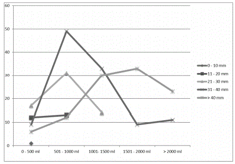
Figure 3: Surgical removal bleeding in the last five years, regarding size and blood loss in milliliters, considering patients in the last five years.
Nerve injury incidence was of 23% (Figure 4), major vascular injury of 25% with an overall mortality of 2%, consisting of deaths due to internal carotid artery thrombosis and cerebrovascular accident mostly.
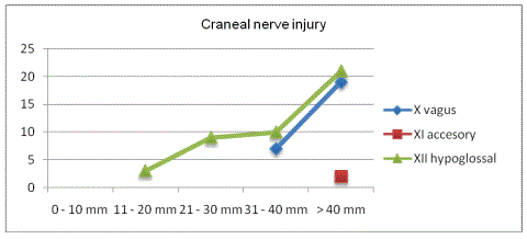
Figure 4: Number of patients and the association between size and cranial nerve injury, considering patients in the last five years.
Due to the volume of referred patients, previous embolization was elected in eight patients; one of them had a 10 × 15 × 15 cm tumor (Figure 5) that had several complications and died within the first day after embolization procedure.
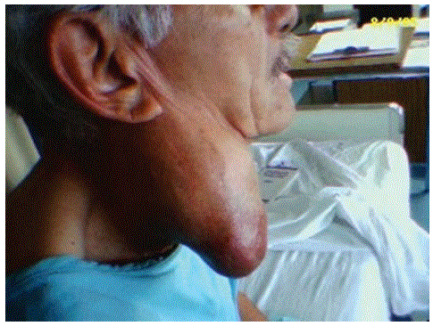
Figure 5: Male patient with Shamblin III right carotid body tumor of 10 × 15 × 15 cm.
Regarding management of carotid artery structures, in ten patients, a stent graft was placed from the common carotid artery to the internal carotid artery to exclude external carotid artery and its branches to reduce blood supply and in other six patients, a double procedure took place, consisting of tumor resection and carotid dolichoectasia correction, and finally in three patients, a simultaneous carotid endarterectomy was performed.
Of our records, six patients ended with external carotid artery closure, one patient required vascular reconstruction with PTFE (Polytetrafluoroethylene) graft, with an overall of 3% cerebrovascular accidents.
Considering medical history 8% had familiar records and 20% were bilateral.
One patient had several syncope episodes that improved after tumor resection, and another patient had severe dysphagia associated with an 80% esophagic stenosis due to tumoral compression. In our medical records, only one patient has had a bilateral surgical removal of CBT with bilateral recidivist tumor after 5 years (Figure 6).
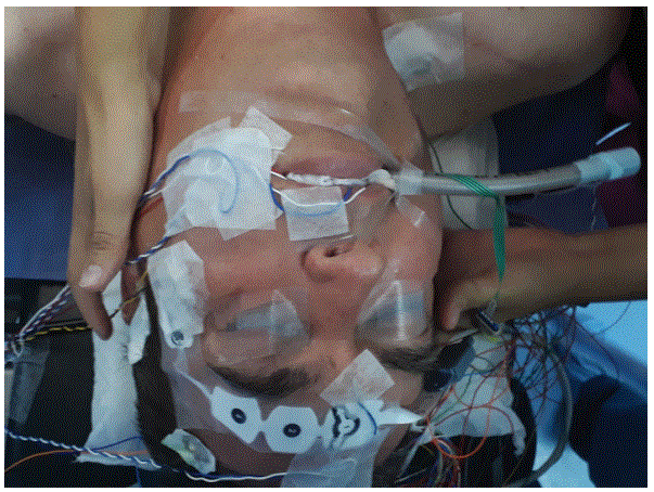
Figure 6: Anesthetic approach for carotid body tumor surgery with intraoperative cerebral monitorization and general anesthesia.
Clinical manifestations mostly involve a painless, slow-growing neck mass anterior to the sternocleidomastoid muscle, which is usually misdiagnosed as ganglia or airway related infections [9].
Doppler ultrasound is the most used diagnostic tool because of its availability and non-invasive approach; in experienced centers, it can provide anatomical detail, vascularization origin, size, surrounding vessel encapsulation and grade of atherosclerotic disease [17]. According to a comparison with CT o MR, ultrasound and color Doppler imaging failed to detect up to five of 60 CBT, scoring a sensitivity of 92% and specificity of 100% [18].
Regarding CT and MRI, the classic finding is described as the “lyre sign” due to the separation of both internal and external carotid arteries because of tumor growth in between. CT scan has a sensibility of 77 to 98% and a specificity of 92% and MRI has a 90-100% and 95% respectively [18]. In our center, a sub-analysis of our population, considering the geometrical findings of CT scan regarding internal carotid artery involvement with tumor showed a media of involvement of 220 degrees, being associated to a rate 30% of neurological injury during surgery and a most frequent classification of Shamblin III tumors; similar analysis are discussed by Kaddah RO, et al. [19].
Surgical removal is accepted as the best choice of treatment. Bleeding and nerve injury have an association with size and carotid vessels involvement, being directly associated with the Shamblin classification. In our center, those tumors considered as Shamblin III had an association of neurological injury up to 30%, with predominance of the mandibular branch of the facial nerve, the glossopharyngeal nerve and the hypoglossal nerve in decrease order.
The usage of LigaSure TM by Medtronic is being used as an approach to reduce bleeding and surgical time, especially in large carotid body tumors, although carotid surgery is not in the manufacturer specifications, and nerve injury continues to be an important postoperative complication [16].
Embolization is an endovascular technique that aims to starve carotid body tumors of their blood supply, reduce the size, induce necrosis and in some cases, attempt an easier second-timed surgical approach; coils are the most used material.
Several have questioned the need to perform a previous embolization, considering risks, costs, nerve injury and a low significant impact [20].
Radiation therapy and stereotactic radiosurgery are proposed as other options for treatment, especially in high-risk patients, extensive tumor, advanced age or other specific conditions considering airway, bleeding risk, religion (example given, Jehovah’s Witnesses and blood transfusion refusal) or no acceptance of surgery [21].
They are at least four genomic mutations that increase the risk of paraganglioma development; hereditary paraganglioma is usually diagnosed around 30 years of age [22,23].
The Succinate Dehydrogenase Enzyme (SDH) it is coded by the genes SDHB, SDHC and SDHD, and it play an important role on energy metabolism in mithocondrias. The enzyme performs as an oxygen sensor, and it has been described that hypoxia can trigger an SDHB mutation that has an important association with solid tumors [22].
The fact that p81L mutation of the SDHD gene is present in the Mexican population, in a greater proportion than in the American overall population, could explain our higher incidence, although, more data is required [14].
Regarding our center approach to treatment, data showed no significant benefit from other therapies such as embolization, stentgraft placement at the external carotid artery or radiotherapy, probably associated to a low number of cases managed by these therapies. The usage of LigaSure or Cell Saver also had no impact on the overall bleeding average.
We observe that in younger patients, with rapidly growing tumors and mostly in men, tumors more firmly adhere to the arteries and surrounding structures making the surgical approach more challenging, as well as surgery in those patients with thyroid impairment.
In terms of growth, a 1 to 2 mm yearly increase of volume is accepted as norm, but in our population, we observe that 20% of the patients refer a greater growth in a two to five years lapse [10,11].
The fact that Shamblin III tumors are as common as Shamblin II tumors it is probably due to the idiosyncrasy of our population (of do not ask for medical advice) and the great demand for medical services in our center. As for the association of Shamblin III tumors and the need for external carotid artery repair, we observe a dual cause, first due to the difficulty of the surgical removal and second due to overconfidence of our surgical team as being the center with the most cases.
In the last years, there is an increase in the Shamblin III type tumors.
In average our center provides 25 medical appointments daily, and in the last 19 years, there is an average of yearly surgical CBT removal or 45 patients, being a total of 461 from 1987 to 2007, 144 tumors removed from 2008 to 2013 and 357 from 2013 to 2019 [14,17].
Its combination with embolization is proposed as a first-choice treatment in some centers, although youngest patients, catecholamine secreting paragangliomas, or those rapidly progressing tumors may be more appropriate for open resection [21].
In our center experience, surgical removal is the first treatment of choice; there is no benefit from using other therapies such as embolization, usage of stent-graft in the external carotid artery and only certain chosen patients could benefit from radiotherapy. Malignancy is less than 1%, severe neurological complications in about 3%. Genetic factor in our population, associated with the area height can be determinant.
Authors declare no conflict interest.
Download Provisional PDF Here
Article Type: RESEARCH ARTICLE
Citation: Bizueto-Rosas H, Gutiérrez-Vogel S, Hernández-Salgado R, López-Silva J, Enríquez-Vega ME, et al. (2020) Carotid Paraganglioma. Experience of a Reference Center, UMAE Hospital De Especialidades Del Centro Médico Nacional “La Raza”, 964 Cases in 32 Years. J Surg Open Access 6(3): dx.doi.org/10.16966/2470-0991.211
Copyright: © 2020 Bizueto-Rosas H, et al. This is an open-access article distributed under the terms of the Creative Commons Attribution License, which permits unrestricted use, distribution, and reproduction in any medium, provided the original author and source are credited.
Publication history:
All Sci Forschen Journals are Open Access