Abstract
Objective: We present a case report of an 89 years old male with a diagnosis of incarcerated left inguinal hernia, with surgical findings of a complicated sigmoid diverticulum and a tubular structure of 0.5 mm attached to the sigmoid colon, fixed to the inguinal ligament with a true lumen that could correspond to a duplicated appendix B4 type.
Introduction: Colon diverticular disease is more frequent in western countries, present in 30% of 60 years old patients and in more than 50% in 80 years old patients, with 95% of the times located at sigmoid colon. The most frequent complication is the acute diverticulitis (25%) and in 30% of these patients, surgery is required.
Discussion: Acute, complicated, diverticular disease is an infrequent pathology, even more, a colonic diverticulum included within the hernia sac. Of the appendix congenital malformations, a duplicated appendix is the rarest; usually found in asymptomatic adults as an incidental find during abdominal surgery or during contrasted abdominal scans.
Conclusion: Awareness must be raised within first-contact health care professionals about diagnosing surgical entities with high mortality rates, in asymptomatic patients with unclear clinical findings, especially with the occurring of a demographic transition in most countries. In this specific patient, Hartman surgical procedure and a second time inguinal plasty were the best surgical options for this patient; even considering the mortality rates (25%) of a perforated diverticulum, considering added surgical site infection.
Keywords
Incarcerated hernia; Complicated sigmoid colon diverticulum; Duplicated appendix
Introduction
A demographic transition is occurring in most countries towards having elderly patients requiring medical attention. Acute abdominal pain is still a very frequent entity among elderly patients, and correct diagnosis and initial management is of critical importance, us thus forcing to have better treatment guidelines.
Within the causes of acute abdominal pain, diverticulitis it is not considered the most common diagnosis, with only about 30% of patients requiring an emergency surgical approach. Regarding appendix congenital malformations, a duplicated appendix is the rarest, with an incidence of 0.0004% and can be located within all digestive tube. We present the case of an 89 years old male patient with acute abdominal pain associated with a left inguinal bulge, considered to be an incarcerated left inguinal hernia but with surgical findings corresponding to be a sigmoid colon gigantic diverticulum and a duplicated appendix. As a consequence, we analyze the action of performing a precocious surgical laparotomy approach in this type of patients (elderly, acute abdominal pain, no clear initial diagnosis) considering its low mortality
Case Report
We present an 89 years old admitted at the Emergency Department due to left inguinal tumour, abdominal pain, nausea, abdominal distension and vomit, establishing an initial diagnosis of acute abdomen due to incarcerated left inguinal hernia. A surgical procedure consisting of abdominal laparotomy, with findings of severe inflammatory reaction and sigmoid colon induration, at the antimesenteric border a diverticulum of 2.5 × 12 cms, within hernia sac, with abundant purulent material and a punctate perforation; also findings of infarctions of the greater omentum and of the epiploic appendages. Distal to the diverticulum, 2 cms, a tubular structure of 5 mm diameter and 8 cms length, with a true lumen, was firmly attached to the abdominal transverse muscle aponeurosis (Figure 1). Hemicolectomy with Hartman procedure was performed, and within hernia defect a nylon 1-0 suture plasty from inside the peritoneum, with a mesh inguinal plasty differed. After surgery, the patient develops pulmonary disease requiring Intensive Care Unit admission and after 10 days of postoperative care is discharged. Histopathological study of surgical obtained pieces showed,
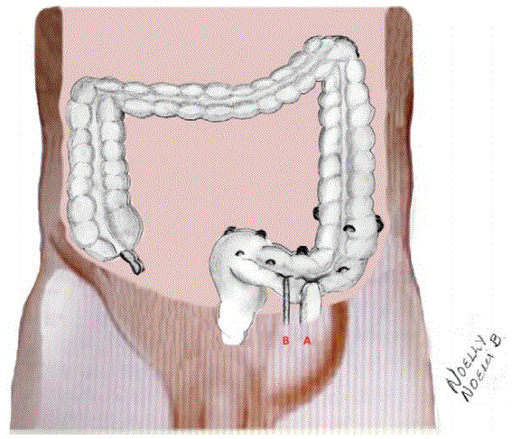
Figure 1: Schematic presentation of surgical findings.
A-Antimesenteric border diverticulum of 2.5 × 12 cms.
B-A tubular structure of 5 mm diameter and 8 cms length, with a true lumen and firmly attached to the abdominal transverse muscle aponeurosis.
(1) Diverticular sigmoid disease with ischemia, vascular congestion and dilatation, inflammatory infiltrate, edema, necrotic areas, acute and chronic serositis, lymph node hyperplasia and peritonitis and
(2) Punctuate perforated diverticulum of 2.5 × 9 cms, smooth wall and fibrotic adherence; after cut, with 6 mm wall and mucous edema
(3) 6 × 0.5 piece with interior mucosa atrophy.
Four months later intestinal continuity was re-established with good postoperative evolution, so it was decided 5 days later, to perform a left inguinal mesh plasty (Lichtenstein technique), with the patient being discharged home 10 days after admission.
Literature Review
Within inguinal hernia entity, a man of white race, with chronic coughing or constipation and medical family history is up to 8 times more prone to develop it. Male predominance may be due to anatomical differences of the inguinal region [1].
Of all abdominal hernias, inguinal ones represent up to 75% of all (Nyhus) to 92% (Watson) [2]. The incarcerated hernia has an incidence of 12.5-15 cases every 100,000 habitats and has no vascular impairment. A strangulated hernia interrupts both, intestinal transit and the vascular flow, with a prevalence of 1-3% in adults [3]. Even though it is an infrequent pathology, it is still a mortal disease [4]. There are reports describing an inguinal approach and inguinal mesh plasty within the same surgical procedure, with several reports addressing that meshes can be used in contaminated surgical areas [5,6]. Hartman procedure comes with high mortality and high rates of surgical site infection, up to 25%, but it is still highly recommended by several authors [7,8].
About colon diverticulum, defined as a mucosa protrusion through weak zones, that only contain a layer of mucosa and submucosa, some authors consider the term “pseudo diverticulum” more accurate. Colon diverticulum are most frequent at the sigmoid colon, 65-95%, and have an acute diverticulitis presentation in about 25% of patients, being the most frequent complication, requiring in 30% of cases an emergency surgical approach due to peritonitis or sepsis [7].
Colon diverticulum are more frequent in men, up to the 80 years old, when after that become more frequent in women, with obesity considered a risk factor in 95% of patients [7,8].
In the weakened gut zones where there is a mucosa protrusion, there is usually an association of penetrating small blood vessels between 5 to 10 mm of diameter [9].
Diverticula disease includes diverticulosis, diverticulitis (inflammatory disease) and diverticulum bleeding. In about 75% of patients, it is not complicated, and in the other 25%, there is a presentation of abscess, fistulae, gut obstruction, peritonitis or sepsis [9,10].
In Mexico, diverticular disease incidence is about 4.1% [11], affecting 65% of cases 80 years old or older. Typical presentation form is a “pseudodiverticulum” (incomplete wall layers).
The gigantic colonic diverticulum was first described in France by Bonvin and Bonte in 1946; it is called when it’s greater than 4 cms of diameter; being rare, it is thought to occur due to a progressive dilatation, with a unidirectional valve effect [10-13].
A diverticulum within hernia sac was first described by Skandalakis, who defined the so-called Littré hernia as a Meckel diverticulum within any hernia orifice (inguinal, crural or diaphragmatic) [14].
Amyand hernia is described as an inflammated or perforated appendix within a reducible inguinal hernia [15]. Duplicated appendix has been described more commonly in adults, and without symptoms. It has been described as an incidental finding during abdominal surgeries or during contrasted abdominal scans [16].
Duplicated appendix is the rarest appendix congenital malformation, being the positional anomalies the most common. Its incidence is about 0.004%, and can be located within all digestive tube, or even, being triplicated [16], it can cause small intestine obstruction, simulate a colon adenocarcinoma and it is generally associated to other genital or urinary anomalies [16,17].
One of the most accepted theories considers that it origins due to an anomaly of the embrionary intestine development, becoming a diverticulum, a cyst or a doubling of a portion of the intestine within ambient factors such as trauma, hypoxia [16-18]. There are 3 types, depending on its implantation, and another group, being in total 4 different types:
Type A, the 2 duplicated appendix origins from a common base (partial duplication), and it has no other congenital associated anomaly
Type B, one normal positioned appendix and another wrongly positioned appendix with several subtypes:
- B1, wrongly positioned being just above the ileocecal valve
- B2, located within cecum taenia coli
- B3, located along hepatic flexure taenia
- B4, located along splenic flexure
Type C, cecum duplication, each one with one appendix
Type D, horseshoe appendix
Discussion
The gigantic colon diverticulum is rare, there are about 113 literature case reports up to the year 2004 [11,13]. According to the McNutt classification [12], there is a type II gigantic colon diverticulum, consisting in a perforated diverticulum, with abscess formation, that keeps communication within intestinal lumen and has progressive dilation and corresponds to the presented case report. It could be considered as gigantic or pseudo gigantic diverticulum, according to different reports.
Due to histopathological analysis of diverticular sigmoid colon disease with ischemia, punctuate perforated diverticulum and findings of a tubular structure of 5 mm diameter and 8 cms length, with a true lumen and firmly attached to the abdominal transverse muscle aponeurosis, surgical findings are considered to be a colon diverticulum and a B4 type of duplicated appendix [18].
The presented case had the medical history of being a 80 years old male patient with an inguinal bulge and symptoms to be considered as an incarcerated hernia or one of the other more common differential diagnosis entities, considering that the above-mentioned anatomical malformations are rare. A late diagnosis can be fatal due to necrotizing fasciitis, necrosis of hernia content, intestinal obstruction or sepsis [3,4].
Awareness must be raised within first-contact health care professionals about diagnosing surgical entities with high mortality rates, in asymptomatic patients with unclear clinical findings, especially with the occurring of a demographic transition in most countries that forces to have better guidelines of treatment. As Dr. Martínez Serrano stated “even when incarcerated or strangulated hernia are considered to be rare, in the middle of the 21st century, it is still a mortal disease” [4] and the duplicated appendix can be associated with the colon adenocarcinoma [17,19].
As above-mentioned, it exists the so-called Litreé hernia, Meckel diverticulum within any hernia orifice, Amy and hernia, appendix within inguinal hernia, to our knowledge there is no case report that describes the surgical finding of sigmoid colon diverticulum within a left inguinal hernia accompanied by a duplicated appendix.
It is important to state that, due to gastrointestinal malformations, obscure clinical presentation, rare abdominal pain symptoms and its mortality repercussion, the action of performing a precocious surgical approach can be supported with the low mortality of a negative abdominal laparotomy.
Conclusion
Awareness must be raised within first-contact health care professionals about diagnosing surgical entities with high mortality rates, in asymptomatic patients with unclear clinical findings, especially with the occurring of a demographic transition in most countries that forces to have better treatment guidelines. In the case of rare abdominal surgical entities, the action of performing a precocious surgical approach can be supported with the low mortality of a negative abdominal laparotomy.
