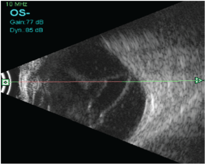
Figure 1: Retinal detachment seen in cases corneal trauma.

Shreya Thatte* Satender Singh
Department of Ophthalmology, Sri Aurobindo Medical College, India*Corresponding author: Shreya Thatte, Department of Ophthalmology, Sri Aurobindo Medical college, India, E-mail: shreyathatte@gmail.com
Purpose: To assess the efficacy of B-scan as diagnostic and prognostic tool in cases of opaque ocular media.
Materials and Methods: A prospective study to evaluate eyes with various media opacities. 112 patients with different media opacities where posterior segment examination was not possible by ophthalmoscopy were divided in four groups depending on the site of involvement Group A (Corneal Pathology – Leucomatous opacity, healed ulcer, corneal melting, chemical burn, corneal degeneration) Group B (Aqueous – Hyphaema due to trauma) Group C (Lens – Mature cataract, hypermature cataract, Morgagnian cataract, hard brown cataract, and traumatic cataract) Group D (Vitreous-Vitreous haemorrhage). Detailed examination followed by B Scan ultrasonography prior to any intervention was done to rule out gross posterior segment pathologies.
Results and Observations: Total 112 cases, aged between 1 to 80 years, both male 64 (57.16%) and female 48 (42.84%) were taken. 81 (72.32%) cases were of cataract and 31 (27.62%) cases of other media opacities, which involved cornea, anterior chamber and vitreous. In group A, i.e. cornea group (n=19), RD with VH was observed in 10.55%. These patients were counselled for probable visual outcome of treatment and explained the need of secondary intervention. In group B, anterior chamber group (n=10), having hyphaema due to blunt trauma, 30%cases had RD on echography and were referred to retina clinic for further management. In group C, cataract group (n=81), was further divided in non-traumatic (70.37%) and traumatic group (29.62%). Patients with vitreous degenerations (21.05%), asteroid hyalosis (5.26%), posterior staphyloma (3.50%) underwent cataract surgery with GVP, whereas in traumatic cataract group, RD (16.66%) and RD with VH (16.66%), a change in management strategy was seen in these cases as they underwent cataract surgery followed by PPV and RD surgery. Group D i.e. the vitreous group 1.76% cases had dense VH and were managed medically.
Conclusion: Good visual outcome in cases with media opacities is often challenging and incomplete evaluation leads to unexplained poor visual outcome in many such cases. Along with anterior segment a detailed posterior segment evaluation is equally important to decide an intervention plan as well explains the probable prognosis to patient. B-scan ultrasonography is a simple, safe, non-invasive, and quick investigative technique which is highly sensitive and specific in diagnosing gross posterior segment pathologies in cases of media opacities and it is inevitable to proceed to treatment especially surgical management in such cases. By performing a routine B scan in all cases of media opacities we can easily explain the patient a gross prognosis and safeguard our position as well and makes us medicolegally sound. It would be justifiable to mention that the future of ophthalmic echography especially B Scan in bright and it will help in better understanding of ocular tissue and various ocular pathological conditions.
B Scan; Ultrasonography; Opaque ocular media; Advanced cataract
AH: Asteroid Hyalosis; GVP: Guarded visual prognosis; RD: Retinal detachment; VH: Vitreous Hemorrhage; PPV: Pars plana vitrectomy
The ocular media, which is formed anteriorly by the tear film, cornea, aqueous humor, lens and posteriorly by vitreous humor [1] is an important factor for maintaining good visual acuity. Any alteration in the clarity of this media is responsible for the decrease in visual acuity whereas a complete opacification not only leads to gross visual deterioration but also, averts us from detailed ocular examination. It is very important to diagnose the posterior segment pathologies in such cases so that a treatment strategy can be planned, and the patient can be explained the probable prognosis. Any intervention in such cases without performing a thorough ocular examination and evaluation of posterior segment may lead to the suboptimal outcome.
There are various modalities for evaluation of posterior segment [1].
In 1956 ultrasound was first used by Mundt and Hughes for ocular diagnosis [1,2]. Later Baum and Greenwood developed the first twodimensional B-scan for use in ophthalmology with immersion technique [1,3,4]. The first commercially available contact B-scan, with a hand-held probe which could be directly applied to the closed lid, was introduced by Nathaniel Bronson in 1972 [1,5]. B Scan works on the principle of ultrasonography i.e. high-frequency sound waves (10 MHz) are used to produce a two-dimensional image of the minute structures on a screen [1]. In these advancing years, it has proven to be a safe, noninvasive, and affordable tool for detection and differentiation of various ocular pathologies [1,6]. It has become an important and accurate diagnostic device for evaluating posterior segment lesions in cases where a clinical examination and ophthalmoscopy is less informative, due to media opacity which may be caused by various pathologies like corneal opacity, hyphaema, dense cataract, and vitreous hemorrhage, inflammatory conditions etc.[7,8]. Although B- Scan is a very handy and indispensable tool in ruling out gross pathologies of the posterior segment of the eye, but it has few limitations, as exact retinal pathologies like retinopathy and various types of maculopathy etc. cannot be ruled out based on echography. Our aim in this study was to diagnose gross posterior segment pathologies with media opacities of various types with the help of B Scan and decide the plan of management accordingly. It also helped in explaining the probable prognosis of visual outcome prior to management.
Present study is a randomized prospective study for assessment of posterior segment pathologies in cases of eyes with all types of media opacities coming to the outpatient department form the duration of November 2015 to March 2016
A total of 112 eyes of patients with various types of opacities of ocular media where posterior segment assessment was difficult by routine examination on slit lamp by 90D and direct and indirect ophthalmoscopy were taken for study. They were divided into different groups depending on the site of involvement and evaluated by B scan to note gross posterior segment pathology.
Group A (Corneal Pathology – Leucomatous opacity, healed ulcer, corneal melting, chemical burn, corneal degeneration)
Group B (Aqueous – Hyphaema due to trauma)
Group C (Lens – Mature cataract, hypermature cataract, Morgagnian cataract, hard brown cataract, and traumatic cataract)
Group D (Vitreous – Vitreous hemorrhage)
Detailed ophthalmic examination, which included a detailed history of the patient, visual acuity, and best corrected visual acuity(BCVA), tonometry and thorough slit lamp examination. Pre-and post-operative visual acuity was noted and grouped accordingly to assess the prognosis of intervention.
Patients and their relatives were explained, and consent was taken. B-Scan was done using the Optos Scan 3000 B scan machine with a handheld probe with a transducer using ultrasound frequency of 10 MHz. The examination was performed with the patient lying down in supine position over the couch, a water-soluble coupling jelly was applied on the head of the probe and it was gently placed over the closed eyelid. B scan was performed once the patient is comfortably lying down in three different views.
In cases with blunt trauma B scan was done with utmost care, avoiding any harm to the affected eye, whereas in cases with penetrating trauma it is advisable to do B scan only after primary repair and with aseptic precaution to prevent intense spread of infection and such cases were excluded from our study, with the help of B scan, it is possible to evaluate the amount of damage to the ocular tissue and the exact site of any intraocular foreign body can be located [8-10].
Postoperatively visual acuity, best corrected visual acuity was noted, and fundus examination was done in all cases.
Out of total 112 cases, aged between 1 to 80 years, both male 64 (57.16%) and female 48 (42.84%) were taken. 81 (72.32%) cases were of cataract and 31 (27.62%) cases of other media opacities, which involved cornea, anterior chamber and vitreous (Tables 1,2).
| Age (yrs.) | Cornea | Anterior Chamber | Lens | Vitreous | Total | Percentage (%) | Percentage | Percentage |
| Male | Female (%) | |||||||
| (%) | ||||||||
| 10-Jan | - | - | 2 | - | 2 | 1.78 | 0.89 | 0.89 |
| 20-Nov | - | - | 12 | - | 12 | 10.71 | 6.25 | 4.46 |
| 21-30 | 1 | 3 | - | - | 4 | 3.57 | 3.57 | - |
| 31-40 | 4 | 2 | - | - | 6 | 5.35 | 4.49 | 0.89 |
| 41-50 | 3 | 3 | 8 | 1 | 15 | 13.39 | 8.92 | 4.46 |
| 51-60 | 4 | - | 25 | 1 | 30 | 26.78 | 11.6 | 15.17 |
| 61-70 | 3 | 2 | 26 | - | 31 | 27.67 | 16.07 | 11.6 |
| 71-80 | 4 | - | 8 | - | 12 | 10.71 | 5.37 | 5.37 |
Table 1: Age wise distribution of various media opacity
| No of cases | Pathology | B scan | Treatment Given |
| 5 | Corneal melting | Normal posterior segment | Scerlo-corneal Keratoplasty following Medically management |
| With Visual prognosis explained | |||
| 5 | Leucomatous corneal opacity | Normal posterior segment | Penetrating Keratoplasty under GVP |
| 2 | Corneal scar following trauma | RD | Referred to retina clinic |
| 4 | Healed corneal ulcer | Vitreous Degeneration | Penetrating Keratoplasty under guarded visual prognosis |
| 3 | Corneal degeneration | Normal posterior segment | Lamellar Keratoplasty |
Table 2: Cases with corneal pathology (n=19).
B Scan was performed to evaluate the posterior segment, in 16.96% (19) cases with various corneal pathologies Cases of corneal melting following chemical injury (26.31%) echography showed normal posterior segment and all patient underwent sclero-corneal keratoplasty. Other cases of leucomatous opacity (26.31%), also had normal posterior segment and were further planned for penetrating keratoplasty, patients with healed corneal ulcer (21.05%) with vitreous degeneration underwent optical keratoplasty, patient with superficial corneal degeneration (15.77%) with normal posterior segment, lamellar keratoplasty was done, all these cases were operated under guarded visual prognosis. Whereas 2(10.55%) cases of corneal scar following trauma, had retinal detachment (Figure 1) and were referred to retina clinic for an opinion. Patients were advised keratoplasty followed by pars plana vitrectomy with endolaser and silicon oil injection. Based on echographic findings patients were counseled and were explained the need for secondary intervention. It was seen that only in 10.55% cases of corneal pathology group, B scan influenced the change of management strategy
Figure 1: Retinal detachment seen in cases corneal trauma.
Another group of patients with anterior chamber involvement following blunt trauma with hyphaema i.e. in 8.92% (10) cases out of which 70% (7) cases with hyphaema had normal retina and were medically managed whereas 30% (3) cases with hyphaema had retinal detachment (Figure 2) were referred to retina clinic and underwent pars plana vitrectomy with endo laser and silicone oil injection (Table 3).
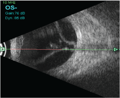
Figure 2: Retinal detachment seen in a case hyphema due to blunt trauma.
| No of cases | % of cases | B Scan finding | Management |
| 40 | 70.17 | Normal posterior segment | Cataract Surgery under Guarded visual prognosis |
| 12 | 21.05 | Vitreous Degeneration | Cataract Surgery under Guarded visual prognosis |
| 3 | 5.26 | Asteroid Hyalosis | Cataract Surgery under Guarded visual prognosis |
| 2 | 3.5 | Posterior Staphyloma | Cataract Surgery under Guarded visual prognosis |
Table 3: Non-traumatic cataract (Mature, Hyper-mature, Hard brown, Morgagnian) n= 57.
Out of 81 cases of various types of cataract patients were divided into two broad groups a) Non-traumatic 57 (70.37%) cases and b) Traumatic group 24 (29.62%) cases. In this group, higher numbers of cases were of the senile age group, 67 (82.71%) cases aged between 41 to 80 yrs. and remaining 14 (17.28%) were of pediatric age group i.e. 1 to 20 yrs. In the non-traumatic group of 57 patients, on pre-operative echographic evaluation, 40 (70.17%) cases had normal posterior segment, 12 (21.05%) had vitreous degenerations (Figure 3), 3 (5.26%) had asteroid hyalosis, 2 (3.50%) had posterior staphyloma (Figure 4) all the cases underwent cataract extraction with IOL implantation under guarded visual prognosis. Detailed fundus examination was done to confirm B scan findings first follow up (Table 4).
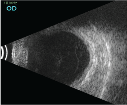
Figure 3: Vitreous degenerations in case of mature cataract.
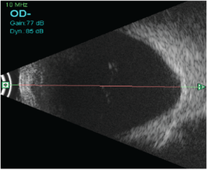
Figure 4: Posterior staphyloma seen in mature cataract.
| No of cases | % of cases | B scan finding | Management |
| 9 | 37.5 | Normal posterior segment | Cataract surgery under Guarded visual prognosis |
| 4 | 16.66 | Retinal Detachment | Cataract surgery followed by PPV with endolaser and silicon oil injection. |
| 4 | 16.66 | Retinal Detachment with vitreous hemorrhage | Cataract surgery followed by PPV and endolaser and silicon oil injection. |
| 7 | 29.16 | Complete Posterior Vitreous detachment (PVD) | Cataract surgery under guarded visual prognosis |
Table 4: Traumatic cataract group (n= 24)
Whereas in the traumatic group of 24 patients, 14 (58.33%) patients with traumatic cataract belonged to the pediatric age group of 1- 20 yrs., which correlated well as they are more involved in outdoor activities. 9(37.50%) patients had a normal posterior segment, 7 (29.16%) had complete posterior vitreous detachment following blunt trauma all of them underwent cataract extraction under guarded visual prognosis. 4 (16.66%) had retinal detachment (RD) (Figure 5) and remaining 4 (16.66%) had retinal detachment with vitreous hemorrhage (VH) these cases were referred to a vitreoretinal surgeon and underwent cataract extraction with pars plana vitrectomy (PPV) along with retinal detachment surgery.
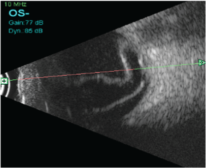
Figure 5: Retinal detachment seen in a case of traumatic cataract.
1.76% (2) cases on ophthalmoscopy had dense vitreous hemorrhage (Figure 6) which obstructed the view of the retina, B scan ultrasonography revealed normal posterior segment. They were medically managed with regular follow up.
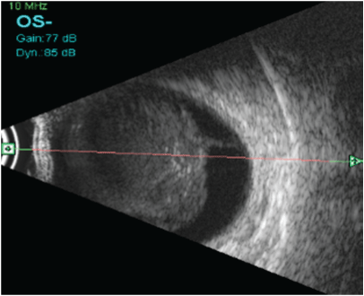
Figure 6: Dense organized vitreous hemorrhage.
In this study, 86.60% patients underwent surgical intervention as planned. Whereas in 11.60% (13 of 112) cases, there was a change in strategy and they underwent cataract surgery followed by retinal surgery (Table 5).
| Visual Acuity | Pre-Operative | Post- Operative |
| % Patients | % Patients | |
| >1/60 | 41.07% (46) | 1.78% (2) |
| 1/60 to 3/60 | 55.35% (62) | 2.67% (3) |
| 4/60 to 6/60 | 3.57% (4) | 7.14% (8) |
| 6/36 to 6/24 | - | 14.28% (16) |
| >6/24 | - | 74.10% (83) |
Table 5: Showing visual acuity pre-and post-operatively
Pre-operatively most of the patients (96.42%) had visual acuity 3/60 or less including all the groups. We observed an improvement in postoperative visual acuity in 88.38% cases of which 74.10% had a visual acuity better than 6/24 on Snellen’s chart. There were 1.78% cases of chemical injury that had minimum improvement postoperatively.
The sensitivity and specificity of B Scan were statistically derived and was found to 100% along with 100% positive and negative predictive value.
B scan has proven to be a very handy device for the accurate evaluation of posterior segment pathologies in cases with all types of opaque media. This study has included all types of media opacities under one heading to diagnose posterior segment lesions and observe the influence of B scan on the management strategy. A total of 112 patients were divided into four different groups based on site of involvement i.e. Group A - Corneal pathology (16.96%), Group B- Anterior chamber involvement (8.92%), Group C- various types of advanced cataract (72.32%) and Group Dvitreous opacities (1.76%). Such that influence of B scan on treatment plan can be understood in each group individually.
Age group for the present study was 1 to 80 years [8,9,11-13] including both genders male (57.16%) and females (42.84%) and we observed a male predominance in the current study which correlated well with other studies by Chanchalani et al [13], Rekha Agrawal et al [9], Daxa Chavda et al [11] where males were 58%, 76% and 66% and females 42%, 24%, and 33% respectively. Contrastingly in the study by Jatin et al [14] and Meenakshi et al [8], female predominance was reported i.e. 51.89% and 63.15% females and 48.10% and 36.84% males respectively.
In a study conducted by Ramadhas et al [15] 100% specificity and sensitivity of B scan was reported in cases with normal posterior segment, this correlated well with the current study, but slightly decreased sensitivity was reported in vitreous haemorrhage (95.23%) with 100% specificity whereas it was 100% sensitive and specific in all groups in the present study (Table 6).
| Parameter |
B-Scan findings |
Fundus findings | Total | Chi-square Value | P value | Sensitivity | Specificity | PPV | NPV | |
| Negative | Positive | |||||||||
| Non-traumatic cataract (Positive finding: Vitreous degeneration) | Negative | 46 100.0% |
0 0.0% |
46 80.7% |
57.00, df=1 | 0.000* | 100% | 100% | 100% | 100% |
| Positive | 0 0.0% |
11 100.0% |
11 19.3% |
|||||||
| Non-traumatic cataract (Positive finding: Posterior staphyloma) | Negative | 54 100.0% |
0 0.0% |
54 94.7% |
57.00, df=1 | 0.000* | 100% | 100% | 100% | 100% |
| Positive | 0 0.0% |
3 100.0% |
3 5.3% |
|||||||
| Non-traumatic cataract (Positive finding: Asteroid hyalosis) | Negative | 54 100.0% |
0 0.0% |
54 94.7% |
57.00, df=1 | 0.000* | 100% | 100% | 100% | 100% |
| Positive | 0 0.0% |
3 100.0% |
3 5.3% |
|||||||
| Traumatic cataract (Positive finding: Retinal detachment) | Negative | 20 100.0% |
0 0.0% |
20 83.3% |
24.00, df=1 | 0.000* | 100% | 100% | 100% | 100% |
| Positive | 0 0.0% |
4 100.0% |
4 16.7% |
|||||||
| Traumatic cataract (Positive finding: Complete Posterior vitreous detachment) |
Negative | 17 100.0% |
0 0.0% |
17 70.8% |
24.00, df=1 | 0.000* | 100% | 100% | 100% | 100% |
| Positive | 0 0.0% |
7 100.0% |
7 29.2% |
|||||||
| Traumatic cataract (Positive finding: Retinal detachment vitreous hemorrhage) | Negative | 20 100.0% |
0 0.0% |
20 83.3% |
24.00, df=1 | 0.000* | 100% | 100% | 100% | 100% |
| Positive | 0 0.0% |
4 100.0% |
4 16.7% |
|||||||
| Corneal Involvement (Positive finding: Vitreous degeneration) | Negative | 15 100.0% |
0 0.0% |
15 78.9% |
19.00, df=1 | 0.000* | 100% | 100% | 100% | 100% |
| Positive | 0 0.0% |
4 100.0% |
4 21.1% |
|||||||
| Corneal Involvement (Positive finding: Retinal detachment) | Negative | 17 100.0% |
0 0.0% |
17 89.5% |
19.00, df=1 | 0.000* | 100% | 100% | 100% | 100% |
| Positive | 0 0.0% |
2 100.0% |
2 10.5% |
|||||||
| Anterior Chamber Involvement (Positive finding: Retinal detachment) | Negative | 7 100.0% |
0 0.0% |
7 70.0% |
10.00, df=1 | 0.000* | 100% | 100% | 100% | 100% |
| Positive | 0 0.0% |
3 100.0% |
3 30.0% |
|||||||
Table 6: Showing sensitivity, specificity, positive predictive value (PPV) and negative predictive value(NPV) of B scan in present study.
In corneal pathology group 16.96% (19), 68.42% had a normal posterior segment on B scan ultrasonography, 21.05% in this category had vitreous degeneration all of them underwent keratoplasty surgery as per plan. But 10.52% cases had retinal detachment along and were referred to a retinal surgeon for opinion and further planning. A study by Ramadhas et al [15], 10.88% cases of corneal opacities was reported but specific echographic findings have not been declared. There were no such studies that incorporated the management plan for corneal opacities. Maximum patients (89.47%) of this group in our study underwent planned modality and were followed up accordingly.
Advanced cataract (72.32%) was reported as the most common media opacity leading to visual impairment and the most common indication of diagnostic B scan in the present study and other studies [13,15,16]. In the traumatic group, 10.71% cases were of 11 to 20 years in the present study which correlated well with their age as they are more prone to trauma due to involvement in outdoor activities and this trend was seen in other studies as well [9,14,17]. Maximum cases (52.67%) in non-traumatic cataract group belonged the senile age group of 41 to 70 years which correlated well with other studies [14-17].
About 60.49% cases of advanced cataract (both traumatic and nontraumatic) had normal posterior segment on echography which did not correlate well with the studies by Ramadhas et al. [15] and Meenakshi et al. [8] where a lower number (41.58% and 46% respectively) had normal posterior segment, which could possibly be due to a wide variation in their sample size i.e. 65 and 490 respectively. Contrastingly Jatin Garg et al. [14] reported a higher number of cases (83.55%) with normal echography in a sample size of 158 eyes.
Cataract group was broadly divided into the traumatic and nontraumatic group in the study by Jatin et al. [14], Meenakshi et al. [8] and in the present study as well. In the traumatic group, cases of RD were 22.72% 2.85% and 16.66% followed by VH 22.72% and 2.09% and 1.76% respectively. RD with VH (4.54% and 16.66%) cases were seen in the current study and by Jatin et al. [14] only. We reported a higher number of cases with PVD (29.16%) in comparison to Meenakshi et al. [8] (1.22%). The most common finding was RD in this category [8,14].
Posterior segment pathologies were much lower in the non-traumatic group. Asteroid hyalosis was reported by Jatin et al. [14] (0.73%), Meenakshi et al. [8] (0.8%) and in the present study (5.26%) as well. Only the current study and Meenakshi et al. [8] had reported posterior staphyloma (3.50% and 0.8% respectively). Jatin et al. [14] in his study reported PVD (2.94%), RD (2.20%), VH (1.47%) whereas no such cases were seen in the present study. Vitreous degeneration (29.10%) was reported as most common finding in this group which did not correlate with any of the studies. Both Jatin et al. [14] and Meenakshi et al. [8] were restricted to cases of advanced cataract only and did not emphasize on how B scan led to modification in their plan of management.
The other group wherein vitreous was the primary cause of media opacity, we observed a very low number (1.76%) which on B scan had no retinal pathology and were managed medically. Whereas Agrawal R and Ahirwal S [9], found vitreous hemorrhage (56.41%) as the most common pathology, although their study was a diagnostic study which included patients with the clear ocular media and opaque media.
We also recorded the pre- and postoperative visual acuity to assess the prognosis and found that 88.38% patient had a visual acuity better than 6/24 post-operatively. Although, patients with chemical injury, corneal scar with RD, posterior staphyloma and traumatic cataract with RD (11.60%) showed improvement, postoperative visual acuity less than 6/60. These patients were counseled beforehand about the probable prognosis prior to surgical intervention based on the diagnosis made on echography
To the best of our knowledge, there are a lot of studies performed on the diagnostic role of B scan ultrasonography in various types of media opacities [7-19] but there is no study on the prognostic role of B scan. In the present study, we not only diagnosed pathologies on B scan but also explained the gross prognosis of the visual outcome of the planned modality to the patient and their relatives which other studies did no emphasize upon [8,9,14]. We also studied the influence of B scan on our management strategy and found to have changed in management strategy in 11.60% cases in total from all the groups.
We concluded that two-dimensional B-scan ultrasonography is an excellent diagnostic tool for evaluation of posterior segment in cases with opaque ocular media which is non-invasive, cost-effective, easily available quick investigative technique which is sensitive and specific and almost accurate in detecting gross posterior segment pathologies. B scan should be routinely done in all cases of media opacities as it helps to explain the gross visual prognosis and secondary procedures if required. It serves a mode of documentation and makes us medicolegally sound by safeguarding our position.
With the advent of better and sophisticated technology, the present B scan technique will be upgraded to next level where with the use of better piezoelectric crystals and broadband transducers a good imaging technique. A three and four-dimensional imaging in ophthalmic ultrasound would be possible and new doppler modes are under research with the help of which retinal blood flow can be visualized. A newer technique known as elastographic imaging, with the help of which not only ocular but periocular tissue can be visualized [1]. It would be justifiable to mention that the future of ophthalmic echography especially B Scan in bright and it will help in better understanding of ocular tissue and various pathological conditions.
Download Provisional PDF Here
Article Type: Research Article
Citation: Thatte S, Satender S (2017) B- Scan an Innocent Key to the Dynamic Lock. J Ophthalmic Stud 1(1): doi http://dx.doi.org/10.16966/2639-152X.103
Copyright: © 2017 Thatte S, et al. This is an open-access article distributed under the terms of the Creative Commons Attribution License, which permits unrestricted use, distribution, and reproduction in any medium, provided the original author and source are credited.
Publication history:
All Sci Forschen Journals are Open Access