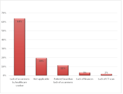
Figure 1: Factors for late diagnosis.

John Trizah Tracey1* Mwang’ombe NMJ2 Akuku OP3
1Department of Surgery, University Of Nairobi, Kenyatta National Hospital, Ministry of Health, Kenya*Corresponding author: John Trizah Tracey, Department of Surgery, University Of Nairobi, Kenyatta National Hospital, Ministry of Health, Kenya, E-mail: traceyjhn@yahoo.com
Background: Brain tumors are the second commonest tumors after leukemia and the most common solid tumors in children. Childhood Brain Tumors (CBT) are the most common cause of cancer-related deaths in children. Delayed diagnosis is associated with increased morbidity and mortality. The development of guidelines for the early diagnosis will provide better outcomes.
Objective: To develop guidelines for early diagnosis of childhood brain tumors.
Methods: Cross-sectional study involving 61 children 0-12 years with informed consent. Results were used to formulate statements for the Delphi survey, a questionnaire was presented to Consultant Neurosurgeons and Pediatricians. Statements reaching consensus were retained for final guidelines.
Results: 25 signs and symptoms were recorded of which headache (75.4%) was most common. The Pre-Diagnostic Symptomatic Interval (PSI) ranged from one week to 3 years with a median of 3 months and a mean of 7.7 months. The predominant reason for delayed diagnosis was a lack of healthcare worker awareness (64%). In the Delphi survey, eighteen (72%) statements achieved consensus and formed the final guideline. Seven (18%) of the statements did not meet consensus and were excluded from the guidelines.
Conclusion: The guidelines will aid healthcare workers by providing the varied presentation pattern of Childhood brain tumors and imaging recommendations. The guidelines are the first in Kenya and the region.
Childhood brain tumors; Early diagnosis; Guidelines; Kenya
Childhood Brain Tumor (CBT) comprises 15-20% of all brain tumors [1]. Brain tumors are the second most common tumors after leukemia and the most common solid tumors in children [2]. The overall annual incidence of CBT is 1.2-5.14 per 100,000 children [3]. The management of CBT remains a great challenge to clinicians. CBT is the most common cause of cancer-related deaths in children with a five-year survival rate of 75%. [4]
Despite the neuroimaging advancement, the timely diagnosis of CBT remains difficult. This is mainly due to the varied presentation and perceived rarity of CBT [5]. The variable presentation is dependent on the type and location of the CBT, as well as the age of the patient. CBT initially presents like other common but less serious illnesses [6]. This leads to delayed diagnosis which is associated with increased morbidity and mortality at presentation; disabling neurological complications as well as increased cognitive impairment in survivors [2]. Delayed diagnosis has also been associated with psychological distress for patients and caregivers as well as less trust in the health care system by the healthcare seekers [7].
The Pre-diagnostic Symptom Interval (PSI) of an illness is defined as the period between symptom onset and diagnosis. The mean PSI in children with Central Nervous System tumors reported in studies published over 15 years ranges from 1.8 to 9.8 months and a median of 1 to 3 months [7]. The factors associated with the delayed diagnosis can be grouped into three categories that include; patient and or caregiver factors, health professional factors, and factors related to the healthcare system.
Early diagnosis of CBT is a fundamental goal to allow for timely treatment when the disease is still at its early stages. The recognition that certain combinations of symptoms and signs indicate a focal brain lesion is crucial to the diagnosis of CBT. Increasing awareness of the variable and complex symptomatology that occurs with CBT will help tumor diagnosis and reduce the prolonged PSI [8]. This can be achieved through the development of clinical guidelines for the identification and imaging of children who have brain tumors [5].
Kenyatta National Hospital (KNH) is a tertiary facility and the largest hospital in the country with a bed capacity of about 2000 beds. The hospital hosts the largest neurosurgical unit in the country and receives referrals from the entire nation.
The broad objective of the study was to develop guidelines for the early diagnosis of CBT. The specific objectives were; to determine the pattern of signs and symptoms of CBT, to determine the Pre-diagnostic symptomatic interval of CBT, to establish reasons for delayed diagnosis of CBT, and to determine the specificity of symptoms and signs of CBT.
The design was a cross-sectional study and a Delphi Survey.
The study was conducted in KNH, a tertiary level six facility (which is a teaching and referral hospital) in the units which host patients with childhood brain tumors. The Institutional Research Ethics Committee (IREC) approved the commencement of the study.
The study population comprised all patients between 0-12 years who presented to the tertiary facility with childhood brain tumors and met the inclusion criteria. Pediatric patients in the country are classified as those between the ages of 0-12 years.
1. Patients between the ages 0-12 years with a brain tumor confirmed either radiologically with CT scan and or MRI of the brain and or histologically.
2. Patients who met criteria [1] above between the ages 7-12 years who assented to participate in the study
3. Patients who met criteria [1] above and whose caregivers gave informed consent to participate in the study.
Patients/caregivers who declined to give consent/assent or opted out during the study Patients with a brain tumor who’s radiological and or histological diagnosis records were unavailable.
The selection method was non-randomized consecutive sampling until the desired sample size was achieved. CBT constitutes 15- 20% of all brain tumors [1]. Using Fisher’s formula for sample size determination [9].
n=Z2P(1-P)/E2
Where,
n=Desired sample size
Z=value from standard normal distribution corresponding to desired confidence level (Z=1.96 for 95% CL)
P=prevalence/proportion (0.2)
E=Margin of error (+/- 10%)
Minimum number of patients 61
This was done to provide information on childhood brain tumor presentation, establish the PSI, and determine factors associated with delayed diagnosis.
The study period was 2016/2017.
This was done by the principal investigator.
The caregivers of patients and those patients between the ages of 7-12 years who meet the inclusion criteria were taken through the purpose of the study.
The patients’ caregivers were asked to make an informed choice to participate in the study by signing the consent form. In addition, those patients between the ages of 7-12 years were asked to assent by signing the assent form.
A questionnaire was used to collect the required data through an interview and physical examination of the patient. Medical records were reviewed and information was recorded in the questionnaire. The data collected included; General information; the patient’s age and gender.
Clinical data; the patient and or caregiver were interviewed about symptomatology, the duration of symptoms, and signs of the illness before diagnosis. A neurological examination was carried out to elicit signs of the illness. The patient’s notes, radiological images, and histological results were reviewed. The patient’s clinical notes were reviewed from the point of first contact with the healthcare worker to the point of diagnosis of a brain tumor. The type of tumor, symptomatology, PSI, and the reasons for delayed diagnosis were recorded in the questionnaire. The completed questionnaires were submitted to the statistician for analysis.
Questionnaires were coded and data entered into a passwordprotected database. The data were analyzed using the Statistical Package for Social Scientists (IBM SPSS) [10]. Analysis was done using descriptive statistics (mean, median) and summarized using frequencies and percentages. Bar graphs, tables, and pie charts were used for the presentation of results.
This was done to provide professional expertise through professional consensus. The expertise helped to determine the specificity of symptoms and signs associated with childhood brain tumors and to advice on appropriate indications for imaging.
The results of the cross-sectional study were used to formulate a Delphi Questionnaire. 25 statements were formulated into a questionnaire which was presented to Consultant Neurosurgeons, Pediatricians, Lecturers, Professors from the School of Medicine, University of Nairobi a premier University in the country, and the tertiary teaching and referral hospital, KNH. The purpose of the study was explained by the principal investigator. The nine respondents ranked their agreement to each statement using the Likert scale and returned the questionnaire. The feedback was analyzed by the statistician. The rankings for each statement were collated.
During the study period, a total of 61 patients presented with childhood brain tumors, of which 38 (62.3%) were male, and 23 (37.7%) were female, with a male to female ratio of 1.7:1. Patient ages ranged from as low as 1 month to 12 years with a median age of 8.2 years. The mean age was 7.3 years.
Seventeen 17 (27.9%) of the patients had craniopharyngioma, this was followed by medulloblastoma and pilocytic astrocytoma which had 10 (16.4%) patients each, PNET from 6 (9.8%) patients, ependymoma from 4 (6.6%) patients. Brainstem glioma, GBM, meningioma, and thalamic glioma each had 2 (3.3%) patients while choroid plexus papilloma, colloid cyst, optic glioma, pineal gland, SEGA, and tectal glioma had 1 (1.6%) patient each (Table 1).
| Type of Tumor | Frequency n(%) |
| Craniopharyngioma | 17(27.9) |
| Medulloblastoma | 10(16.4) |
| Pilocytic astrocytoma | 10(16.4) |
| PNET | 6(9.8) |
| Ependymoma | 4(6.6) |
| Brainstem glioma | 2(3.3) |
| GBM | 2(3.3) |
| Craniopharyngioma | 17(27.9) |
| Meningioma | 2(3.3) |
| Thalamic glioma | 2(3.3) |
| Choroid plexus papilloma | 1(1.6) |
| Colloid cyst | 1(1.6) |
| Optic glioma | 1(1.6) |
| Pineal gland tumor | 1(1.6) |
| SEGA | 1(1.6) |
| Tectal glioma | 1(1.6) |
| Total | 61(100.0) |
Table 1: Type of tumor.
Pre-Diagnostic Symptomatic Interval (PSI) ranged from as low as 1 week to 3 years with a median PSI of 3 months. The mean PSI was 7.7 months. Eleven (18%) of the patients had a PSI of less than a month while 50(82%) had a PSI longer than one month (Table 2).
| Months | Frequency n(%) |
| .2 (6 days) | 2(3.3) |
| .5 (15 days) | 8(13.1) |
| .7 (21 days) | 1(1.6) |
| 1.0 | 9(14.8) |
| 1.5 | 1(1.6) |
| 2.0 | 9(14.8) |
| 3.0 | 5(8.2) |
| 4.0 | 3(4.9) |
| 5.0 | 1(1.6) |
| 6.0 | 1(1.6) |
| 7.0 | 1(1.6) |
| 8.0 | 2(3.3) |
| 11.0 | 1(1.6) |
| 12.0 | 4(6.6) |
| 16.0 | 1(1.6) |
| 18.0 | 2(3.3) |
| 19.0 | 1(1.6) |
| 24.0 | 6(9.8) |
| 28.0 | 1(1.6) |
| 36.0 | 2(3.3) |
| Total | 61(100.0) |
Table 2: Pre-Diagnostic Symptomatic Interval (PSI).
On the factors for late diagnosis, the study found out that for 39 (64%) patients it was due to lack of awareness by the health worker, 7 (11%) respondents lacked awareness of the illness, 2 (3%) patients lacked finances and, 1 (2%) patient there was the unavailability of CT scan (Figure 1).
Figure 1: Factors for late diagnosis.
The study identified 25 signs and symptoms. The top two signs and symptoms were headache in 46 (75.4%) patients, followed by nausea and vomiting in 43 (70.5%) patients. The other prevalent signs and symptoms were lethargy and school difficulties from 24 (39.3%) patients, focal motor weakness from 20 (32.8%) patients, abnormal gait from 18 (29.5%) patients, seizures from 17 (27.9) of the patients, and reduced visual acuity from 16 (26.2%) patients. Other signs and symptoms were alterations in or loss of consciousness 11 (18%), abnormal co-ordination and cranial nerve palsies 10 (16.4%) patients each, nystagmus 5 (8.2%) patients, abnormal handwriting, diplopia, endocrine and growth anomalies 4 (6.6%) patients each, abnormal tone, squint, papilledema, and optic atrophy 3 (4.9%) patients each, abnormal speech and exophthalmia 2 (3.3%) patients each while abnormal reflexes, reduced visual fields, eye pain, unequal pupils and sunset eyes had 1 (1.6%) patient each (Table 3).
| List of signs and symptoms | Frequency n(%) |
| Headache | 46(75.4) |
| Nausea and vomiting | 43(70.5) |
| Lethargy and school difficulties | 24(39.3) |
| Focal motor weakness | 20(32.8) |
| Abnormal gait | 18(29.5) |
| Seizures | 17(27.9) |
| Reduced visual acuity | 16(26.2) |
| Alteration in or loss of consciousness | 11(18.0) |
| Abnormal co-ordination | 10(16.4) |
| Cranial nerve palsies | 10(16.4) |
| Nystagmus/Other abnormal eye movements | 5(8.2) |
| Abnormal handwriting | 4(6.6) |
| Diplopia | 4(6.6) |
| Endocrine and growth abnormalities | 4(6.6) |
| Abnormal tone | 3(4.9) |
| Squint | 3(4.9) |
| Papilloedema | 3(4.9) |
| Optic atrophy | 3(4.9) |
| Abnormal speech | 2(3.3) |
| Exophthalmia | 2(3.3) |
| Abnormal reflexes | 1(1.6) |
| Reduced visual fields | 1(1.6) |
| Eye pain | 1(1.6) |
| Unequal pupils | 1(1.6) |
| Sunsetting eyes | 1(1.6) |
Table 3: List of signs and symptoms.
A total of 25 statements formed the Delphi questionnaire. The level of agreement to the statements was rated using Likert’s Scale with the least score as 1 (strongly disagree) and the highest score of agreement at 7 (strongly agree). A score of 5-7 in or more than 80% of respondents constituted consensus while a score of 5-7 in less than 80% of the respondents was lack of consensus (Figures 2-5).

Figure 2: General statements.
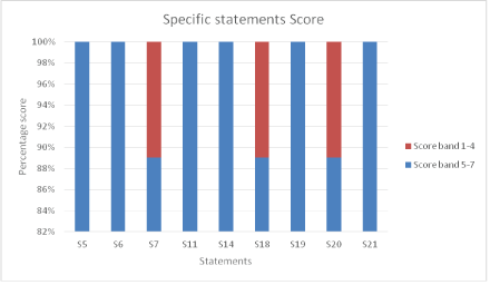
Figure 3: Specific statements.
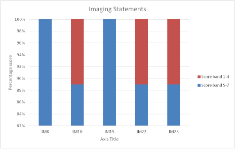
Figure 4: Imaging statements.
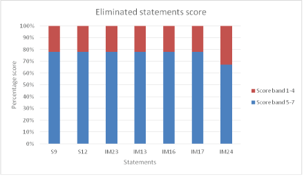
Figure 5: Eliminated statements.
Statements on General Considerations: The following General statements achieved consensus.
G1: The initial symptoms of a brain tumor may resemble symptoms that occur with other more common and less serious childhood conditions.
G2: Symptoms occurring with brain tumors may fluctuate in severity.
G3: The absence of neurological abnormalities does not exclude a brain tumor.
G4: Children aged 3 years and under with a brain tumor may present differently from older children.
Statements on Specific considerations: The following Specific statements achieved consensus.
S5: A symptomatic child with a brain tumor will have one or more of the following symptoms and/or signs:
• Headache
• Nausea and vomiting
• Focal motor abnormalities
• Abnormal vision, eye movements, and fundoscopy findings
• Altered consciousness
• Abnormal gait and co-ordination
• Seizures
• Abnormal behavior including lethargy
• Abnormal growth
S6: A child presenting with any of the symptoms and signs listed in 5 above requires the following:
• A detailed history including specific inquiry for associated symptoms
• A complete neurological assessment
• Assessment of height, weight & head circumference in a child aged <2 year
• Assessment of developmental stage in a child <5 years
S7: A continuous or recurrent headache lasting more than 4 weeks should be regarded as persistent.
S11: Nausea and/or vomiting for longer than 2 weeks should be regarded as persistent and the possibility of a brain tumor considered.
S14: Visual assessment of a child with a different diagnosis of a brain tumor must include assessment of:
• Visual acuity
• Eye movements
• Pupil responses
• Optic disc appearance
• Visual fields (in children >5 years)
S18: History should enquire into minor changes in motor skills e.g. change of hand or foot preference, loss of learned skills.
S19: Assessment of the gross motor skills of a child who may have a brain tumor should include observation of:
• Sitting or crawling in infants
• Walking or running
• Gross motor coordination.
S20: Assessment of a child’s fine motor skills should include observations of:
• Handling of small objects e.g. cup, spoon
• Handwriting in older children
S21: Abnormal balance or gait is not an indication of inner ear disease in the absence of confirmatory history, examination, or investigation findings.
Imaging Statements: The following statements on imaging achieved consensus.
IM8: Persistent headache requires Brain imaging.
IM10: A child with headache and episodes of confusion requires Brain imaging.
IM15: Brain imaging is required in papilledema, optic atrophy, proptosis, and reduced visual field.
IM22: Brain imaging is required for any child with
• Regression in motor skills.
• Abnormal gait or co-ordination unless there is an indication of a non-neurological cause .
• Focal motor weakness.
IM25: If MRI is not available a contrast-enhanced CT scan should be performed in a child who may have a brain tumor.
Eliminated statements: The following statements did not achieve consensus and were eliminated.
S9: A young child who is unable to complain of headache may demonstrate head pain by holding their head, lethargy, or withdrawal.
S12: Persistent nausea and/or vomiting in the absence of confirmatory history, examination, or investigation findings should not be associated with a gastrointestinal or other systemic infective cause.
IM13: Persistent vomiting on awakening requires Brain imaging.
IM16: Brain imaging is needed in new-onset non-paralytic squint and nystagmus.
IM17: Brain imaging is required in reduced visual acuity not caused by a refractive error.
IM23: A child with impaired growth with no identifiable psychosocial or physical cause should have brain imaging.
IM24: MRI is the imaging modality of choice for a child who may have a Brain tumor.
Eighteen (72%) of the statements achieved consensus; Seven (28%) statements did not meet the consensus threshold and were eliminated (Figures 6 and 7).
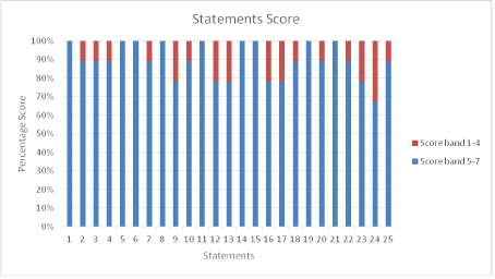
Figure 6: Statements score.
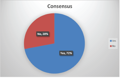
Figure 7: Consensus.
The 18 statements that achieved consensus were outlined into the guideline in three categories: A) General considerations B) Specific considerations and C) Imaging considerations.
A) General Considerations
1. The initial symptoms of a brain tumor may resemble symptoms that occur with other more common and less serious childhood conditions.
2. Symptoms of brain tumors in children may fluctuate in severity.
3. The absence of neurological abnormalities does not exclude a brain tumor
4. Children aged 3 years and below with a brain tumor may present differently from older children.
B) Specific Considerations
5. A child with a brain tumor who is symptomatic will have one or more of the following symptoms and or signs:
I. Headache.
II. Nausea & Vomiting.
III. Focal motor abnormalities.
IV. Abnormal vision, eye movements, and fundoscopy findings.
V. Altered consciousness.
VI. Abnormal gait and co-ordination.
VII. Seizures.
VIII. Abnormal behavior including lethargy.
IX. Abnormal growth.
6. A continuous or recurrent headache lasting more than 4 weeks should be regarded as persistent.
7. Nausea and/or vomiting for longer than 2 weeks should be regarded as persistent and the possibility of a brain tumor should be considered.
8. History should enquire into minor changes in motor skills e.g. change of hand or foot preference, loss of learned skills.
9. A child presenting with any of the symptoms and signs listed in 5 above requires the following:
i) A detailed history including specific inquiry for associated symptoms.
ii) A complete neurological assessment.
iii) Assessment of height, weight & head circumference in a child aged <2 years.
iv) Assessment of developmental stage in a child <5 years.
10. Visual assessment of a child with a differential diagnosis of a brain tumor should include assessment of:
i) Visual acuity.
ii) Eye movements.
iii) Pupil responses.
iv) Optic disc appearance (Fundoscopy).
v) Visual fields (in children >5 years).
11. Assessment of the gross motor skills in a child who may have a brain tumor should include observation of:
i) Sitting or crawling in infants.
ii) Walking or running.
iii) Gross motor coordination.
12. Assessment of a child’s fine motor skills should include observation of:
i) Handling of small objects e.g. cup, spoon.
ii) Handwriting in older children.
13. Abnormal balance or gait is not an indication of inner ear disease in the absence of confirmatory history, examination, or investigation findings.
C) Imaging Considerations
14. If MRI is not available a contrast-enhanced CT scan should be performed in a Child who may have a brain tumor (from considerations (A) and (B) above).
15. Persistent headaches require brain imaging.
16. A child with headache and episodes of confusion requires brain imaging.
17. Brain imaging is required in a child with papilloedema, optic atrophy, proptosis, and reduced visual field.
18. Brain imaging is required for any child with:
i) Regression of motor skills.
ii) Abnormal gait or coordination unless there is an indication of a non-neurological cause.
iii) Focal motor weakness.
The study identified 25 signs and symptoms where the two most common signs and symptoms were headaches in 46 (75.4%) patients, followed by nausea and vomiting in 43 (70.5%) patients. The other prevalent signs and symptoms were as shown in table 3. The varied pattern of presentation of CBT was established and was similar to a systematic review and meta-analysis of studies published for 15 years in which the most common presenting symptom was a headache, followed by nausea and or vomiting with a total of 56 signs and symptoms that were recorded [5]. Another study recorded 30 signs and symptoms with a similar presentation pattern [1]. Several other studies also documented headache as the most common symptom followed by nausea and or vomiting [6,8,11-14].
Pre-Diagnostic Symptomatic Interval (PSI) ranged from as low as 1 week to 3 years with a median PSI of 3 months. The mean PSI was 7.7 months. The study findings compared with the upper limit of the mean and median PSI of 13 cohorts and case series of children with central nervous system tumors published for 15 years ranging from 1.8 to 9.8 and 1 to 3 months respectively [7]. Other published studies recorded a median PSI range from 1.0-2.5 [1,6,11-13]. Another study by Coven SL, et al. [8] found a mean PSI of 4.5 and a median PSI of 1.4 months. The PSI in these studies was shorter than the PSI recorded in this study. Wanyoike PK [15] studied children with infratentorial tumors at Kenyatta National Hospital (KNH) and found a mean PSI of 3.7 months and a median PSI of 3.7 months. Edith O [16] conducted a study in childhood cancers at KNH and found the PSI to be comparable to other developing countries but longer compared to more developed countries.
On the factors for late diagnosis, the study found out that the predominant reason was due to lack of awareness by the health worker (64%). Other reasons included the lack of awareness by the caregiver, lack of finances, and unavailability of CT scans for diagnosis. This was similar to other studies. In a study on posterior fossa tumors in children at Kenyatta National Hospital [15], the reasons for late diagnosis included lack of awareness on CBT by the healthcare worker and inadequate diagnostic tools, especially in the rural areas. Other studies documented a lack of awareness by the healthcare professional as the main reason for delayed diagnosis [1,2,7,8,11,17]. Most of the patients with CBT are seen in the lowerlevel facilities where most of the healthcare workers lack awareness of brain tumors before referral to the higher-level facilities that host neurosurgical units in the country.
In the Delphi survey, 18 (72%) of the statements achieved consensus while 7 (28%) did not meet the consensus threshold and were excluded. The 18 statements were consolidated into the final guideline. The professional expertise helped to determine the specificity of symptoms and signs associated with CBT and to advice on appropriate indications for imaging. Guidelines development was based on the quality level of evidence through a combination of a cross-sectional study and a Delphi survey [7]. The guideline is a first in KNH and the low/middle-income country.
The study established the varied pattern of presentation of CBT, the delayed diagnosis of CBT, and the main reason for delayed diagnosis. Therefore, the guidelines will assist the health professionals in the early diagnosis of CBT by guiding the identification of the varied and complex signs and symptoms associated with CBT and indications for brain imaging.
Dissemination of guidelines in the health facilities in the country mainly through the Ministry of Health. Training of healthcare workers on the utilization of the guidelines will be required.
The guidelines will be disseminated in user-friendly materials like wall posters, brochures, and pocket cards both in digital and hard copy formats. The brochures and pocket cards will be issued to individual healthcare workers for use as quick reference materials.
Further studies on CBT to establish the prevalence of different types of tumors in the country especially Craniopharyngioma.
Special acknowledgment to all lecturers/consultant neurosurgeons and pediatricians from Kenyatta National Hospital and School of Medicine, University of Nairobi, who participated in the Delphi Survey.
Download Provisional PDF Here
Article Type: RESEARCH ARTICLE
Citation: John TT, Mwang’ombe NMJ, Akuku OP (2022) Development of Guidelines for Early Diagnosis of Childhood Brain Tumors at Kenyatta National Hospital. J Neurol Neurobiol 8(1): dx.doi.org/10.16966/2379-7150.184
Copyright: ©2022 John TT, et al. This is an open-access article distributed under the terms of the Creative Commons Attribution License, which permits unrestricted use, distribution, and reproduction in any medium, provided the original author and source are credited.
Publication history:
All Sci Forschen Journals are Open Access