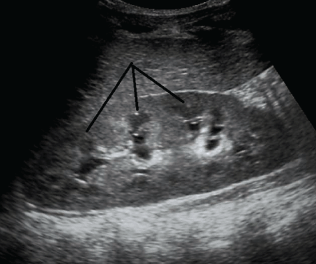
Figure 1: Ultrasound of the right kidney showing mild hydronephrosis

Salah Termos1 Majd Alkabbani1 Mohammad Alali1 Ahmad AlSaleh1 Sami Sanjad2 Jean-Daniel Delbet3 Bilal Aoun2*
1Department of Surgery, Al-Amiri Hospital, Kuwait*Corresponding author: Bilal Aoun, Division of Pediatric Nephrology, Department of Pediatrics, American University of Beirut, Lebanon, E-mail: drbilal77@hotmail.com
Introduction: Posterior urethral valves (PUV) disorder is the most common cause of bladder outlet obstruction in male newborns males. The diagnosis is usually made prenatally and only 10% of the patients manifest during adolescence or adulthood period [1].
Presentation of the case: We report the case of a three-yearold boy, previously healthy presented with abdominal pain and urinary retention. His mother noted a history of balanitis one week prior to the current presentation, which was treated by general practitioner with combination of topical antiseptic medications. Laboratory studies revealed acute kidney injury with elevated serum creatinine level (Cr.) of 1380 µmol/l. No urinary tract infection was detected. The Cr. level went back drastically to its normal value after insertion of a urinary catheter. (Foley’s catheter). Further evaluation confirmed the presence of posterior urethral valve (PUV) requiring cystoscopic resection.
Discussion and conclusion: We present a case of late presentation of PUV that was discovered incidentally during the investigation for balanitis causing obstructive renal failure. Awareness of this entity and its common prevalence is significantly important when evaluating boys at any age presenting with urinary symptoms. According to the literature, we found no direct relation between balanitis and PUV; however, we consider it a possible trigger that detected an insidious renal anomaly that could lead to serious medical problems in the future.
Posterior urethral valves (PUV); Balanitis; Children; Obstructive Renal Failure (ORF)
Posterior urethral valves (PUV) are obstructing membranes in the posterior male urethra. They are the most common congenital lesions that cause bladder outlet obstruction in male neonates with an incidence of one case per 8000 to 25,000 live births [2,3]. This disorder is usually sporadic; some cases have been seen in twins or siblings. It is usually established prenatally or at birth and rarely during adolescence or adulthood, thus making it challenging to be detected in the late onset presentation [3]. Clinically, it varies in degree of severity as it can be asymptomatic or can be presented as a weak urine stream, history of recurrent urinary tract infections and rarely in the adult age group presenting with infertility and enuresis [4].
We describe a case of PUV that was discovered incidentally during work-up for severe balanitis leading to ORF.
A three-year-old uncircumcised boy, who was presented to the emergency department (ED) with abdominal pain, persistent vomiting and loose stool of seven days duration, despite supportive therapy given by his general practitioner. The mother noted a history of balanitis that was managed with topical antiseptic solution (Astringent compresses using dilute vinegar and potassium permanganate) 10 days prior to this current presentation. She also noticed a marked decrease in his urine output. In the ED, the child was pale and hypoactive. Vital signs showed pulse rate of 90/min, blood pressure 125/75 mmHg, and oxygen saturation 98% in room air and temperature of 37°C. Review of the system was unremarkable except for abdominal distension and large palpable urinary bladder. Laboratory investigations revealed severe acidosis with acute renal failure (ARF). Serum Cr 1380 (N: 40-70 µmol), BUN (blood urea nitrogen) 71 (N: 3-7 mmol/L) Potassium (K) 6.2 mmol/L, Sodium (Na) 129 mmol/L and bicarbonate (HCO3) 9 mmol/L. Hemoglobin level was low 6.9 g/dl with MCV (Mean corpuscular volume) 69 fl, and C-reactive protein of 25 mg/l (N: <5). Urine analysis was normal and urine culture showed no bacterial growth.
Foley catheter was easily inserted and the patient was admitted to the pediatric intensive care unit (PICU) for 5 days. The renal function started to improve on his second day of admission with values reaching: serum Cr 42 µmol/l, BUN 6 mmol/l, and K 3.8 mmol/l. His stay was marked with polyuria that required intravenous fluid compensation and severe constipation resistant to laxative treatment. No hemodialysis was needed.
The patient was then transferred to the pediatric nephrology unit (PNU), where the urinary catheter was eventually removed. On the next day after removal of the urinary catheter, the boy developed severe bladder distension and a decrease of urine output with an elevation of serum creatinine to 165 µmol/l. Work-up for urinary retention was conducted, pelvic ultrasound was urgently done and revealed mildly distended calyceal system (mild hydronephrosis) (Figure 1). Voiding cystourethrogram (VCUG) was performed and showed dilatation and elongation of the posterior urethra (the equivalent of the ultrasonographic keyhole sign) (Figure 1 and 2) confirming the diagnosis of PUV. The decision was made to proceed for urgent urethrocystoscopy. The procedure was done under general anesthesia and showed type I PUV, bladder trabeculations and mildly hypertrophied bladder neck. Endoscopic valve ablation using knife urethrotome was performed, and the urethral catheter was kept for 48 hours. After removal of the catheter, the patient voided normally and the urine output was adequate and clear.
Figure 1: Ultrasound of the right kidney showing mild hydronephrosis
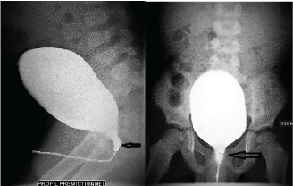
Figure 2: Voiding cystourethrographic study (VCUG). Images in lateral and anteroposterior views demonstrating a dilated and elongated posterior urethra
The child was followed up as an outpatient in the nephrology clinic at 1, 3, 6, 12 and 24 months after PUV resection. The patient had a stable clinical condition with normal values of serum creatinine levels and unremarkable ultrasounds (Absence of dilation or hyperechogenicity).
Posterior urethral valves (PUV) result from the formation of a thick, valve-like membrane from the tissue of Wolffian duct origin (failure of regression of the mesonephric duct) those courses obliquely from the verumontanum to the most distal portion of the prostatic urethra [5]. This is thought to occur in early gestation around 5-7 weeks [6]. The valve is actually a diaphragm with a central pinhole, however, as it is more rigid along its line of fusion, it gradually distends and becomes distended into a bilobed sail-like or windsock-like structure [7,8]. Clinical presentation of PUV depends on the severity of obstruction. In severe obstruction, the diagnosis is usually made antenatally. The fetus will be small for gestational age and ultrasound examination will demonstrate oligohydramnios and associated abnormalities [8]. In mild to moderate cases, the diagnosis is often not apparent until early infancy. UTIs are common in this group. Posterior urethral valves are also seen in association with other congenital abnormalities including chromosomal abnormalities such as Down syndrome, bowel atresia and craniospinal defects [7].
Although our patient showed a late presentation of the disease, he had no previous history of any urinary tract infections or enuresis and his mother reported that he had a normal antenatal ultrasound. He is a 3 years-old-boy with no associated congenital abnormalities, not circumcised, had experienced a single episode of severe balanitis, which in our assumption was incidental but can be the triggering factor for detection of a silent PUV secondary to the extension of the inflammation at the tip of the penis to the urethra leading to its narrowing and exacerbation of the stenosis and manifestation of clinical symptoms. In patients with no underlying urological malformations, balanitis leads to transient dysuria that usually improves with local care, however in patients with PUV, it may lead to urinary retention with bladder distension and subsequent obstructive renal failure (ORF) [9].
When detrusor hypertrophy overcomes obstruction, 10% of PUV cases may remain silent until later in life. Poor or weak stream, dribbling, repeated UTI, hematuria, and ORF are the most common pictures of presentation. Nearly 30 percent of boys with PUV have long-term kidney failure [10]. Late presentation is usually suggestive of lesser degrees of obstruction; the prognosis improves when detected early. According to the mother, this was the first time that her child experiencing these symptoms. The renal function normalized completely after insertion of a bladder catheter in a short period of postobstructive diuresis and patient did not require dialysis. Precise history can lead to a high index of suspicion where ultrasound is a valuable study and advised to be done in expert hands.
Following birth, findings on ultrasound are the same as those on antenatal ultrasound. Bladder is typically thick-walled and trabeculae with an elongated and dilated posterior urethra (keyhole sign) [4] kidneys in most cases are hydronephrotic and they may also be hyperechoic (dysplasia), although it is important to note that in up to 10% of cases they appear normal [5].
Examination of the posterior urethra can be performed through the perineum. The valve may actually be seen as an echogenic line, a diameter of more than 6mm is considered abnormal [5]. The Differential diagnosis includes urethral atresia, which is far less common [7].
PUV are associated with vesicoureteral reflux (VUR) in 50% of patients [5]. Voiding cystourethrogram (VCUG) is the best imaging technique for the diagnosis of posterior urethral valves. The diagnosis is best made during the micturition phase in lateral or oblique views, such that the posterior urethra can be imaged adequately [6,11]. Findings include dilatation and elongation of the posterior urethra (the equivalent of the ultrasonographic keyhole sign), bladder trabeculation or diverticula [7].
Endoscopic valve ablation is the mainstay of treatment. Means of ablation are variable and include hot loop resectoscope, cold knife urethrotome, hook diathermy electrode, bug bee electrode and less frequently used Fogarty catheter. The reported complications of this procedure include urethral stricture, urethral bleeding, urinary retention and urinary extravasation, however, the procedure is considered safe, as most of the complications can be managed conservatively [12,13]. Other treatment modalities, which are considered if primary valve ablation cannot be done, include vesicostomy and upper tract diversion whether by cutaneous ureterostomy or pyelostomy.
It is recommended to consider circumcision in patients with PUV, as it reduces the incidence of urinary tract infection (UTI) by 83%, especially that UTI can rapidly progress to pyelonephritis and sepsis in those with PUV [14].
According to the literature, circumcision may decrease the incidence of balanitis. In the uncircumcised male, the space between the prepuce and the phallus must be cleaned regularly. Proponents of circumcision argue that it is difficult for uncircumcised boys and men to maintain proper hygiene. Our patient had severe balanitis 10 days prior to presentation; we assume that this could have been the cause of aggravation of his medical condition.
Follow up with these patients is important to ensure good bladder function and to avoid any delay in detecting progressive or residual renal complications.
Silent PUV with late presentation (later than 2 years) associated with higher risk of developing chronic insufficiency and worse long-term outcome as well as decreased improvement in voiding capacity after treatment compared to those who present earlier [15,16].
Early detection of PUV is the key for better prognosis; although we found no direct link in the literature we think that balanitis was a red herring or a possible trigger that aggravated the preexisting urinary obstruction causing acute renal failure. The aim of this article is to highlight the need to investigate male children of any age with any urologic manifestation.
Download Provisional PDF Here
Article Type: CASE REPORT
Citation: Termos S, Alkabbani M, Alali M, AlSaleh A, Sanjad S et al. (2018) Posterior Urethral Valves, Balanitis and Acute Renal Failure: Any Link? Int J Nephrol Kidney Fail 4(1): dx.doi. org/10.16966/2380-5498.153
Copyright: © 2018 Termos S, et al. This is an open-access article distributed under the terms of the Creative Commons Attribution License, which permits unrestricted use, distribution, and reproduction in any medium, provided the original author and source are credited.
Publication history:
All Sci Forschen Journals are Open Access