Nanomedicine and Nanosurgery-Sci Forschen

Full Text

Review Article
Nanotechnology-Dermatological Perspective

  Tasleem Arif1*       Mohammad Adil2   

1Assistant Professor, Postgraduate Department of Dermatology, STDs and Leprosy, Jawaharlal Nehru Medical College (JNMC), Aligarh Muslim University (AMU), Aligarh, India
2Senior Resident, Postgraduate Department of Dermatology, STDs and Leprosy, Jawaharlal Nehru Medical College (JNMC), Aligarh Muslim University (AMU), Aligarh, India

*Corresponding author: Tasleem Arif, Assistant Professor, Postgraduate Department of Dermatology, STDs and Leprosy. Jawaharlal Nehru Medical College (JNMC), Aligarh Muslim University (AMU), Aligarh, India, Tel: +919557480345; E-mail: dr_tasleem_arif@yahoo.com


Abstract

Nanotechnology is the application of the science dealing with the study of small particles with dimensions of less than a hundred nanometers. Nanoparticles, owing to their size, have special physicochemical properties that are distinct from the derived bulk material. These nanoparticles have recently been employed for various diagnostic and therapeutic purposes and are particularly suited for use in the field of dermatology. This article discusses in brief the types of nanoparticles, the current and potential uses of nanoparticles in dermatological and cosmetic medicine. An account of potential hazards of indiscriminate use of nanoparticles has also been added to the review.

Keywords

Dermatology; Medicine; Nanoparticles; Nanotechnology

Introduction

Nanoscience, as the name suggests (nanos, Greek=dwarf), is the scientific study of small particles, with at least one dimension in the range of 1 to 100 nanometers. Nanotechnology is the application of this scientific knowledge for various diagnostic and therapeutic purposes [1]. This technology has kick started an exponentially expanding industry, still considered in its infancy by experts, that promises to revolutionize medicine, in terms of diagnosis and treatment, as nanoparticles are believed to be more specific, efficacious, customizable and cost effective. Nanoparticles may show physical and chemical properties quite distinct from similar material with particles of a larger scale, opening the exciting gateway for novel uses of already existing substances [2]. Nanodermatology is the use of nanotechnology in the prevention, diagnosis and treatment of various skin disorders and in the field of cosmetology.

Richard Feynman, considered the father of nanotechnology, was the first to conceptualize that molecules and atoms can be manipulated to form small components, not visible to the unaided eye [3]. The term nanotechnology was given by Professor Norio Taniguchi in 1974, referring to “process of separating, consolidating and deforming materials atom by atom or molecule by molecule” [4]. Nanotechnology has since then emerged as a fast growing field with great interest shown by pharmaceutical industries, medical professionals and patients alike.

Skin Penetration by Topical Formulations

The skin is one of the largest organs of the body and provides ample opportunities for nanoparticles to act. However any topical delivery system intended for systemic effects has to traverse the barrier formed by surface lipids, stratum corneum and other epidermal layers to reach the dermis where blood vessels are situated. Substances use one of the three possible pathways to traverse the epithelial barrier. Trans cellular permeation, where substances need to pass through cells of stratum corneum and intercellular lipids [5]; trans-appendageal permeation, where solute passes through hair follicles and sweat ducts [6]; and intercellular permeation where solutes pass tortuously via the extracellular lipids between stratum corneum cells [7]. Particles of size greater than 500 Daltons cannot pass through intact skin [8]. Hair follicles can act as micro-channels and are utilized as a conduit for the entry of such larger particles [9]. It has been demonstrated that particles of size as large as 10 micrometers can penetrate the hair follicle orifice [10]. Nanotechnology aids in drug permeation by releasing active substances at specific sites, increasing stability, ensuring adequate contact, enhancing stability and reducing the need of chemical enhancers [11].

Types of Nanoparticles

Nanoparticles can be classified on the basis of size, shape structure, physical and chemical properties. Nanoparticles can be spherical with a phospholipid covering, called liposomes or vesicle with a polymeric membrane, termed nanocapsules [12]. Lipid particles in an aqueous base termed solid lipid nanoparticle have hydrating photoprotective properties [13]. Drugs can be conjugated with lipids for increasing drug loading and targeted release [14]. Semipolymeric nanoparticles with a branched tree like structure are called dendrimers, while those packed in a cube like shape are cubosomes. Fullerenes are carbon atoms arranged in the form of a hollow tube, sphere or ellipse [15]. Nanocrystal is the crystalline arrangement of 10-400 nm size particles used for delivering poorly soluble drugs [16]. Gold and silver nanoparticles are utilized for strong antiseptic properties. Nonionic surfactant vesicles called niosomes have high penetration and stability [17]. Microsponges are microporous beads that have controlled drug releasing property, particularly responding to rubbing, temperature, pH and moisture [18]. Virosomes are viral proteins in liposomes and utilized in vaccines [4].

Applications

The various uses of nanotechnology in dermatology and cosmetology include (Table 1):

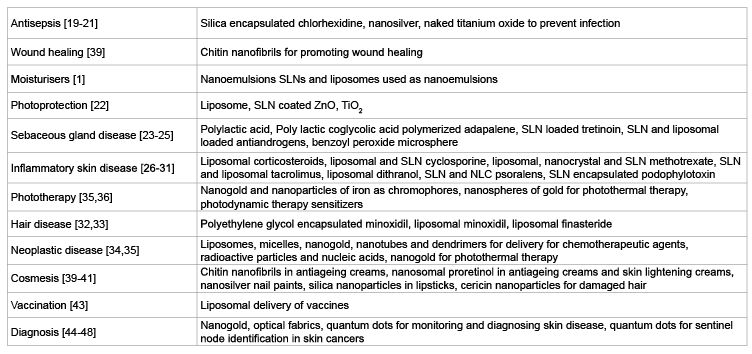
Table 1: Nanoparticles and their applications in dermatology and cosmetology

  • Antiseptics: Chlorhexidine nanoemulsion has a long lasting effect due to slow release of the drug from the core of the particle [19]. Nanosilver and titanium oxide nanoparticles act as antiseptics by being toxic to cell constituents [20,21].
  • Photoprotectors: Nanoparticles of zinc and titanium do not leave white residue on the skin and are cosmetically pleasing, while they still absorb and reflect UV rays [22]. Liposomes and SLNs enhance penetration and improve stability of the oxides of titanium and zinc used in sunscreens.
  • Sebaceous gland diseases: Adapalene when polymerized with nanoparticles like polylactic acid shows better efficacy [23]. Retinoids show less irritation when delivered by SLNs [24]. Benzoyl peroxide in microspheres is available in the market and is readily acceptable to the patient due to less irritation [25].
  • Inflammatory skin diseases: Liposomal formulations of steroids have a lesser chance of side effects like atrophy of skin [26]. Oil in water nanoemulsions has smaller size of the emulsified material and this facilitates replacement of ceramides in patients with disturbed skin barrier (as in atopic dermatitis) with the added advantage of a non-greasy feel and sensory texture enhancement. Tacrolimus has shown better penetration and enhanced efficacy and greater safety when incorporated in SLNs [27]. Incorporation of cytotoxic drugs like methotrexate and cyclosporine in forms like SLNs, plectin nanoparticles, nanocrystals etc., has been done in a bid to reduce their toxicity and to enhance therapeutic outcome [28,29]. The delivery of topical psoralens has been demonstrated to be greater if the drug is incorporated in SLNs and nanostructured lipid carriers (NLC) [30]. SLNs have also been used for encapsulation of podophyllotoxin to treat genital warts [31].
  • Hair diseases: Liposome encapsulated minoxidil and finasteride have a better penetration and lesser side effects [32,33].
  • Neoplastic diseases: Nanotechnology based chemotherapeutic agents are better delivered, needing lower dosages and decreasing adverse effects. Liposomes, micelles, carbon nanotubes, nanogold particles and dendrimers have been used for the same [34]. Agitated nanoparticles combined with monoclonal antibodies and hormones have been used in photothermal therapy to target tumor cells and with minimal damage to the normal adjoining tissue [35].
  • Lasers: Nanogold particles are used as specific chromophores that cause microscopic tissue damage [36].
  • Cosmetics and anti-ageing products: Nanoparticles have been used in products like deodorants, perfumes, shampoos, conditioners, lipsticks, eye shadows and after shave lotions etc [37,38]. Chitin nanofibrils are made from natural polysaccharides and are safe due to easy metabolization by enzymes. These particles are proliferators of fibroblasts and regulators of collagen synthesis and inflammatory cells. These are useful to decrease wrinkles and photoaging, and are believed to promote wound healing [39]. Retinoids containing nanosomes are being marketed for reduction of wrinkles by causing skin tightening, reducing photodamage and skin lightening [40]. Sericin nanoparticles have been reported to repair damaged hair cuticle [41]. Nanoparticle based hair straighteners cause less severe damage to hair. Nanoparticle based nail paints, polishes and lacquers prevent scratching and cracking. Nanosilver particles such as nail paint might be used to treat onychomycosis [2]. Silica nanoparticles for lipstick cause homogenous pigment dispersal and better cosmesis.
  • Vaccines: Activation of immunity by topical delivery of a vaccine holds promise in the treatment of dermatological and systemic infections and various neoplasias [42]. Liposomes with follicular route of administration have been proposed [43].
  • Diagnostic uses: Diagnostic tools based on nanoparticles are supposed to have greater sensitivity, specificity and require lesser tissue. Fabrics made of optical fibres can be used for mapping nevi and measuring dimensions of lesions while calorimetric assessments can detect inflammation [44]. Gold nanoparticles are used as labels for sensors [45]. Semiconductor nanoparticles with florescence property called quantum dots can be used to identify sentinel lymph nodes [46]. Nanopunch is a new method for collection of biopsy material from sites like nail matrix [47]. Newer diagnostic uses of nanotechnology include Raman spectroscopy, optical coherence tomography, 7-T magnetic resonance imaging, thermography, and multi photon laser scanning microscopy [48].
Risks of Nanotechnology

A large population is being increasingly exposed to nanoparticles, with percutaneous entry, inhalation and ingestion being the common routes of exposure [1,49]. The skin is a large organ and acts as an important portal of entry of nanoparticles; the penetration greatly increases when the skin barrier is disrupted due to wounds or some dermatitis, allowing larger molecules to pass through [50]. The respiratory route is involved during the use of aerosolized cosmetic products like perfumes or during production of such products. Entry into brain via olfactory nerves after inhalation has been documented [51]. Cosmetics like lip colour and lip gloss may gain entry in the body after ingestion. The toxicity of nanoparticles has been explained to be due to their small size, leading to exponential increase in surface area to volume ratio [52]. Other determinants of toxicity include chemical composition, charge, concentration, surface coating, shape and solubility. Toxicity may occur when the host has defective excretory mechanisms, best exemplified by the occurrence of nephrogenic systemic fibrosis in renal failure patients due to accumulation of gadolinium [53]. Sunscreens with nanoparticles of oxides of zinc and titanium accumulate in stratum corneum and their application to skin with abnormal barrier seems risky [1,54]. Titanium oxide generates free radicals on UV exposure and can lead to DNA damage and chromosomal mutations apart from damage to cell constituents [55]. Several in vivo studies regarding safety of nanoparticles in sunscreens have been done with no toxic effects as these particles are not able to penetrate the intact skin barrier [56,57]. Carbon nanotubes have been reported to cause granuloma in laboratory animals [58]. Quantum dots nanoparticles have the potential to penetrate epidermal keratinocytes of neonates and induce inflammation [59]. Concerns are expressed about nanosilver particles after it was demonstrated that the concentration that is toxic to bacteria also damages fibroblasts and keratinocytes [60,61]. Cosmetics with nanoparticles have the potential to induce foreign body granuloma and granulomatous cheilitis [62]. Nanoparticles are highly active particles and have the potential to act as allergens or haptens by reacting with proteins, forming a complex with MHC embedded selfpeptides, T cell recognition and an immune response [10,63]. Teratogenic effects of metal oxides have been shown in human fetal lung fibroblasts in in-vitro studies [64], cranial nerves and genital system in murine models [65] and carbon nanoparticles have shown to decrease sperm counts in murine models [66]. Nanoparticles are released in water, air, soil and food during manufacture and after use by consumers, also posing potential hazards to the environment [67].

Conclusion

Nanotechnology is a new branch of science that is believed to have a great potential for diagnostic and therapeutic purpose in the medical field. Dermatology and cosmetology is particularly more likely to benefit from the immense promise it holds. However, certain concerns regarding the safety of these particles need to be addressed before it is employed on a greater scale.

Competing Interest

The authors declare that they have no competing interests.

References
  1. Arif T, Nisa N, Amin SS, Shoib S, Mushtaq R, et al. (2015) Therapeutic and diagnostic applications of nanotechnology in dermatology and cosmetics. J Nanomedine Biotherapeutic Discov 5: 134. [Ref.]
  2. Saraceno R, Chiricozzi A, Gabellini M, Chimenti S (2013) Emerging applications of nanomedicine in dermatology. Skin Res Technol 19: 13-19. [Ref.]
  3. Drexler KE (2001) Machine-phase nanotechnology. Sci Am 285: 74-75. [Ref.]
  4. Antonio JR, Antônio CR, Cardeal IL, Ballavenuto JM, Oliveira JR (2014) Nanotechnology in dermatology. An Bras Dermatol 89: 126-136. [Ref.]
  5. Lawrence VA (2006) Development and evaluation of chitosan microparticles for the placement of dimethylaminoethanol (DMAE) in skin. Ribeirao Preto (SP): Universidade de Sao Paulo 117. [Ref.]
  6. Gupchup GV, Zatz J (1997) Target delivery to pilosebaceous structures. Cosmet Toiler 12: 79-88.
  7. Abraham MH, Chadha HS, Mitchell RC (1955) The factors that influence skin permeation of solutes. J Pharm Pharmacol 47: 8-16. [Ref.]
  8. Bos JD, Meinardi MM (2000) The 500 Dalton rule for the skin penetration of chemical compounds and drugs. Exp Dermatol 9: 165-169. [Ref.]
  9. Ptzelt A, Lademann J (2013) Drug delivery to hair follicles. Expert Opin Drug Deliv 10: 787-797. [Ref.]
  10. Toll R, Jacobi U, Richter H, Lademann J, Schaefer H, et al. (2004) Penetration profile of microspheres in follicular targeting of terminal hair follicles. J Invest Dermatol 123: 168-176. [Ref.]
  11. Gupta S, Basal R, Gupta S, Jindal N, Jindal A (2013) Nanocarriers and nanoparticles for skin care and dermatological treatments. Indian Dermatol Online J 4: 267-272. [Ref.]
  12. Papakostas D, Rancan F, Sterry W, Blume-Peytavi U, Vogt A (2011) Nanoparticles in dermatology. Arch Dermatol Res 303: 533-550. [Ref.]
  13. Pardeike J, Hommoss A, Müller RH (2009) Lipid nanoparticles (SLN, NLC) in cosmetic and pharmaceutical dermal products. Int J Pharm 366: 170-184. [Ref.]
  14. Schwarz C, Mehnert W, Lucks JS, Muller RH (1999) Solid lipid nanoparticles (SLN) for controlled drug delivery: I. Production, characterization and sterilization. J Contro Release 30: 83-96. [Ref.]
  15. Chirico F, Fumelli C, Marconi A, Tinari A, Straface E, et al. (2007) Carboxyfullerenes localize within mitochondria and prevent the UVBinduced intrinsic apoptotic pathway. Exp Dermatol 16: 429-436. [Ref.]
  16. Keck CM, Müller RH (2006) Drug nanocrystals of poorly soluble drugs produced by high pressure homogenisation. Eur J Pharm Biopharm 62: 3-16. [Ref.]
  17. Lohani A, Verma A, Joshi H, Yadav N, Karki N (2014) Nanotechnology based cosmeceuticals. ISRN Dermatology 2014: 843687. [Ref.]
  18. Panwar AS, Yadav CS, Yadav P, Darwhekar GN, Jaina DK, et al. (2011) Microsponge: a novel carrier for cosmetics. Journal of Global Pharma Technology 3: 15-24. [Ref.]
  19. Lboutounne H, Faivre V, Falson F, Pirot F (2004) Characterization of transport of chlorhexidine-loaded nanocapsules through hairless and wistar rat skin. Skin Pharmacol Physiol 17: 176-182. [Ref.]
  20. Chen X, Schluesener HJ (2008) Nanosilver: a nanoproduct in medical application. Toxicol Lett 176: 1-12. [Ref.]
  21. Tsuang YH, Sun JS, Huang YC, Lu CH, Chang WH, et al. (2008) Studies of photo killing of bacteria using titanium dioxide nanoparticles. Artif Organs 32: 167-174. [Ref.]
  22. Choksi AN, Poonawalla T, Wilkerson MG (2010) Nanoparticles: a closer look at their dermal effects. J Drugs Dermatol 9: 475-481. [Ref.]
  23. Rolland A, Wagner N, Chatelus A, Shroot B, Schaefer H (1993) Sitespecific drug delivery to pilosebaceous structures using polymeric microspheres. Pharm Res 10: 1738-1744. [Ref.]
  24. Castro GA, Oliveira CA, Mahecha GA, Ferreira LA (2011) Comedolytic effect and reduced skin irritation of a new formulation of all-trans retinoic acid-loaded solid lipid nanoparticles for topical treatment of acne. Arch Dermatol Res 303: 513-520. [Ref.]
  25. Wiesenthal A, Hunter L, Wang S, Wickliffe J, Wilkerson M (2011) Nanoparticles: small and mighty. Int J Dermatol 50: 247-254. [Ref.]
  26. Santos Maia C, Mehnert W, Schaller M, Korting HC, Gysler A, et al. (2002) Drug targeting by solid lipid nanoparticles for dermal use. J Drug Target 10: 489-495. [Ref.]
  27. Pople PV, Singh KK (2010) Targeting tacrolimus to deeper layers of skin with improved safety for treatment of atopic dermatitis. Int J Pharmaceutics 398: 165-178. [Ref.]
  28. Muller RH, Runge S, Ravelli B, Mehnert W, Thunemann AF, et al. (2006) Oral bioavailability of cyclosporine: Solid lipid nanoparticles (SLN®) versus drug nanocrystals. Int J Pharmaceutics 317: 82-89. [Ref.]
  29. Seo D-H, Jeong Y II, Kim DG, Jang M-J, Jang M-K, et al. (2009) Methotrexate-incorporated polymeric nanoparticles of methoxypoly(ethylene glycol)-grafted chitosan. Colloids Surf B Biointerfaces 69: 157-163. [Ref.]
  30. Fang JY, Fang CL, Liu CH, Su YH (2008) Lipid nanoparticles as vehicles for topical psoralen delivery: Solid lipid nanoparticles (SLN) versus nanostructured lipid carriers (NLC). Eur J Pharm Biopharm 70: 633-640. [Ref.]
  31. Chen H, Chang X, Du D, Liu W, Liu J, et al. (2006) Podophyllotoxinloaded solid lipid nanoparticles for epidermal targeting. J Control Release 110: 296-306. [Ref.]
  32. Jain B, Singh B, Katare OP, Vyas SP (2010) Development and characterization of minoxidil-loaded liposomal system for delivery to pilosebaceous units. J Liposome Res 20: 105-114. [Ref.]
  33. Kumar R, Singh B, Bakshi G, Katare OP (2007) Development of liposomal systems of finasteride for topical applications: design, characterization, and in vitro evaluation. Pharm Dev Technol 12: 591-601. [Ref.]
  34. Wei C, Wei W, Morris M, Kondo E, Gorbounov M, et al. (2007) Nanomedicine and drug delivery. Med Clin North Am 91: 863-870. [Ref.]
  35. Lu W, Xiong C, Zhang G, Huang Q, Zhang R, et al. (2009) Targeted photothermal ablation of murine melanomas with melanocytestimulating hormone analogue conjugated hollow gold nanospheres. Clin Cancer Res 15: 876-886. [Ref.]
  36. Pitsillides CM, Joe EK, Wei X, Anderson RR, Lin CP (2003) Selective cell targeting with light-absorbing microparticles and nanoparticles. Biophys J 84: 4023-4032. [Ref.]
  37. Epstein HA (2011) Nanotechnology in cosmetic products. Skinmed 9: 109-110. [Ref.]
  38. Hougeir FG, Kircik L (2012) A review of delivery systems in cosmetics. Dermatol Ther 25: 234-237. [Ref.]
  39. Mattioli-Belmonte M, Zizzi Z, Lucarini G, Giantomassi F, Biagini G, et al. (2007) Chitin nanofibrils linked to chitosan glycolate as spray, gel and gauze preparations for wound repair. J Bioact Compat Polym 22: 525-538. [Ref.]
  40. Choi M, Berson DS (2006) Cosmeceuticals. Semin Cut Med Surg 25: 163-168. [Ref.]
  41. Carmen MD, Pereda V, Polezel A (2012) Sericin cationic nanoparticles for application in products for hair and dyed hair. US Patent 20120164196A1. [Ref.]
  42. DeLouise LA (2012) Applications of nanotechnology in dermatology. J Invest Dermatol 132: 964-975. [Ref.]
  43. Jung S, Patzelt A, Otberg N, Thiede G, Sterry W, et al. (2009) Strategy of topical vaccination with nanoparticles. J Biomed Opt 14: 021001. [Ref.]
  44. Eden JG, Park SJ, Ostrom NP, Chen KF (2005) Recent advances in microcavity plasma devices and arrays: a versatile photonic platform. J Phys D Appl Phys 38: 1644-1648. [Ref.]
  45. Zuo L, Wei W, Morris M, Wei J, Gorbounov M, et al. (2007) New technology and clinical applications of nanomedicine. Med Clin North Am 91: 845-862. [Ref.]
  46. Kim S, Lim YT, Soltesz EG, De Grand AM, Lee J, et al. (2004) Nearinfrared fluorescent type II quantum dots for sentinel lymph node mapping. Nat Biotechnol 22: 93-97. [Ref.]
  47. Nasir AA (2010) Nanodermatology: a bright glimpse just beyond the horizon - part I. Skin Therapy Lett 15: 1-4. [Ref.]
  48. Boixeda P, Feltes F, Santiago JL, Paoli J (2015) Future prospects in dermatologic applications of lasers, nanotechnology, and other new technologies. Actas Dermosifiliogr 106: 168-179. [Ref.]
  49. Yah CS, Simate G, Iyuke SE (2012) Nanoparticles toxicity and their routes of exposures. Pak J Pharm Sci 25: 477-491. [Ref.]
  50. Yah CS, Iyuke SE, Simate G (2011) A review of nanoparticles toxicity and their routes of exposure. Iranian J Pharmaceutical Sci 8: 299-314. [Ref.]
  51. Paul JAB, Roel PFS (2006) Toxicological characterization of engineered nanoparticles. In: Gupta RB, Kompella UB (Eds) Nanoparticle Technology for Drug Delivery. Taylor & Francis, New York, USA.
  52. Auffan M, Achouak W, Rose J, Chane´C, Waite DT, et al. (2008) Relation between the redox state of iron based nanoparticles and their cytotoxicity towards Escherichia coli. Environ Sci Technol 42: 6730-6735. [Ref.]
  53. High WA, Ayers RA, Chandler J, Zito G, Cowper SE (2007) Gadolinium is detectable within the tissue of patients with nephrogenic systemic fibrosis. J Am Acad Dermatol 56: 21-26. [Ref.]
  54. Dussert AS, Gooris E, Hemmerle J (1997) Characterization of the mineral content of a physical sunscreen emulsion and its distribution onto human stratum corneum. Int J Cosmet Sci 19: 119-129. [Ref.]
  55. Hidaka H, Kobayashi H, Koike T, Sato T, Serpone N (2006) DNA damage photoinduced by cosmetic pigments and sunscreen agents under solar exposure and artificial UV illumination. J Oleo Sci 55: 249-261. [Ref.]
  56. Nohynek GJ, Dufour EK, Roberts MS (2008) Nanotechnology, cosmetics and the skin: is there a health risk? Skin Pharmacol Physiol 21: 136-149. [Ref.]
  57. Nohynek GJ, Dufour EK (2012) Nano-sized cosmetic formulations or solid nanoparticles in sunscreens: a risk to human health? Arch Toxicol 86: 1063-1075. [Ref.]
  58. Nasir A (2010) Nanodermatology: A glimpse of caution just beyond the horizon part II. Skin Therapy Lett 15: 4-7. [Ref.]
  59. Zhang WL, Yu WW, Vicki LC, Monteiro-Riviere NA (2008) Biological interactions of quantum dot NPs in skin and in human epidermal keratinocytes. Toxicol Appl Pharmacol 228: 200-211. [Ref.]
  60. Poon VK, Burd A (2004) In vitro cytotoxity of silver: implication for clinical wound care. Burns 30: 140-147. [Ref.]
  61. Basavaraj KH (2012) Nanotechnology in medicine and relevance to dermatology: Present concepts. Indian J Dermatol 57: 169-174. [Ref.]
  62. Lansdown AB, Taylor A (1997) Zinc and titanium oxides: promising UV absorbers but what influence do they have on the intact skin? Int J Cosmet Sci 19: 167-172. [Ref.]
  63. Hirai T, Yoshioka Y, Ichihashi K, Mori T, Nishijima N, et al. (2014) Silver nanoparticles induce silver nanoparticle specific allergic immune responses (HYP6P.274). J Immunol 192: 118.19. [Ref.]
  64. Zhang XQ, Yin LH, Tang M, Pu YP (2011) ZnO, TiO2 , SiO2 , and Al2 O3 nanoparticles-induced toxic effects on human fetal lung fibroblasts. Biomed Environ Sci 24: 661-669. [Ref.]
  65. Takeda K, Suzuki K-I, Ishihara A, Irie M-K, Fujimoto R, et al. (2009) Nanoparticles transferred from pregnant mice to their offspring can damage the genital and cranial nerve systems. J Health Sciences 55: 95-102. [Ref.]
  66. Yoshida S, Hiyoshi K, Oshio S, Takano H, Takeda K, et al. (2010) Effects of fetal exposure to carbon nanoparticles on reproductive function in male offspring. Fertil Steril 93: 1695-1699. [Ref.]
  67. Borm PJ, Robbins D, Haubold S, Kuhlbusch T, Fissan H, et al. (2006) The potential risks of nanomaterials: a review carried out for ECETOC. Part FibreToxicol 3: 11. [Ref.]

Download Provisional PDF Here


Article Information

Article Type: Review Article

Citation: Arif T, Adil M (2016) NanotechnologyDermatological Perspective. Int J Nanomed Nanosurg 2(2): doi http://dx.doi.org/10.16966/2470- 3206.112

Copyright: © 2016 Arif T, et al. This is an openaccess article distributed under the terms of the Creative Commons Attribution License, which permits unrestricted use, distribution, and reproduction in any medium, provided the original author and source are credited.

Publication history: 

  • Received date: 18 Feb 2016

  • Accepted date: 26 Apr 2016

  • Published date: 29 Apr 2016
  •