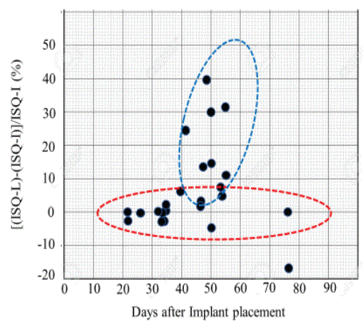
Figure 1: ISQ changes as a function of time after implant placement.

Takashi Miyazaki*
Director, Miyazaki Dental Clinic, Kashiba-City, Nara Prefecture, Japan*Corresponding author: Takashi Miyazaki, Director, Miyazaki Dental Clinic, Kashiba-City, Nara Prefecture, Japan, E-mail: miyarin3366@gmail.com
Purpose: The aim of this retrospective study was to evaluate the feasibility of dual effects of UV photofunctionalized implant, which is placed into piezosurgery-prepared site.
Methods: Total 35 cases were subjected to this study. All placed implants were made of commercially pure titanium (grade IV), which were originally surface treated by sandblasting followed by an acid etching. Diameter was ranged from 3.3 mm to 5.0 mm, while length varied from 7.0 mm to 11.5 mm. These original implants were UV photofunctioned and were placed into previously prepared by piezosurgery technique. ISQ scale was measured at implant placement (prior to suturing). ISQ scale was also measured at initial loading after certain days from the implant placement. Changes in the differences between these two ISQ scale readings were related to days between implant placement and loading (in other words, healing time).No statistic studies were made.
Results: There appears to be two distinctive relations between changes in ISQ scale and healing time; namely, one trend indicates that there are not noticeable changes and remain initial ISQ scale measured at the implant placement and the other relation exhibits a remarkable increase in ISQ during the healing process.
Conclusion: It was found that UV surface alteration and enough blood supply by piezosurgery preparation exhibited synergistic effects on improvement of ISQ scales, indicating that these dual techniques appears applicable to implant treatments.
Commercially pure titanium grade IV implant; SA treatment; ISQ scale; Early loading; UV photofunctioning; Piezosurgery
Implant stability is a prerequisite characteristic of osseointegration. This is true for both dental implants and orthopedic implants. During the survival period of placed implants, there are a variety of challenging factors leading to osteolysis and/or loosening implants, which clinically characterized by increased mobility with or without pain. Adverse factors for loosening implants should include biotribology (wear and/or friction actions in biological environment), fatigue, biomechanisms (e.g., nonaxial loading) and biocorrosion (including biodegradation) [1-5]. Other factors that might affect implant loosening are risk of bacterial infections associated with open surgery, osteoporosis, steroid medication, and diabetes mellitus [6- 10]. As to loosening of dental implants, it was mentioned that (i) soon after the implant placement, it is primarily due to surgical trauma, overheating of the osteotomy, complicated wound healing, insufficient primary stability and/or initial overload, and (ii) intermediate or late loosening of a dental implant more commonly results from marginal infection (peri-implant mucositis) and/or biomechanical overload, influenced largely by host characteristics [11]. Directly or indirectly surface characteristics and designs of implant could also result in the loosening implants [5,12]. Hence, continuous monitoring in a quantitative and objective manner is important to determine the status of implant stability [13,14].
Osseointegration as a measure of implant stability can occur in two stages: primary and secondary [15]. Primary stability mostly occurs from mechanical engagement with cortical bone. A key factor for the implant primary stability is the Bone-to-Implant (BIC) interaction [16], therefore, the primary stability is affected by bone quality and quantity, surgical technique and implant geometry (length, diameter, surface characteristics). Secondary stability offers biological stability through bone regeneration and remodeling [17-20], which should be affected by the primary stability [19,21]. The primary mechanical stability gradually decreases as time goes on, whilst the secondary biological stability increases, so that these two curves should intersect each other at some point during the bone healing process [21]. It is estimated that such transition point occurs roughly 2~3 weeks after implant placement when osteoclastic activity decreases the initial mechanical stability of the implant but not enough new bone has been produced to provide an equivalent or greater amount of compensatory biological stability [16,22-24]. It was mentioned that the transition period will be about 4 weeks in the post-operation for implant placement [25], indicating that timing of the transition appears to not agree among various studies. According to the author’ experiences of implant treatments with more than 2,000 implants (up to now), the transition period seems to exhibit in a range from 10 to 15 days after the implant placement. And in many cases, loading can be allowed about 3 weeks. However, with taking consideration of soft tissue healing, one month till the first loading has been normally practiced at the author’s clinic.
The stability is related to the biologic reaction of the bone to surgical trauma during the initial bone remodeling phase; bone and necrotic materials resorbed by osteoclastic activity is reflected by a reduction in Implant Stability Quotient (ISQ) value. This process is followed by new bone apposition initiated by osteoblastic activity, therefore leading to adaptive bone remodeling around the implant [26]. The ISQ scale is further subdivided into three zones: (1) low stability zone (ISQ<60), (2) medium stability (60<70) and (3) high stability (ISQ>70) [27]. The ISQ scale makes it possible to establish a standard clinical range of 1 to 100. More than 500 studies have been conducted based on RFA (Resonance Frequency Analysis) measurements and the ISQ scale [27]. In summary, it is true that implant stability is one of the most important factors for the success of implant treatments. Although most studies showed a correlation between bone density and implant stability, some studies suggest the opposite; due to the differences in the methods used. Recent studies suggest that implant stability during the healing process only increases for implants with low initial stabilities; meanwhile, loss of stability during the healing can be observed in implants with high initial stabilities [28].
For making metallic implant surfaces active, there are variety of surface modifications available [29]. Surface modification can be achieved by either surface concave shaping or surface convex forming. The former surface concave forming should include (i) mechanical modification (mostly, sandblasting or shot peening) and (ii) electrochemical or chemical treatment (such as acid etching or electrochemical etching); whilst the latter surface convex formation can include (iii) physical modification (typified by plasma-spray coating, for example, of hydroxyapatite) and (iv) thermal modification (such as MAO: microarc oxidation) [29]. In this study, as explained later, we have employed ultraviolet energy to activate implant surface, called as an ultraviolet photofunctionalization [30], which is defined as an overall phenomenon of modification of titanium surfaces by UV treatment. Outcome of the treatment should include the alteration of physicochemical properties and the enhancement of biologic capabilities of metallic (in particular, titanium) implants. It was also mentioned that bone morphogenesis around UV-treated titanium implants is distinctly improved when compared to that seen around untreated control implants, leading to rapid and complete establishment of osseointegration with nearly 100% bone-to-implant contact (BIC) in an animal model, as opposed to less than 55% for untreated implants [30]. Since the surface layer is positively charged by the UV photo action, early osseointegration can be expected due to the biological action by that osteoblastic cells and others might condense on implant surface zone. As a downside of this technique, UV treatment is a contraindicated to HA (hydroxyapatite)-coated implant since UV photoenergy generates deteriorating high temperature (ca. 70°C) on coated HA itself, resulting in possible debonding or weakening bonding strength to the substrate (such as titanium).
As to the implant site preparation, although a drilling is a dominant method, it possesses several problematic issues and challenges to surrounding tissues. In the case of rotating cutting instruments, internal watering can’t be achieved at satisfactory level and cutting debris of bone pieces might be pressed against the inner boner wall, causing insufficient blood supply for subsequent implant placement and possible overheating at localized bottom portion of preparation sites. Hence, the piezosurgery was employed [31-33], which is a process that utilizes piezoelectric vibrations in the application of cutting bone tissue to enable a precise cutting hard tissue, while precluding injury to soft tissue and the minimal heat is generated during cutting, thus maintaining vitality of adjacent tissue [34,35].
Although individual effect of UV photofunctionalization and piezosurgery has been reported separately, yet dual (synergistic) effects of these two techniques has not been reported and the combination possesses a high potential and clinical applicability, for particularly implant treatment. Based on the above background, specific aim of this retrospective study was to evaluate dual effects of UV photofunctionalized implant, which is placed into piezosurgeryprepared site with the ISQ (implant stability quotient) scale as a measure of the implant stability. ISQ scale at implant placement and ISQ scale at early loading are measured and the differences between these scales could provide a promising indicator for healthy bone healing and the onset timing of loading.
Total 35 implants (made from either Straumann or Osstem) were placed at sites (which are listed in table 1). The diameters of placed implants were 1 implant of 3.3 mm (S for Straumann), 3 implants of 3.5 mm (O for Osstem), 11 of 4.0 mm (O), 15 of 4.8 mm (S), 1 of 4.8 mm (S), and 4 of 5.0 mm(O). The lengths of placed implants were 1 implant of 7 mm long (O), 1 of 8.5 mm (S), 19 of 8.5 mm (O) 11 of 10 mm (O), and 3 of 11.5 mm (O). All implants were made of commercially pure titanium (CpT)-grade IV.
| No. | Implant | Loc | Size (mm) | ISQ@I | Load | ISQ@L | Days | ΔISQ | ΔISQ | ISQ-A |
| 2014 | bet. I/L | rate (%) | ||||||||
| 1 | 03/14 | 45 | 4.5 × 7.0 | 28-Apr | 77-77 | 34 | 78-78 (05/09) | |||
| 2 | 03/27 | 44 | 4.0 × 10.0 | 58 | ||||||
| 3 | 03/27 | 46 | 4.0 × 11.5 | 58 | ||||||
| 4 | 03/27 | 46 | 4.5 × 8.5 | U | 24-Jun | 62-59 | 66 | |||
| 5 | 03/28 | 45 | 4.5 × 8.5 | 71-71 G | 12-Jun | 58-58 | 76 | -13 | -18.3 | 18-18 (05/20) |
| 6 | 03/28 | 46 | 4.5 × 8.5 | 83-83 | 12-Jun | 83-83 | 76 | 0 | 0 | 75-75 (05/20) |
| 7 | 03/29 | 44 | 4.0 × 10.0 | 24-May | 75-75 | 57 | ||||
| 8 | 03/29 | 46 | 5.0 × 10.0 | 57 | ||||||
| 9 | 04/03 | 26 | 5.0 × 8.5 | 68-68 U | 30-May | 70-69 | 57 | 1 | 5.9 | |
| 10 | 04/05 | 25 | 5.0 × 8.5 | 48-52 S | 24-May | 71-70 | 49 | 20 | 40 | 80-80 (06/28) |
| 11 | 04/05 | 22 | 3.5 × 10.0 | 6-Jun | 64-64 | 62 | ||||
| 12 | 04/11 | 21 | 4.0 × 10.0 | 67-67 | 26-May | 74-74 | 55 | 7 | 10.4 | |
| 13 | 04/11 | 44 | 4.5 × 8.5 | 75-75 | 13-May | 75-75 | 32 | 0 | 0 | |
| 14 | 04/11 | 46 | 4.5 × 8.5 | 72-72 | 13-May | 72-72 | 32 | 0 | 0 | |
| 15 | 04/17 | 46 | 4.5 × 8.5 | 37 | ||||||
| 16 | 04/18 | 35 | 4.0 × 10.0 | 80-79 | 22-May | 77-77 | 34 | -3 | -3.8 | |
| 17 | 04/18 | 36 | 4.5 × 8.5 | 84-80 | 22-May | 80-80 | 34 | 0 | 0 | |
| 18 | 04/24 | 33 | 4.0 × 8.5 | 81-81 | 9-Jun | 83-83 | 46 | 2 | 2.4 | |
| 19 | 04/24 | 35 | 4.0 × 8.5 | 69-69 | 9-Jun | 73-73 | 46 | 3 | 4.3 | |
| 20 | 05/13 | 37 | 5.0 × 8.5 | 80-80 | 21-Jun | 77-82 | 21 | 0 | 0 | |
| 21 | 05/15 | 46 | 4.8 × 8.0 | 77-82 | 24-Jun | 84-85 | 40 | 5 | 6.3 | |
| 22 | 05/16 | 24 | 4.5 × 8.5 | 70-71 | 18-Jun | 70-72 | 33 | 0 | 0 | 71-71 (07/02) |
| 23 | 05/29 | 23 | 4.0 × 10.0 | 61-61 | 10-Jul | 76-76 | 42 | 15 | 24.6 | |
| 24 | 05/31 | 35 | 4.0 × 11.5 | 79-79 | 21-Jun | 75-78 | 21 | -2 | -2.5 | |
| 25 | 06/07 | 14 | 4.5 × 11.5 | 75-76 | 5-Jul | 75-71 | 33 | -1 | -3.9 | |
| 26 | 06/12 | 16 | 4.5 × 8.5 | 69-70 S | 5-Jul | 63-70 | 33 | -1 | -1.4 | |
| 27 | 06/12 | 45 | 4.5 × 8.5 | 62-61 | 8-Jul | 61-68 | 26 | 0 | 0 | 72-72 (07/14) |
| 28 | 06/12 | 46 | 4.5 × 8.5 | 82-82 | 8-Jul | 82-82 | 26 | 0 | 0 | 82-82 (07/14) |
| 29 | 06/28 | 31 | 3.5 × 10.0 | 44-49 G | 22-Aug | 62-62 | 55 | 15 | 31.9 | |
| 30 | 07/03 | 44 | 4.0 × 8.5 | 80-80 | 22-Aug | 75-75 | 50 | -5 | -6.3 | |
| 31 | 07/03 | 46 | 4.5 × 8.5 | 71-70 | 22-Aug | 82-82 | 50 | 10 | 14.1 | |
| 32 | 07/05 | 22 | 3.5 × 10.0 | 63-60 G | 21-Aug | 70-70 | 47 | 8 | 13 | |
| 33 | 07/10 | 22 | 3.3 × 10.0 | 20-Aug | 63-63 | 41 | ||||
| 34 | 07/17 | 24 | 4.0 × 10.0 | 64-65 | 21-Aug | 65-65 | 35 | 1 | 1.5 | |
| 35 | 07/17 | 26 | 4.5 × 8.5 | 59-61 UP | 9-Sep | 65-65 | 54 | 5 | 8.3 |
Table 1: Summary of obtained data in terms of loading date, implant dimension and location, ISQ at placement and loading date and ISQ at loading.
Note:
U: Remaining bone thickness is less than 4 mm
G: Bone grafting material was applied
S: Socket floor elevation was performed
P: Platelet-rich fibrin was applied
For treating implant surfaces, the commercially available TheraBeamAffiny UV system was utilized for 15 minutes for an automatic program of UV exposure, followed by immediate implant placement.
Mectron Piezosurgery 2 systems were employed (Mectron SPA, Carasco, Italy). The average operation time was about 10 minutes including exchanging times of 4 to 5 insert chips. The load that patients can feel is so light that this operation normally does not provide any unnecessary anxiety on patients.
The implant placement was performed by routine procedure which should be common practice regardless of surface conditions of implants and implant placement site preparation. Tooth extractions were performed as atraumaticas possible, followed by an immediate placement of implants. Local anesthesia was conducted through the local infiltration with articaine (with adrenaline).
The ISQ (implant stability quotient) scale is the value, indicating the level of stability and osseointegration in dental implants and is obtained using resonance frequency analysis (RFA) [36]. In this study, the OSSTELL ISQ system was employed. The ISQ scales were measured at two directions, namely lingual and buccal sides. In table 1, a pair (for example, 76-77) indicates (76 measured at lingual side77 obtained at buccal side) ISQ scale data. ISQ scale measurements were conducted at immediate after implant placement (even before the suturing), which we designated as ISQ@I, and at the time of first loading, designated as ISQ@L, respectively.
Table 1 summarizes all obtained results. Blank spaces in table 1 indicate that we could not measure ISQ scales. However, since all 35 cases have been treated under a combination of UV-treated implant which was placed into piezosurgery-prepared sites during a period from March 14th through July 17th, we decided to present all cases involved.
In first three columns, implantation date, placement locations (with tooth number) and dimensions (diameter × length in mm) are listed.
For ISQ scales, the following data are listed in the table 1:
1) ISQ@I: ISQ scale was measured at immediately after implant placement before suturing.
2) Load: after certain days (as marked under “Days bet. I/L), loading was conducted.
3) ISQ@L: ISQ scale was measured upon loading.
4) ΔISQ: the differences of ISQ scales between two readings: ΔISQ=ISQ@L-ISQ@I.
5) ΔISQ rate: for normalizing baseline, the ΔISQ rate was obtained by [ΔISQ/ISQ@I] × 100 (%).
6) ISQ-A: additionally, ISQ scales were measured for clinical reasons.
Figure 1 illustrates ΔISQ rate in terms of days after implant placement till the initial loading. It appears to be that there are two distinctive groups in this graph; (i) a group which did not exhibit remarkable changes in ISQ scales, marked with red, and (ii) a group shows an increasing trend of ISQ scales during the healing period, marked with blue.
Figure 1: ISQ changes as a function of time after implant placement.
A pair reading of ISQ scale appears to be same or closely similar, so that there are no differences in ISQ scale readings between lingual and buccal sides, as expected.
Various studies can be found on predictability of short implants [37,38]. For example, Vazouras K, et al. [37] studied the predictability of short implants (≤ 6 mm) and found that short implants in function for more than 3 years presented higher failure rates compared to short implants in function for less than 3 years. Moreover, Gultekin BA, et al. [39] evaluated the stability during healing and before loading of implants placed at two different supracrestal levels with various implant diameters and placement sites. It was reported that (i) submerging implant more inside bone may only influence primary stability, and (ii) implant diameter and site of placement influence primary and secondary stabilities before loading, whereas the implant length does not when its diameter is not accounted for. From the biomechanics standpoint, the physical contact total surface area (which should be a function of both diameter and length) of placed implants affects the resultant osseointegration. Accordingly, majority of placed implants in table 1 was medium (in a range from 7 to 10mm) and long (>11 mm) implants.
Cases 4,9 and 35 indicate that the remaining bone thickness at the bottom of implantation socket was less than 4 mm, after more than 2 months later, the satisfactory level of ISQ scales were obtained. Particularly, cases 4 and 9 were not subjected to any additional bone enriching treatment (such as bone grafting treatment). In this study, total 35 implants were placed, in which 33 implants were Osstem TS3 SA and 2 implants were Straumann Bone Level SLA. SA indicates that the implant surface was SAed (sandblasted with alumina particle and acid etched), while SLA means that implant surface layer was subjected to sandblasted with large alumina grits, followed by acid etching. By a simple calculation, when a particle size of 120 µm (with an assumption of 20 µm of each indentation) and coverage rate of blasting is 100%, it was estimated that original flat surface area can increase up to about 200%, indicating that effective surface area can increase two-fold. As a result, both surfaces are almost identical surface feature exhibiting a hybrid surface roughness, which is combined of macroscopic roughness due to sandblasting and microscopic roughness by chemical etching (which is reacting in nano-scale order) [29]. Such featured surfaces, in this study, were further treated by additional UV photofunctionalization. The evidence that bone morphogenesis around UV-treated titanium implants was improved (i.e., 100% BIC value in animal models) compared with that seen around untreated control implants (less than 55% of BIC value) was clearly reasoned by the alteration of physicochemical properties and the enhancement of biologic capabilities by UV treatment [30]. This UV surface modification alters surface physiochemical nature from hydrophobicity to hydrophilicity (in other words, improved wettability), removes unavoidably contaminated hydrocarbons, and exhibits a unique electrostatic status and act as direct cell attractants without the aid of ionic and organic bridges, which imparts a novel physicochemical functionality to titanium [30]. There would be no meaning to compare between Straumann implant system and Osstem implant system, since their original surface conditions (sandblasted and acid-etched) were altered by further UV treatment.
Bone grafting was performed for cases 5, 29 and 32, and sinus floor elevation was performed for cases 10, 26. Although these additional treatments improved ISQ scales (except case 5) to maintain in the acceptable zone (60<70), the UV treatment should further enhance the placed implant stability. It should be noted that, even after several days after the loading, in cases 1, 10, 27 and 28, ISQ scales increased as indicated in the last column in the table 1. As mentioned by Stanley M, et al. [40], the acceptable level of ISQ scale at the loading should lead to the high survival and success rates.
Piezosurgery is a relatively new and modern technique of bone surgery in implantology. This surgery technique exhibits therapeutic features, including a micrometric cut (precise and secure action to limit tissue damage, especially to osteocytes), a selective cut (affecting mineralized tissues, but not surrounding soft tissues, so that saving vital anatomical structures such as Schneiderian membranes or nerve tissues), and a clear surgical site (the result of the cavitation effect created by an irrigation/cooling solution and oscillating tip). As a result, implantology surgical techniques such as bone harvesting (chips and blocks), crestal bone splitting, and sinus floor elevation can be performed with greater ease and safety. Overall, piezosurgery enables critical operations in simple and fully executable procedures; and effectively, areas that are difficult to access have less risk of soft tissue and neurovascular tissue damage via piezosurgery [41,42].
In all cases presented in this study, and particularly case Nos. 4, 9 and 35 in which there were not enough remaining bone at the socket bottom, UV surface alteration and enough blood supply by piezosurgery preparation exhibits synergistic effects on improvement of ISQ scales. About the case No. 5, this female patient was 74 years old when she was treated March 38, 2014. When the implant was placed under relatively high torque value and ISQ@I scale was about 70 or higher, the subsequent reading (which was May 20, 2014) dropped down to ISQ scale of 20 or lower. Although this is not usual, it could happen, since the implant was placed at mandibular jaw under high applied torque value, the enough blood supplied was interrupted. However, later on (on June 12, 2014) at the loading time, the ISQ scale recovered to 58 scale which was at the satisfactory level. This evidence invites an attention on potential risk of the immediate loading.
Figure 1 illustrates ΔISQ rate in terms of days after implant placement till the initial loading. The transition point from primary mechanical stability to secondary biological stability takes place about 2-3 weeks, which also shows the lowest ISQ scale on the total stability curve. The earliest loading was conducted on case Nos. 20 and 24 at 3 weeks (21 days) from implant placement and both ISQ scale readings indicate satisfactory levels (for case 20, 80-80 at ISQ@I and 77-82 at ISQ@L; for case 24, 79-79 at ISQ@I and 75-78 at ISQ@L). We can consider ISQ scales measured as ISQ@I as the starting point, and the subsequent ISQ scales should stay the same level or lower than ISQ@I values, in order to assess the onset timing of initial loading. Nevertheless, there are many cases which show higher ISQ scales during the bone healing stage and even after loading (see Table 1 a in ISQ-A column). Figure 1 is another presentation suggesting that there should be remarkable synergistic effects of UV photofunctioning and piezosurgery.
For implant placement, there are immediate postextraction placement and delayed placement. On the other hand, for loading strategy, there could be an immediate loading and a delayed loading. Hence, taking all possible treatment plans, four possible combinations might exist, as seen in table 2.
| Immediate placement | Delayed placement | |
| Immediate loading | A | B |
| Delayed loading | C | D |
Table 2: Possible combination of timing for implant placement and subsequent loading timing.
There are several reports on an immediate placement and immediate loading [43-45] (A in Table 2). Guida L, et al. [43] concluded their case report that immediate loading did not seem to impair osseointegration of an immediate postextraction implant compared to an unloaded postextraction one, although further studies with a larger number of samples are needed to confirm these preliminary results. The effect of the timing of loading was investigated on bone-to-implant contact (BIC) following immediate placement of implants with a hydrophilic sandblasted, large-grit and acid-etched surface into fresh extraction sockets in a mini pig model [45]. It was concluded that (i) an immediate implant placement and loading showed similar BIC with immediate placement and delayed loading when implants with an SLA surface were used, and (ii) both procedures showed similar buccal bone crest levels, which presented some resorption irrespective of the treatment modality. Although Del Fabbro M, et al. [46] mentioned that an immediately placed and immediate loading implants are more predictable and successful than before, this approach cannot be applied to every immediate implant patient [47].
As to a combination B (delayed placement and immediate loading), Esposito M, et al. [48] compared the effectiveness of immediate postextractive single implants with delayed implants, placed in preserved sockets after 4 months of healing, and concluded that there are more complications with immediate postextractive implants in comparison to delayed implants. A similar study was made and it was concluded that no significant differences were observed between the two procedures, although the only two implant failures were for immediate postextractive implants [49]. Susarla SM, et al. [50] estimated one-year implant survival for delayed versus immediately loaded implants and identify risk factors for implant failure and concluded that immediately loaded implants were 2.7 times (after adjusting) more likely to fail at one-year compared to delayed loaded implants.
A combination D (delayed implant placement and delayed loading) is a case when bone quality as well as quantity is poor and bone grafting is mandatory, so that there are very few cases reported.
A healing period without early loading is currently still considered a prerequisite for implant integration, so that there should be a sinequo-non time between implant placement and initial loading. There is no clear differentiation on definition between immediate loading and early loading and their effects on implant stability as well as osseointegration neither [51-53]. Nevertheless, it is clearly stated that both share an early functional rehabilitation [54-57]. As a result, majority of cases should fall in a combination C (immediate placement and delayed loading).
Usually, early loading is conducted less than 60 days from implant placement and delayed loading is performed later than 60 (normally 90) days post-implantation. Majority (about 90%) of implant treatments can be found in a combination of immediate placement and early loading. As Singh A, et al. [57] mentioned, the immediate placement and early loading of dental implant provides advantages like fewer surgical procedures, shorter treatment time, and improved aesthetic and psychological confidence. Several studies were done to compare immediate postextraction placement and delayed placement [58-60]. Delayed placement is normally conducted in a range from 3 to 4 weeks after the tooth extraction. Rathor K, et al. [59] reviewed the predictability of success of immediate implant over delayed implant. It was concluded that (i) immediate implant placement following tooth extraction has been found to be viable and predictable solution to tooth loss, and (ii) advantage of immediate implant over delayed is the crestal bone loss was found to be minimal, suggesting that immediate placement of implant is better when compared to the delayed implant placement. On the other hand, Tonetti MS, et al. [60] compared need for bone augmentation, surgical complications, periodontal, radiographic, aesthetic and patient reported outcomes in subjects receiving implant placement at the time of extraction (Immediate Implant) or 12 weeks thereafter. It was mentioned that immediate implant placement should not be recommended when aesthetics is important. In summary, through this retrospective analysis in addition to valuable reports and studies cited in the above, a combination of immediate implant placement and early (delayed if necessary) loading should be recommended. Besides, further surface modification of UV photofunctioning and site preparation by piezosurgery are highly recommended to assure implant stability and assess the initial loading timing.
Although individual effect of UV photofunctionalization and piezosurgery has been recognized separately, dual effects (synergistic) effect has not been reported yet. Within limited number of presented cases in this retrospective study, we can conclude this study with (1) in all cases presented in this study, UV surface alteration and enough blood supply by piezosurgery preparation exhibited synergistic effects on improvement of ISQ scales, making the assured loading timing, and (2) even lower the ISQ scale during the bone healing stage, they are still acceptable level for pursuing early loading.
Since bone metabolism (or bone healing) is slow among aged patients. In addition, bone density level could differ between male and female patients and this difference could be emphasized among aged patients. Case analyses on these aspects are in progress.
As a final note, a portion of this study was presented at the Osstem World Meeting (Oct 19, 2014), Busan Korea.
Download Provisional PDF Here
Article Type: CASE SERIES
Citation: Miyazaki T (2020) Early Osseointegration Attained by UV-Photo Treated Implant into Piezosurgery-Prepared Site. Report I. Retrospective Study on Clinical Feasibility. Int J Dent Oral Health 7(1): dx.doi.org/10.16966/2378-7090.344
Copyright: © 2020 Miyazaki T. This is an open-access article distributed under the terms of the Creative Commons Attribution License, which permits unrestricted use, distribution, and reproduction in any medium, provided the original author and source are credited.
Publication history:
All Sci Forschen Journals are Open Access