Introduction
In the permanent dentition tooth agenesis has a prevalence of 2.5-9.6% [1-3]. The prevalence of non-syndromic oligodontia (six or more permanent teeth missing [4]) varies between 0.08 and 0.3 [3,5].
Several genes involved in tooth development have been described such as MSX1, AXIN2 and PAX9 [1,6]. Mutations in these genes, which are responsible for the regulation of the initial stages of odontogenesis, may result in oligodontia.
Denervation studies on animals have shown that unilateral removal of neurovascular structures will stop tooth eruption [7,8]. A theory has therefore been presented that insufficient innervation could be correlated to tooth agenesis [9-11].
It has been found that the inferior alveolar nerve develops in the mandible from three individual nerve paths, why it has been suggested that the pattern of tooth agenesis is related to the threeincisor, canine/premolar and molar-fields for neural development in the mandible [12]. The region within each of these fields, where innervation occurs last, is the tooth positions most often affected by agenesis.
Jacobsen J, et al. [13] presented an analysis of a mandible from the medieval period with unilateral absence of teeth, where the mandibular canal could not be detected on a panoramic radiograph on the side affected by total tooth agenesis, while the mandibular canal could be clearly identified on the opposite side.
The main purpose of this study was to evaluate if the previously suggested correlation between innervation and tooth agenesis [9- 11] in the mandible would result in a different appearance of the mandibular canal, in accordance to previously presented results, where unilateral absence of teeth and mandibular canal was seen in a historical material [13], which was suggested to support the theory of correlation between innervation and tooth agenesis. We, therefore, wanted to find out if the innervation to the mandible was affected to such an extent in individuals with oligodontia, that the differences in proportions of the mandibular canal height as part of the mandibular height could be identified on panoramic radiographs.
The hypothesis was that the mandibular canal’s vertical proportion (%) of the mandibular height at the first lower molar is smaller when comparing individuals with oligodontia to an age-and gender-matched control group of individuals without tooth agenesis.
Materials and Methods
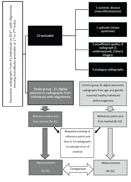
A flow chart of the study is presented in figure 1.

Figure 1: Flow chart for the study. Inclusion and exclusion of panoramic radiographs from individuals with oligodontia. The study group (N=31) was compared to an age-and gender-matched control group (N=31) of healthy, non-syndromic, non-medicating individuals. Error of method was evaluated after 15 duplicate measurements.
*DS4: All permanent teeth (excluding 3rd molars) erupted.
Materials
Forty-one adolescents from the county of Östergötland, Sweden, who were diagnosed with oligodontia, were available for the evaluation. Either analog, digitized (scanned) analog radiographs or digital images were available for the retrospective study. Only one panoramic radiograph from each patient was evaluated. If more than one panoramic radiograph was available, the one of best quality where all permanent teeth (excluding the third molars) had erupted was chosen.
Inclusion criteria for the study group were: oligodontia including agenesis of at least one mandibular premolar or first or second permanent molar, available panoramic radiograph of sufficient quality for diagnosis of the mandibular canal (not over-or under-exposed radiograph) and a dental stage DS4 according to Björk A, et al. [14], where all permanent teeth (with exception for the third molars) had erupted.
Exclusion criteria were: history of systemic disease or syndrome, missing or insufficient quality of panoramic radiograph and unerupted permanent teeth (third molars excluded).
Of the initial 41 patients diagnosed with oligodontia, 10 were excluded due to the exclusion criteria: one subject had a history of systemic disease (neurofibromatosis), one had Down syndrome, the available panoramic radiograph of one subject was underexposed and the panoramic radiographs from another two subjects were not sharp enough to enable analysis of the defined details for the study and five subjects did not possess any digital panoramic radiograph. Panoramic radiographs of the remaining 31 subjects (21 girls, 10 boys) with the mean number of 8.3 ± 3.4 missing teeth were evaluated. In the mandible the mean number of missing teeth was 4.0 ± 2.1. On the evaluated side of the mandible patients in the oligodontia group missed 1-6 permanent teeth (third molars excluded). The control group was a sample of 31 healthy, non-syndromic patients with full dentition undergoing orthodontic treatment in the county of Östergötland. All included patients were without history of Severe Medical Disorder (ASA 1) according to physical status classification by American Society of Anesthesiologists [15] and without medication according to anamnestic information in the dental records. Each oligodontia patient was matched with one control patient according to gender and age. Matching according to date of exposure of the panoramic radiograph was made to avoid possible bias from different panoramic radiographic methods used. Characteristics of the study material are presented in table 1.
| |
Oligodontia Group |
Control Group |
Difference |
|
N |
Mean age ± SD [Range] (years) |
N |
Mean age ± SD [Range] (years) |
P-value |
Boys
|
10 |
19.8 ± 1.9 [17.2-22.9] |
10 |
18.8 ± 1.7 [15.4-21.5] |
0.23 |
| Girls |
21 |
17.5 ± 2.3 [12.8-21.9] |
21 |
17.6 ± 3.2 [13.5-27.9] |
0.90 |
| Total |
31 |
18.3 ± 2.4 [12.8-22.9] |
31 |
18.0 ± 2.8 [13.5-27.9] |
0.70 |
Table 1: Age and gender of the participants.
The study was approved by The Regional Ethical Board at Linköping University, Sweden (Dnr 02-312).
Measurements
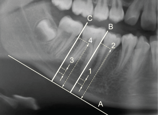
Each panoramic radiograph was transferred to the Adobe Photoshop CC image editor version 14.0 × 64 trial (Adobe Systems Incorporated, Copyright 1990-2013), where a mandibular base line was drawn bilaterally tangential to the most inferior points at the mandibular angle and the lower border of the mandibular body. Lines perpendicular to the mandibular base line were drawn through the contact points distally and mesially to the first mandibular molars. The height of the mandibular canal in relation to the total mandibular height was measured on the lines distally and mesially to the first mandibular molar and expressed as the proportional height of the mandibular canal in percent (Figure 2).

Figure 2: Reference lines and measured distances on the panoramic radiograph.
A-mandibular base line drawn bilaterally tangential to the most inferior points at the mandibular angle and the lower border of the mandibular body.
B-Line perpendicular to the mandibular base line drawn through the contact point mesially to the lower first molar.
C-Line perpendicular to the mandibular base line drawn through the contact point distally to the lower first molar.
1-Height of the mandibular canal mesially to the lower first molar.
2-Total mandibular height mesially to the lower first molar.
3-Height of the mandibular canal distally to the lower first molar.
4-Total mandibular height distally to the lower first molar.
The first mandibular molar was chosen as reference tooth since it is one of the mandibular teeth least frequently affected by agenesis [2,5]. If the first molar was missing the measurements were performed at the approximal sites of the second molar, which was the case in two of the subjects in the oligodontia group.
All measurements were made separately for the right and the left side of the panoramic radiograph. In cases where the radiographic details were impossible to distinguish on one side, the measurements were made on the opposite side.
Pictures were edited according to contrast and brightness to facilitate the measurements. Digital measurements were made using Image Tool version 3.0 (UTHSCSA, San Antonio, TX, USA) on a LCD screen Iiyama Pro Lite H481S. The distances were measured in pixels. All the image analyses were made by the same examiner (AP).
The measurements were performed in both the oligodontia group and the control group. The mean proportional height of the mandibular canal in relation to the total mandibular height on each side was compared between the two groups.
Method error
Repeated measurements were performed after 2 weeks in the panoramic radiographs from fifteen of the patients to calculate the error of the method using the Dahlberg’s formula [16]:

Where d is the difference between the first and the second measurement, and N is the sample size describing the number of individuals where the panoramic radiographs were re-measured.
There were no significant differences (P>0.05) regarding the repeated measurements for any of the evaluated sites (Table 2).
|
Measurements |
Dahlberg’s error of the method |
Systematic Error* |
Mean
difference |
p-value right side |
p-value left
side |
| MC 6 mes |
1.43 |
0.843 |
0.145 |
0.109 |
| MC 6 dist |
1.04 |
0.130 |
0.642 |
0.798 |
| MH 6 mes |
1.00 |
-0.081 |
0.778 |
0.960 |
| MH 6 dist |
0.96 |
0.003 |
0.992 |
0.456 |
Table 2: Calculation of the error of the method was performed using
the Dahlberg’s formula [16] comparing repeated measurements after 2
weeks in the panoramic radiographs from fifteen of the patients.
*Systematic error was assessed using paired t-tests.
MC 6 mes.-height of the mandibular canal mesially to the lower first molar [pixels]
MC 6 dist.-height of the mandibular canal distally to the lower first molar [pixels]
MH 6 mes.-total mandibular height mesially to the lower first molar [pixels]
MH 6 dist.-total mandibular height mesially to the lower first molar [pixels]
Statistical methods
Statistical analyses were performed with Stata/MP (version 12.1, StataCorp LP, College Station, TX 77845, USA). Paired t-test was used for evaluation of measurement error and two sample t-test for equal variances was used for comparison between the groups.
Results
Proportional height
The proportional height of the mandibular canal in relation to the total mandibular height is presented in table 3. The calculated proportional height of the mandibular canal was larger in the oligodontia group as compared to the control group on both the right (P<0.05) and the left side (mesially P<0.05 and distally P<0.001).
| |
Oligodontia Group |
Control Group |
Difference |
| N |
Mean ± SD (%) |
N |
Mean ± SD (%) |
|
| MC/MH 46 mesa) |
28 |
13 ± 3 |
28 |
11 ± 2 |
0.0126 |
| MC/MH 46 distb) |
30 |
14 ± 3 |
30 |
12 ± 3 |
0.0213 |
| MC/MH 36 mesc) |
23 |
13 ± 2 |
23 |
11 ± 2 |
0.0015 |
| MC/MH 36 distd) |
28 |
13 ± 2 |
28 |
11 ± 2 |
0.0004 |
Table 3: Proportional analysis of the height of the Mandibular Canal
(MC) in relation to the total Mandibular Height (MH) in subjects with
oligodontia as compared to a control group. Measurements performed
according to Figure 2.
a) MC/MH 46 mes-proportion between the height of the mandibular canal and the total mandibular height mesially to the lower first right molar
b) MC/MH 46 dist-proportion between the height of the mandibular canal and the total mandibular height distally to the lower first right molar
c) MC/MH 36 mes-proportion between the height of the mandibular canal and the total mandibular height mesially to the lower first left molar
d) MC/MH 36 dist-proportion between the height of the mandibular canal and the total mandibular height distally to the lower first left molar
Discussion
Study material
The syndromic patients were excluded due to a possible relationship between tooth agenesis and facial skeletal abnormalities in the syndromes [17-19].
Since the patients in the oligodontia group and the control group were matched according to gender and age at the time of radiographic examination (Table 1), these factors are not expected to have influenced the results. The limited number of patients included in the study should be considered when interpreting the results.
Image quality and measurements
The study material, which was collected from several clinics, had been exposed with different radiological equipment providing various image resolutions and was therefore not homogenous. Due to different enlargement factors in evaluated radiographs it was not relevant to compare the measurements in millimeters or in pixels. Instead, the proportion of the height of the mandibular canal as part of the total mandibular height was calculated. The region of the first permanent molar was used for the measurements, since the lower mandibular first molar is one of the teeth least frequently affected by agenesis [2,5], and since the contour of the mandibular canal is easier to distinguish in this region.
The quality of the performed measurements was satisfying since the measurement error was not significant between the repeated measurements. In our study 2 patients were excluded due to inferior quality of panoramic radiographs, where the needed details were not distinctly recognizable around the mandibular first molar.
The panoramic radiograph has limitations since it may be affected by distortion and since it only gives a two-dimensional picture of a three-dimensional object. Furthermore, the quality depends on the position and possible movement of the patient’s head during the radiographic procedure. Other factors, such as over-or underexposure, will affect the quality of the final radiograph and can lead to misinterpretation of the radiograph and cause difficulties to perform adequate measurements. On the other hand, the risk for distortions and defects in the panoramic radiographs was the same for both the oligodontia group and the control group. Therefore, we regard the results as reliable.
The presence of the mandibular canal was possible to detect in all the examined adolescents from both the oligodontia group and the control group.
No individuals in the oligodontia group missed all of the teeth on one side of the mandible as Jakobsen A, et al. [13] described in the medieval material. Even if Jakobsen A, et al. [13] found no grounds to assume that the absence of teeth on the side where the mandibular canal was missing was caused by disease or inbred, we find it possible that the lack of all teeth on one side of the mandible was associated to previous trauma or extractions due to infection rather than a consequence of oligodontia. The theory behind this could be that infection might cause changes in the bone structure and in the appearance of the mandibular canal.
Proportional height
Our finding that the proportional height of the mandibular canal (as part of the mandibular height) was larger in the oligodontia group than in the control group can be attributed either to a wider mandibular canal or a lower alveolar bone height in the oligodontia group. It is anyhow, possible that the size of the mandibular canal could have been the same in both groups if the mandibular height was different in the two groups. If the mandible did not develop vertically as much as a consequence of agenesis, this could be one reason for such a condition.
Tooth agenesis is one of the most common developmental anomalies in the human dentition [1,3,20] and is caused by multiple co-working factors [1,6]. Several studies support the theory of influence from innervations on tooth development [21,22] and some authors have found a correlation between the lack of innervations and tooth agenesis [9-11].
The hypothesis (that the mandibular canal’s vertical proportion (%) of the mandibular height at the first lower molar is smaller in individuals with oligodontia than in an age-and gender-matched control group of individuals without tooth agenesis) was rejected, since we found that the proportion of the mandibular canal’s height was larger in the individuals with oligodontia than in the control group.
The chosen method for analysis has several limitations. The use of a two-dimensional panoramic radiograph does not give the full picture of dimensional changes. A strictly standardized technique for radiographic examination and analysis would increase the possibilities to evaluate actual differences in terms of size, which was not possible to perform in the present study due to the different methods used over the time of the study and at the different clinics involved. It would be beneficial for further studies if 3D-visualisation techniques, such as CBCT or micro-CT, could be used for more detailed evaluation of possible divergent appearance of the mandibular canal in individuals with oligodontia.
Conclusion
The height of the mandibular canal constitutes a larger part of the mandibular height on panoramic radiographs in individuals with oligodontia than in an age-and gender-matched control group without tooth agenesis. The previously suggested correlation between innervations and agenesis could not be verified by the present study. Further research is necessary to verify such correlation. Further studies could possibly verify if a possibly wider mandibular canal in patients with oligodontia may influence orthodontic tooth movement or implant surgery in positions close to the mandibular canal, which thereby would influence decision-making in therapy-planning and clinical performance.
Acknowledgments
Thanks to Mats Fredrikson, Forum Östergötland (FORUMO), Linköping, for statistical consultation, and to Lukasz Pobiedzinski for technical support.
The study was supported by the Public Dental Service (Folktandvården) Östergötland, Sweden.
Disclosure of Interest
The authors report no conflicts of interest.


