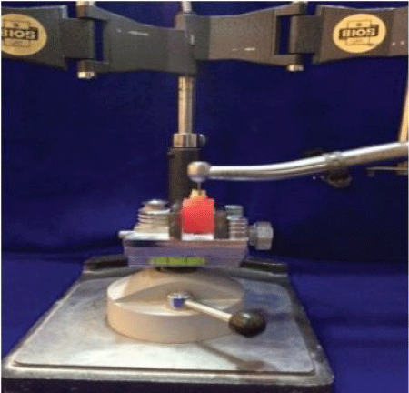
Figure 1: A modified dental surveyor used during tooth preparation.


Lena S Abdullah1* Adel F Ibraheem2
1Master student, Department of Conservative Dentistry, College of Dentistry, University of Baghdad, Iraq*Corresponding author: Lena S Abdullah, Department of Conservative Dentistry, College of Dentistry, University of Baghdad, Iraq, E-mail: lenamaster_88@yahoo.com
Objectives: The purpose of this study was to evaluate and compare the effect of using different finishing line designs (deep chamfer and shoulder) with different occlusal surface reduction schemes (planar and flat) on the vertical marginal fit of full contour CAD/CAM zirconia crown restorations.
Materials and method: Thirty-two sound maxillary first premolar teeth of comparable size and shape freshly extracted for orthodontic purposes were collected to be used in this in vitro study. To minimize confounding variables, One-way ANOVA test was performed for each dimension and no statistically significant difference was found among the four subgroups. Teeth were divided into two main groups according to the design of finishing line used (n=16): Group A: Deep chamfer; Group B: Shoulder. Each group was then subdivided into two subgroups according to the scheme of occlusal reduction used (n=8): (A1, B1) Planar; (A2, B2) Flat. Standardized preparation for full contour zirconia crown restorations was carried out with finishing lines depth 1.0 mm, total convergence angle of 6 degrees and axial height 4 mm (buccally and palatally). The teeth were then scanned directly using digital intra-oral scanner technique (Sirona AC Omnicam camera). Full contour zirconia crowns were then fabricated using Sirona In-Lab MC X5 milling device. Vertical marginal gaps were measured at four points on each tooth surface using a digital microscope at a magnification of (280X).
Results: The results of this study showed that there were statistically highly significant differences (p<0.01) using one- way ANOVA analysis and Student’s t-test.
Conclusions: Deep chamfer with planar occlusal reduction scheme provided better marginal fit compared to that obtained with shoulder. On the other hand, shoulder with flat occlusal reduction scheme provided better marginal fit compared to that obtained with deep chamfer.
Chamfer and shoulder finishing line; Digital impression; Full contour zirconia; Marginal fit; Planar and flat occlusal reduction scheme
The success of all ceramic restorations strongly depends upon marginal adaptation. A well fitted margin is expected to reduce plaque accumulation, recurrent caries which result in damage to the tooth with its supporting periodontium, and potentially decreases the longevity of the restoration [1]. Clinically, it is important that the crown margins fit the prepared tooth precisely to minimize plaque accumulation and, therefore, reduce risk of gingivitis, periodontitis, secondary caries and pulpitis [2]. These defects are common reasons for the failure of restorations [3]. The clinically acceptable limit of the marginal discrepancies is reported to be less than 120 µm [4]. The introduction of computer aided design/computer aided manufacturing (CAD/CAM) technology in dental practice results in more accurate manufacturing of prosthetic frameworks and greater accuracy of restorations [5,6]. One of the CAD/CAM technology is a digital impression which provides speed, accuracy, high quality of restoration as its designing is based on materials characteristics, ability of storing captured information indefinitely and transferring digital images between the dental office and the laboratory [7].
Zirconia ceramics have been extensively studied because of their excellent mechanical properties, which are much greater compared with those of other dental ceramics [8]. In prosthodontics, zirconia has a wider range of possible applications than other all ceramics [9]. It has been used as a framework material for single crowns, FDPs, inlay-retained FDPs and resin bonded FDPs [10]. These restorations can also be fabricated as monolithic restorations without veneering porcelain [11]. Zirconia was also used as a reinforcing material of other materials such as zirconia containing lithium silicate [12] and glass infiltrated alumina [13]. The mechanical properties of zirconia are very similar to those of metals and its color is similar to tooth color after stabilization, high levels flexural strength and fracture toughness as well as good wear resistance can be achieved [14]. In addition, it has versatile properties due to its mechanical, optical and biological properties that accelerated by the development of CAD/CAM system [15]. Partially sintered Y-TZP blocks were used for the production of full contour anatomical crowns. The restoration can be milled from a block of single material as a monolithic which referred to as full contour restoration with no porcelain overlay [16,17]. It can be prepared just like conventional porcelain fused to metal (PFM) crowns using either a shoulder, a chamfer, or a knife-edge finishing line [18]. It was indicated for posterior crowns, crowns over implants, crowns with limited occlusal clearance and full-arch bridges up to 14 units. Primary candidates include bruxers and grinders who do not desire cast gold or metal occlusal PFM restorations and for esthetic reasons, it is recommended that a facial veneer of porcelain be used on any zirconia-based anterior restoration.
However, full contour zirconia may be used in specific anterior cases where a dentist wishes to emphasize the strength of the restoration over its esthetics. It has many properties such as high flexural strength and fracture toughness, resistance to thermal shock (low thermal expansion numbers), improved esthetics and wear compatibility [19].
The designs of tooth preparation can affect the success of the crown restoration [20]. The finishing line designs such as deep chamfer and shoulder have been advocated for all ceramic crown restorations [21]. However, there is no available research dealing with the effect of finishing line designs in combination with different occlusal reduction schemes on the marginal fit of full contour zirconia crowns.
Thirty-two sound human maxillary first premolar teeth of comparable size and shape extracted for orthodontic purposes were selected and collected to be used in this in vitro study. All teeth samples were then embedded in individually cold cure acrylic resin block up to 2 mm apical to the CEJ. Dental surveyor was used to align the long axis of the tooth to be vertical to the horizontal plane of the mold.
To achieve standardization, all teeth preparation was performed with the same operator. Furthermore, high speed turbine hand piece was secured and adapted to a modified dental surveyor to be used all the way during preparation of axial wall for each tooth sample to ensure a consistent degree of taper (Figure 1).

Figure 1: A modified dental surveyor used during tooth preparation.
All the teeth were prepared for full ceramic crown restorations with the following preparation features: A total convergence angle of 6 degrees, the depth for both deep chamfer and shoulder finishing line of 1.0 mm and a standardized axial height of 4 mm (buccally and palatally), these dimensions were checked using a modified digital caliper (Figures 2-5).
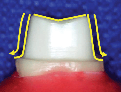
Figure 2: Deep chamfer finishing line with planar occlusal reduction scheme.
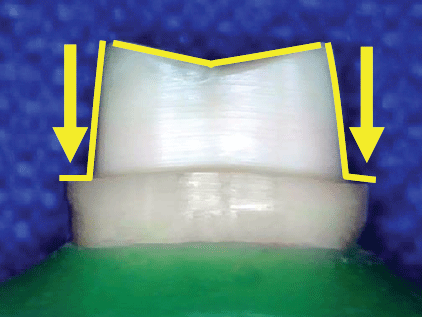
Figure 3: Shoulder finishing line with planar occlusal reduction scheme.
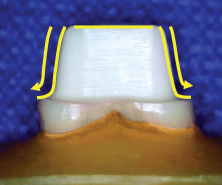
Figure 4: Deep chamfer finishing line with flat occlusal reduction scheme.
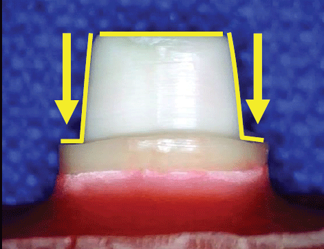
Figure 5: Shoulder finishing line with flat occlusal reduction scheme.
A three dimensional digital image for each tooth sample was taken by AC Omnicam intra-oral scanner (Sirona Dental Systems, Bensheim, Germany) (powder free) (Figure 6). Full contour zirconia crown restorations were then fabricated using In-Lab MC X5 milling device used (Sirona InCoris TZI C blank) (Figure 7).
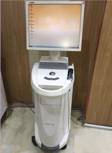
Figure 6: CEREC AC unit with Omnicam camera (Sirona, Germany).
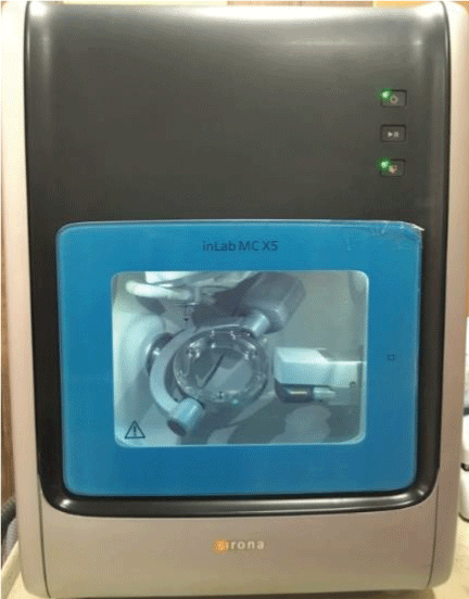
Figure 7: In-Lab MC X5 milling machine.
In this study, a custom made holding device was especially fabricated to be used during seating of the zirconium crown, it serve as a screw that secured the zirconia crowns to the natural tooth sample. Furthermore, it hold the specimens on the horizontal table of the microscope to allow for viewing the references points during measurement of vertical marginal gaps (Figure 8).
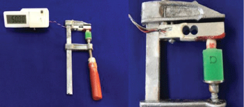
Figure 8: Zirconia crown on natural tooth sample secured with the specimen holding device.
The measurements were made on four points determined on each surface of the tooth (two at the edges of the line, two points drawn in the mid of the surface by permanent marker, while the other two points were at a distance of (1 mm) from the previous one, on both (left and right sides). Sixteen measurements were obtained from each tooth sample; highest one was selected to represent the maximum marginal gap of that sample [22-24] (Figure 9).
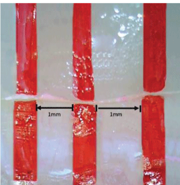
Figure 9: Four points of measurement at a magnification of 50X.
Positioning of the digital microscope was adjusted in such a way so that it’s horizontal plane (long axis) was perpendicular on the long axis of the tooth then the handle was fixed at that position so it couldn’t be moved vertically with no change in horizontal inclination of the microscope (Figure 10).
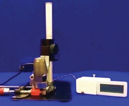
Figure 10: The sample with the holding device was placed under the microscope.
Two images for each surface of the tooth sample were captured and processed by (Dino- capture) software, then the captured images were opened in an image-processing program (Image J 1.50i, U.S. National Institutes of Health, Bethesda, MA, USA), to measure the marginal gap in pixels [25].
In order to convert the measurement to micrometers, a photo of one millimeter of a ruler was captured by the digital microscope at the magnification of (280X) then, the image were opened in (Image J) program and the straight line selection tool was selected to make a line that corresponded to a known distance that was a one millimeter (Figure 11).
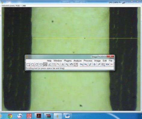
Figure 11: Image of one millimeter at magnification of 280X.
From the main menu (analyze) option was selected at the same calibration and magnification of the microscope, then, the (Set scale) was opened and converted all the calculated readings from pixels to (µm) [26] (Figure 12).
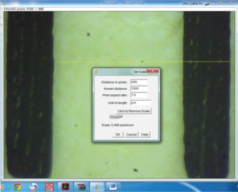
Figure 12: Calibration of the measurements by set scale option.
All measurements were performed by the same investigator [27], they were repeated three times to get precise, accurate readings and decrease the possibility of errors [28].
The SPSS (statistical package of social science) software version 18 for windows XP Chicago, USA was used to perform the statistical analysis. Descriptive statistics were computed for marginal gaps. Statistical methods were used in order to analyze and assess the results included:
A-Descriptive statistic: including mean, standard deviation, statistical tables and graphical presentation by bar charts.
B-Inferential statistics: 1. One-way ANOVA (analysis of variance) test was carried out to see if there were any significant differences among the mean values of the different subgroups.
2. Student’s t-test was carried out to examine the source of significant differences among the different subgroups. Statistical significance according to probability value (P) was determined to be as:
A total of 512 measurements of vertical marginal gap for the four subgroups were recorded, with 16 measurements for each tooth sample.
Table 1 showed the descriptive statistics of vertical marginal gap for the four subgroups measured in µm. It showed that the lowest mean of vertical marginal gap values was scored by subgroup A1 (38.837 ± 9.30) (Deep chamfer finishing line with planar occlusal reduction scheme), while the highest mean of vertical marginal gap values was belonged to subgroup B1 (66.636 ± 8.57) (Shoulder finishing line with planar occlusal reduction scheme) and this is clearly shown in figure 13.
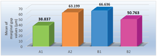
Figure 13: Bar-chart showing the mean values of the marginal gaps for all subgroups in micrometer.
| Types of finishing line | Subgroups | Descriptive Statistics | |||||
| N | Mean | SD | Min. | Max. | |||
| Deep chamfer finishing line | A | A1 | 8 | 38.837 | ± 9.30 | 21.137 | 63.085 |
| A2 | 8 | 63.199 | ± 9.22 | 43.410 | 82.692 | ||
| Shoulder finishing line | B | B1 | 8 | 66.636 | ± 8.57 | 45.684 | 85.829 |
| B2 | 8 | 50.763 | ± 12.88 | 25.684 | 80.615 | ||
Table 1: Descriptive statistics of vertical marginal gaps for the four different subgroups in micrometer.
Table 2 represents One-way ANOVA analysis test that applied to see whether this differences among the four subgroups statistically significant or not. This test showed that there was a statistically highly significant difference in the vertical marginal gap among the four subgroups.
| ANOVA | Sum of Squares | df | Mean Square | F | Sig. |
| Between Groups | 3854.007 | 3 | 1284.669 | 42.615 | 0.000 |
| Within Groups | 844.077 | 28 | 30.146 | ||
| Total | 4698.084 | 31 |
Table 2: One-way ANOVA analysis test for comparison of the marginal gaps among the different four subgroups.
HS: (p<0.01) (highly significant).
Table 3 represents Student’s t-test to examine the source of differences and to detect the significant differences between each two different subgroups.
| Subgroups | Mean ± SD | t-test | P-value |
| A1 vs. A2 | 51.018 ± 9.26 | 9.196 | 0.000 (HS) |
| B1 vs. B2 | 58.700 ± 10.73 | 5.593 | 0.000 (HS) |
| A1 vs. B 1 | 52.737 ± 8.94 | 15.116 | 0.000 (HS) |
| A2 vs. B2 | 56.981 ± 11.05 | 3.637 | 0.003 (HS) |
Table 3: Student’s t-test for comparison of marginal gaps between each two different subgroups.
HS: (p<0.01) (highly significant).
This test showed that there was a statistically highly significant difference in the vertical marginal gap Thi between each two subgroups.
Many studies showed that the marginal opening of less than 120 µm was considered clinically acceptable with regard to longevity for conventionally cemented crown restoration [29-31]. Furthermore, other study showed that clinical acceptable marginal gaps for CAD/CAM crown restorations were within 100 µm [32].
The results of this in vitro study showed statistically highly significance difference but still within the clinically acceptable limit (<120 µm) for all tested subgroups. It is worth to mention that when reviewing the available literature, no previous studies concerning the effect of both finishing line designs with different occlusal reduction schemes on the marginal fit of full contour CAD/CAM zirconia crown restoration was found.
Statistical analysis of the results of this study showed that the teeth prepared with deep chamfer finishing line and planar occlusal reduction showed less mean marginal gap values than teeth prepared with flat occlusal reduction. This might be due to that the occluso-axial line angle is a right angle in the case of flat occlusal reduction scheme that may impende proper seating of the crown restoration.
However, the phenomenon was the opposite when the finishing line changed to the shoulder design where the mean marginal gap values of the teeth prepared with flat occlusal reduction was less than of that teeth prepared with planar occlusal reduction. This might be due to that the flat occlusal surface might produce smaller surface area than that produced with planar occlusal surface which might lead to even distribution of the load that going to be applied on the crown restoration during seating.
Furthermore, teeth prepared with shoulder finishing line and flat occlusal reduction showed less mean marginal gap values than the teeth prepared with deep chamfer finishing line. This might be due to that the force applied on the axio- gingival angle of the shoulder design was perpendicular on the surface of the margin, while in the chamfer design the force applied at the line angle of that surface was smaller and the applied pressure was thus smaller, so the seating of the crown restoration was not as good as in the shoulder design.
In addition to that, the teeth prepared with deep chamfer finishing line and planar occlusal reduction showed less mean marginal gap values than the teeth prepared with shoulder finishing line. This might be due to that deep chamfer finishing line design has a more round angle between the axial and gingival seat which enables more accurate seat for the crown restoration. Furthermore, the stress concentrated at the area of the finishing line during the crown seating is more evenly distributed. This is in total agreement with what has been stated by Rosenstiel et al. [33] stated that “The occluso-axial line angle of the tooth preparation should be a replica of the gingival margin geometry”. In addition this is total agreement with: Wostmann et al. [34] concluded that the lowest mean value of marginal gap was obtained for the chamfer preparation, while the 90° shoulder finishing line always produced the highest mean value. Comlekoglu et al. [35] reported that the cervical finish line type had an influence on the marginal adaptation of the tested zirconia crown restorations. This is also in agreement with Alzubaidy and Alshamaa [36] stated that the deep chamfer finishing line is more preferable than shoulder finishing line for full contour CAD/CAM zirconia crown restorations.
However, this disagrees Komine et al. [37] and Subasi et al. [38]revealed that the finish line design had no influence on the marginal adaptation of zirconia crown restorations.
On the other hand, the quality of the three dimensional image of a tooth preparation might be a factor that affect the marginal adaptation of the final crown restoration [39]. The scanning accuracy have the limitation of finite resolution; which can result in edges that are slightly rounded and leads to interfering contacts at the incisal/occlusal edges [40]. Furthermore, Reich et al. [41] reported the scanning system that depend on optical impression, experience problems with rounded edges and positive error (which simulates virtual peaks near the edges, so- called ‘over-shooters’). The ‘rounded edges’, “point clouds” and ‘over-shooters’ phenomena have been described for the CEREC intraoral camera. In addition, the scanning process based on the principle of “not at the same plane surface” that is obtained in the scanning area were the scanning process transformed into smooth continuous surface [42].
Kunii et al. [43] reported the anisotropic shrinkage of zirconia blanks during construction procedure might play a role in the vertical marginal gap of different subgroups in this study, as a result, sintering shrinkage in the vertical axis was smaller than that in the horizontal axis due to this shrinkage property.
The luting cement and cementation procedure play an important role in the final accuracy of the marginal fit for all ceramic crown restorations, so, the marginal gap values were increased significantly post-cementation, this is in total agreement with other previous studies: Stappert et al. [44] and Okutan et al. [45].
Based on the findings of this in vitro study, the following conclusions can be derived:
In this in vitro study, when using full contour CAD/CAM zirconia crown restorations we recommend the use of deep chamfer finishing line with planar occlusal reduction scheme rather than the shoulder one. However, when using shoulder finishing line, it is recommend using flat occlusal reduction scheme.
Download Provisional PDF Here
Article Type: Research Article
Citation: Abdullah LS, Ibraheem AF (2017) The Effect of Finishing Line Designs and Occlusal Surface Reduction Schemes on Vertical Marginal Fit of Full Contour CAD/CAM Zirconia Crown Restorations (A comparative in vitro study). Int J Dent Oral Health 4(1): doi http://dx.doi.org/10.16966/2378-7090.247
Copyright: © 2017 Abdullah LS, et al. This is an open-access article distributed under the terms of the Creative Commons Attribution License, which permits unrestricted use, distribution, and reproduction in any medium, provided the original author and source are credited.
Publication history:
All Sci Forschen Journals are Open Access