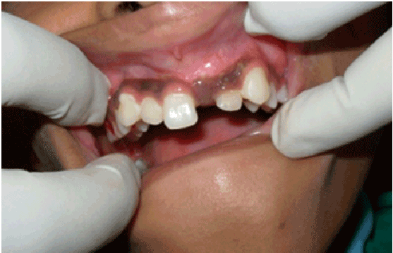
Figure 1: Intraoral photograph showing clinically missing 21

Vijayakumar Rajendran1 Jacob Sam Varghese2 Kavitha Swaminathan1 Selvakumar Haridoss1*
1Department of Pedodontics, Faculty of Dental Sciences S.R.M Dental College, SRM University, Chennai, India*Corresponding author: Selvakumar Haridoss, Reader, Department Of Pedodontics, Faculty of Dental Sciences of Sriramachandra University, Sriramachandra University, Chennai, India, E-mail: Selvakumaarh21@gmail.com
Odontomas are considered to be developmental anomalies which can be seen in any region of the oral cavity. There are two types of commonly occurring odontomas: compound and complex. This case report discusses about twelve years old boy who reported with unerupted upper left central incisor. Radiograph revealed a radiopaque mass in relation to the unerupted tooth which was later confirmed to be compound composite odontoma. A surgical excision was done and around 34 pieces were removed.
Denticles; Compound odontoma; Odontoma
Odontoma is defined as tumour of odontogenic origin [1]. Odontomas are developmental malformations or lesions of odontogenic origin that are non-aggressive, hamartomatous and consists of enamel, dentin, cementum and pulp tissue (hence, they are also called composite i.e. consisting of multiple or more than one type of tissue) constituting 22% of all odontogenic tumours [2]. Paul Broca coined the term ‘odontoma’ in 1867. It is defined as tumours formed by the overgrowth or transitory of complete dental tissues [3]. It may be distributed in numerous combinations and different patterns as soft, calcified or mixed tissues. They are slow growing benign tumours showing non-aggressive behaviour [4]. Though the etiology of odontomas is still unknown, environmental factors such as infection and trauma are considered as causative agents. Genomic changes have also been attributed to the occurrence of odontoma. There is no gender predilection and are most often diagnosed during the first two decade of life on routine radiographic examinations [5]. According to the classification of the World Health Organisation (WHO, 2005) two types of odontomas can be found: complex odontomas and compound odontomas- the latter being twice as common as the former [1]. The purpose of the present article is to a case report with multiple compound composite odontoma with unerupted permanent central incisor.
This case report presentation of a 12 year old male who reported with the chief complaint of missing upper front tooth. There was no relevant medical or dental history and it was the patient’s first dental visit. Any history of trauma was not revealed. On intra oral examination, 21 were found to be clinically missing (Figure 1). An intra oral periapical radiograph was recommended in relation to 21 to verify it was congenitally missing or impacted due to any other reasons. Intra oral periapical radiograph revealed the presence of 21 along with a radiopaque mass overlapping the crown of 21 (Figure 2a). From the clinical and radiographic findings, it was provisionally diagnosed as odontoma. Surgical excision of the radiopaque calcified mass was planned. Oral prophylaxis was done following which local anesthesia was administered in relation to 21 regions. Mucoperiosteal flap was raised both buccally and
palatally in relation to 21 and small tooth like structures were seen after removing the thin bony coverage in relation to 21 (Figure 3). A complete surgical excision was done and there were nearly 34 pieces of tooth-like structures were removed (Figures 4 and 5). A post operative radiograph didn’t show the presence of any denticles (Figure 2b). Once hemostasis was achieved, a suture was placed to close the mucoperiosteal flap that was raised both buccally and palatally. Zinc oxide eugenol pack was placed over the area. Differential diagnosis of odontogenic tumour, impacted tooth and osteoblastoma were considered. Patient was reviewed after one week. Histopathologically the sections revealed tooth like structures containing dentine, pulp and cementum confirming the provisional diagnosis of compound composite odontoma. During decalcification, the enamel cap on the denticles was removed (Figure 6). The patient was advised to seek a review once in 3 months in order to assess the eruption of unerupted central incisor.
Functional differentiation of epithelial and mesenchymal components of odontomas leads to the formation of enamel and dentin. [6] Although the etiology of odontoma is not known, several theories including local trauma, infection, family history, hereditary anomalies (Gardeners syndrome, Hermanns syndrome), odontoblastic hyperactivity and alterations in the genetic components responsible for controlling dental development have been proposed. Another important factor in the etiology of compound or complex odontoma is the persistence of a portion of lamina. [7] Compound odontomas generally occur in the maxillary anterior region, while complex odontomas occur in the posterior mandible region [8]. Tooth eruption disturbances such as delayed eruption of the primary and permanent teeth or over retained primary teeth are usually associated with odontoma in most of the children [9].

Figure 1: Intraoral photograph showing clinically missing 21
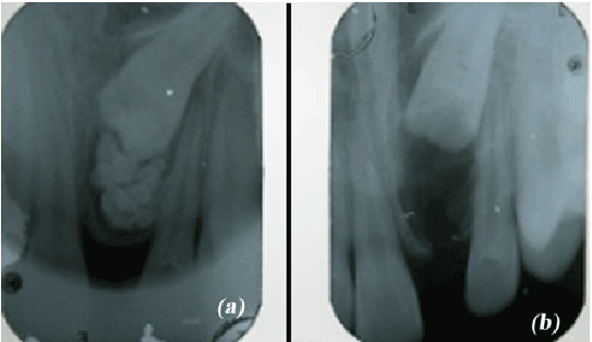
Figure 2: Intraoral periapical radiograph (a) Preoperative radiograph showing unerupted 21 along with calcified radiopaque mass (b) Post operative radiograph showing completely excised compound odontoma
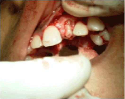
Figure 3: Mucoperiosteal flap elevated and the thin bony covering removed exposing the denticle
In the present case the compound odontoma was located in the anterior region as stated in the literature and the patient had unerupted maxillary central incisor. Clinically odontomas are generally asymptomatic. Occasionally, it increases in size and may produce facial asymmetry due to the expansion of bone. Odontoma most commonly occurs on the right side of the jaw than on the left (Compound 62%, Complex 68%) [1]. In the present case, the odontoma was seen on the left side. Radiographically, compound odontomas are identified as a collection of tooth-like structures that vary in size and shape and have a narrow radiolucent zone surrounding it. The complex odontoma appears as a calcified mass with a radio density of tooth structure, which is surrounded by a narrow radiolucent rim [10].
In a literature review by Amado-Cuesta et al. the number of denticles varied from 4 to 28 [11]. The compound odontoma we encountered in the present case was 34 denticles which is the most striking feature. The extracted denticles display fusion, concrescence and dilaceration. Urvashi Sharma et al. reported a case with 37 compound odontoma denticles [12]. Surgical removal of calcified mass and proper positioning of the impacted tooth in the dental arch with the help of orthodontic traction and (b) extraction of the impacted tooth if it is not in favourable place or require sectioning of tooth are the other treatment options of odontomas with impacted teeth [13].
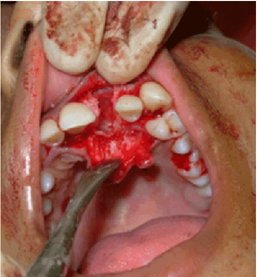
Figure 4: Complete excision of the denticles
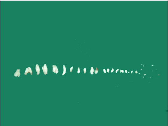
Figure 5: Excised 34 denticles
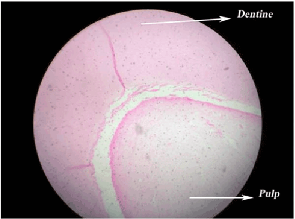
Figure 6: Photomicrograph of the decalcified denticles showing dentine and pulp (H&E X4)
Learning Points
Nil
Download Provisional PDF Here
Aritcle Type: Case Report
Citation: Rajendran V, Varghese JS, Swaminathan K, Haridoss S (2015) Multiple Compound Composite Odontome with Unerupted Permanent Incisor. Int J Dent Oral Health 1(5): http://dx.doi.org/10.16966/2378- 7090.145
Copyright: © 2015 Rajendran V, et al. This is an open-access article distributed under the terms of the Creative Commons Attribution License, which permits unrestricted use, distribution, and reproduction in any medium, provided the original author and source are credited.
Publication history:
All Sci Forschen Journals are Open Access