
Full Text
Nagaveni NB1* Shashikant Katkade2 Poornima P1 Roshan NM1
1Pedodontics, College of Dental Sciences, Davangere, India2Pedodontics, ACPM Dental College, Dhule, India
*Corresponding author: Dr. Nagaveni NB, Reader, Pedodontics, College of Dental Sciences, Davangere, India, E-mail: nagavenianurag@gmail.com
Abstract
Aim: Presentation of case with successful management of impacted maxillary central incisor.
Background: The impaction of permanent central incisors is a well-recognized entity and is usually associated with trauma to one or more primary anterior teeth early in life.
Case Description: We report a case in which a buccally displaced impacted central incisor was brought in the arch by biological tooth movement using a combination of removable and fixed orthodontic traction.
Conclusion: The chances of non-vitality are naturally much lower when the treatment is initiated at a younger age, due to the presence of a wide apical foramen.
Clinical Significance: It is a challenging task for a pediatric dentist to restore esthetics by moving the impacted maxillary incisor back to its natural position. Orthodontic correction, though challenging, is more desirable as the person retains his natural tooth in the arch.
Keywords
Impaction; Incisor; Mixed dentition
Background
Injuries to the primary dentition are among the most common trauma found in the maxillofacial region. Thirty to forty percent of children incur at least one injury to their primary teeth and the incidence is not gender related [1]. The frequency of maxillary incisor impaction has been found to range from 0.006% to 0.2% [2]. The close proximity of developing permanent tooth germ to the roots of primary tooth renders it vulnerable to trauma transmitted through deciduous teeth. The type and severity of disturbance are dependent upon the stage of tooth development, relationship of permanent tooth to the roots of primary tooth and direction and degree of force [3].
Case Description
A 10-year-old girl reported to the Department of Pedodontics, College of Dental Sciences, Davangere, with a chief complaint of an unerupted upper left front tooth. History revealed that the child had a history of trauma at the age of 4 years, following which she lost a few of her upper front teeth. No dental treatment was taken for the same. The patient was asymptomatic and the only concern was regarding the unerupted permanent left upper front tooth. The age of eruption for the contralateral upper permanent central incisor was given as 8 years. Medical history was not contributory
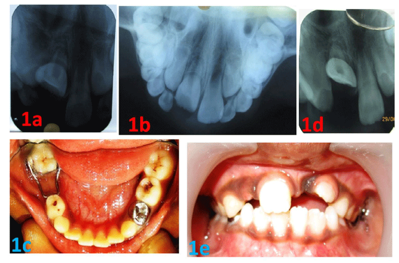
Intraoral examination revealed a buccal bulge in the region of 21. Intra-oral periapical (Figure 1a) and occlusal radiographs (Figure 1b) of the maxillary anterior teeth showed a foreshortened crown and root in relation to 21. The patient had a skeletal Class I malocclusion and a balanced facial pattern. Intraoral examination revealed an early mixed dentition and an Angle Class I molar relationship. Clinical examination showed left maxillary permanent central incisors was missing and no apparent arch length discrepancy in both the maxillary and mandibular arches, grossly carious primary mandibular left second molar and deep caries in relation to primary mandibular right first molar.
Treatment
Extraction of primary mandibular left second molar followed by the band and loop space maintainer was done. Pulp therapy followed by stainless steel crown was done for primary mandibular right first molar (Figure 1c). The unerupted maxillary left central incisor was kept under observation for any spontaneous eruption. However, no significant tooth movement was observed clinically and radiographically over a 6 month period (Figures 1d and 1e).
Several treatment alternatives were explained to the patient and her parents. They agreed for the surgical exposure of the impacted central incisors and alignment of the impacted incisors into the arch by orthodontic traction.
Impacted maxillary central incisor was surgically exposed (Figure 2a). Begg bracket was bonded to the palatal surface. A 0.9 mm hard round stainless steel wire was splinted from canine to canine. A loop was incorporated on the wire to obtain the anchorage for orthodontic traction. Traction was applied by twisting the ligature wire running into the vertical slot of Begg’s bracket and loop on the splinting wire (Figure 2b). Ligature wire activation was done after every 3 weeks. This method proved successful and helped provide the adequate force without any risk of devitalization of the teeth. The 21 gradually moved towards the occlusal plane by 3-4 mm, also uprighted slightly within 2 months (Figure 2c). Since the distance between Begg’s bracket and the hook on the splinted wire reduced further application of traction by twisting of ligature wire was not possible, hence, Nance palatal arch with the incorporation of hook in acrylic button was planed. The E - chain was engaged onto Begg’s bracket at one end and the hook at the other end (Figures 2d and 2e). The E - chain was changed after every one month. The 21 gradually moved toward the occluso- palatable and also uprighted slightly within the next 4 months (Figure 3a and 3b).
After desire palatal movement and uprighting of the tooth, Begg’s bracket was bonded to the labial surface of partially erupted tooth. Orthodontic brackets were also placed on mandibular incisors and orthodontic elastics are used for occlusal movement of upper left central incisor (Figure 3c). Desire tooth movement was achieved within a 2 month period (Figure 3d). After the complete exposure of clinical crown, the tooth was allowed to erupt by means of natural biologic mechanism. The patient is under regular follow up for 6 months after the active tooth movement was stopped, the patient showed normal clinical crown length and gingival contour giving an esthetically pleasing result. Impacted incisor maintained vitality and there was no evidence of root resorption (Figures 4a-4c).
Discussion
The impaction of the maxillary incisor is often clinically and radiologically diagnosed in early ages because the non-eruption of the anterior tooth causes concern to parents during early mixed dentition phase [4]. Clinical signs of an impacted tooth include retention of the primary tooth, space closure, and elevation of the soft tissue of the palatal or labial mucosa [5]. The radiographic findings usually reveal the causes of impaction, such as a supernumerary tooth, an odontoma or a dilaceration; thus the treatment of the impaction is carried out according to the obstacle for eruption [4].
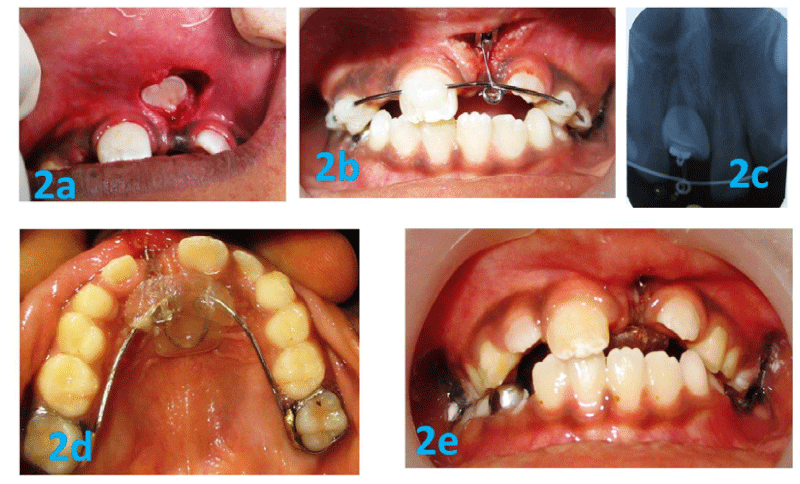
Figure 2: 2a: Surgical exposure of 21, 2b: Begg’s bracket bonded to a palatal surface of 21 and wire and composite splinting, 2c: 2 month follow up IOPA, 2d & 2e: Nance palatal arch with the incorporation of hook
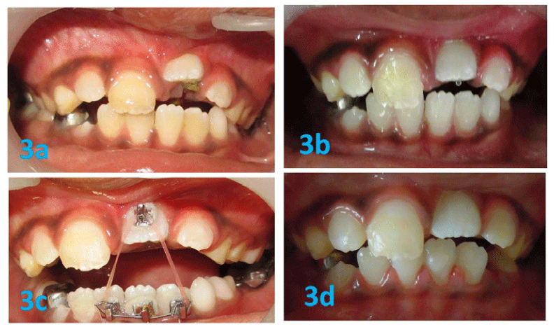
Figure 3: 3a&3b: 4 month follow up, 3c: Orthodontic elastics for incisal movement, 3d: 8 month follow up photograph
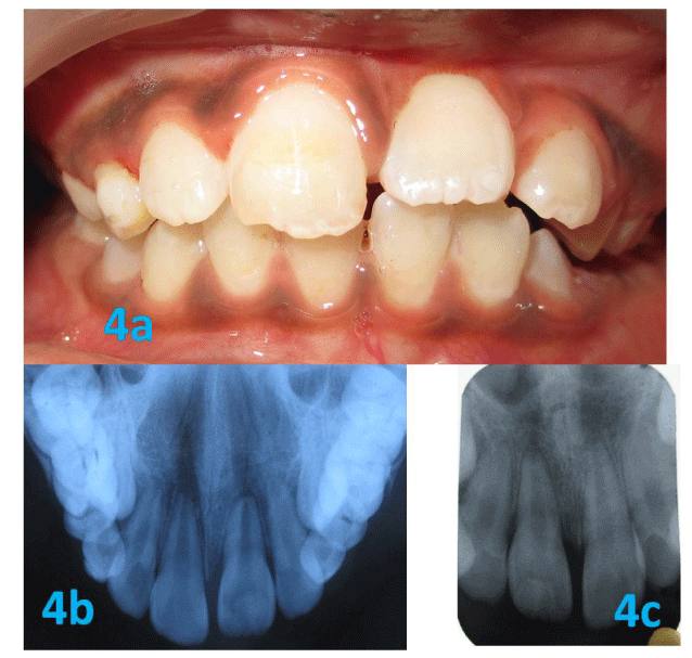
Figure 4: 6 month post treatment photographs
The impaction of permanent central incisors is a well-recognized entity and is usually associated with trauma to one or more primary anterior teeth early in life. Management options for such teeth can be: (i) surgical extraction and moving the lateral incisor to mimic the central incisor and similarly changing the anatomy of other teeth. (ii) Extraction of the impacted tooth followed by an implant. (iii) Surgical repositioning of the impacted tooth, and (iv) orthodontic correction of the impacted tooth [6].
In the first option, modifying a lateral incisor to mimic a central incisor, because of the inherent discrepancy in the mesiodistal width at the cervical area, this compromises esthetics, especially when the person has a short upper lip. The option of extraction and implant placement requires a waiting period of up to 18 years of age and cannot be undertaken at a younger age. Surgical repositioning as a treatment option has a likelihood of failure either due to devitalization, or later replacement resorption; surgical trauma to the child at a young age is another disadvantage. Orthodontic correction, though challenging, is more desirable as the person retains his natural tooth in the arch [6].
In this case report, to bring the impacted tooth into its natural position, a lingual button was initially bonded to the palatal surface of the impacted tooth. Most of the authors, for such alignments have initiated traction from the lingual surface by applying either lingual buttons [4,7,8], Begg brackets [9 ], or edgewise incisor brackets [4,10] on this surface. But in cases where the attachment causes trauma to the labial mucosa, lingual placement would be preferable. The selection of the surface, however, may be decided on an individual basis.
The technique of closed eruption has been highly recommended by most authors [4,7,8] for aligning the impacted tooth compared to methods like excisional gingivectomy and apically positioned flap techniques because of better esthetic results. The extrusion force applied to the impacted central incisor was very light, this may have accounted for the maintenance of vitality of the impacted tooth post-alignment. The chances of non-vitality are naturally much lower when the treatment is initiated at a younger age, due to the presence of a wide apical foramen. The lighter forces did not increase the total treatment time, which was 8 months for bringing the impacted tooth into alignment and an additional 6 months for the final alignment. This is comparable to other reported cases with total treatment times of up to 36 months [10]. This method of orthodontic alignment of impacted central incisors, compared to other methods, is relatively free from treatment associated complications and predictable results; hence, it may become the method of choice/preferred method over extractions or surgical repositioning.
Conclusion
The chances of non-vitality are naturally much lower when the treatment is initiated at a younger age, due to the presence of a wide apical foramen.
Clinical Significance
It is a challenging task for a pediatric dentist to restore esthetics by moving the impacted maxillary incisor back to its natural position. Orthodontic correction, though challenging, is more desirable as the person retains his natural tooth in the arch.
References
- Sennhenn-Kirchner S, Jacobs HG (2006) Traumatic injuries to the primary dentition and effects on permanent successors - a clinical follow - up study. Dent Traumatol 22: 237-241. [Ref.]
- Grover PS, Lorton L (1985) The incidence of unerupted permanent teeth and related clinical cases. Oral Surg Oral Med Oral Pathol 59: 420–425. [Ref.]
- Stewart DJ (1978) Dilacerated unerupted maxillary central incisor. Br Dent J 145: 229-233. [Ref.]
- Lin YJ (1999) Treatment of an impacted dilacerated maxillary central incisor. Am J Orthod Dentofac Orthop 115: 406-409.
- Duncan WK, Ashrafi MH (1983) Management of the nonerupted maxillary anterior tooth. JADA 106: 640–644.
- Kuvvetli SS, Seymen F, Gencay K (2007) Management of an unerupted dilacerated maxillary central incisor: a case report. Dent Traumatol 23: 257–261. [Ref.]
- Uematsu S, Uematsu T, Furusawa K, Deguchi T, Kurihara S (2003) Orthodontic treatment of an impacted dilacerated maxillary central incisor combined with surgical exposure and apicoectomy. Angle Orthod 74: 132-136. [Ref.]
- Paola C, Alessandra M, Roberta C (2005) Orthodontic treatment of an impacted dilacerated maxillary incisor: A case report. J Clin Pediatr Dent 30: 93-97. [Ref.]
- Batra P, Duggal R, Prakash H (2005) Managing morphologically atypical impacted teeth orthodontically. J Clin Pediatr Dent 29: 105-111. [Ref.]
- Keijirou K, Hiroyuki K (2000) Esthetic management of an unerupted maxillary central incisor with a closed eruption technique. Am J Orthop Dentofac Orthop 118: 224-228. [Ref.]
Download Provisional PDF Here
Article Information
Article Type: Case Report
Citation: Nagaveni NB, Shashikant Katkade, Poornima P, and Roshan NM (2015) Management of Impacted Maxillary Central Incisor by Sequential Appliance Therapy- A Clinical Case Report. Int J Dent Oral Health 1(1): doi http://dx.doi.org/10.16966/2378- 7090.118
Copyright: © Nagaveni NB. This is an open-access article distributed under the terms of the Creative Commons Attribution License, which permits unrestricted use, distribution, and reproduction in any medium, provided the original author and source are credited.
Publication history:
SCI FORSCHEN JOURNALS
All Sci Forschen Journals are Open Access
New Journals
Best viewed in Google Chrome | Mozilla Firefox | Microsoft Edge
Copyright © 2023 Sci Forschen Inc., All Rights Reserved