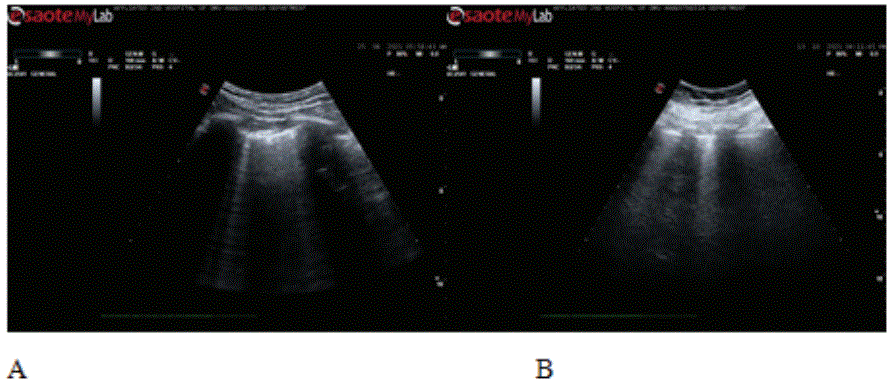
Figure 1: Imagine of edema.

Su Yang1 Yang Haitao1 Xiong Ying2 Zheng Xin2* Zhao Linyan3*
1Department of Anesthesiolgy, the Second Hospital of Dalian Medical University, Zhongshan road, Dalian, Liaoning Province, China*Corresponding author: Zheng Xin, Associate chief physician, Department of Anesthesiolgy, the Second Hospital of Dalian Medical University, No.467 Zhongshan road, Dalian, Liaoning Province, China, E-mail: zhengxinxin008@163.com
Zhao Linyan, Department of critical care, Associate chief physician, the Second Hospital of Dalian Medical University, No.467 Zhongshan road, Dalian, Liaoning Province, China, E-mail: zhaolinyanmail@126.com
Point of care ultrasound has a very good application prospect in perioperative period because of its dynamic, real-time and repeatable advantages. At the same time, it has high accuracy and can quickly find the common causes of hypoxemia, such as pulmonary edema, atelectasis and pleural effusion. It is a good visualization tool. Perioperative combined use of cardiopulmonary ultrasound, on the basis of excluding cardiac dysfunction, rapid diagnosis of pulmonary edema. The author conducted a study on two cases of acute lung injury induced by a relatively large amount of blood transfusion. The patients had no prior cardiopulmonary disease, no abnormal findings were found on cardiac ultrasound, hypoxemia, and signs of lung injury were found on bedside pulmonary ultrasound.
Case presentation: The patient was admitted with acute upper gastrointestinal bleeding, which was large enough to cause hemorrhagic shock. Transfusions were initiated in the ICU, and larger doses were given over a short period of time. The patient subsequently developed significant hypoxemia with tachypnea for first case. The second patient was a Da Vinci robot-assisted cystectomy with excessive bleeding during the operation. A large infusion of red blood cells followed, intraoperative blood gas analysis found oxygenation index below 300, and the patient was admitted to ICU. Cardiopulmonary ultrasound revealed left heart dysfunction and hypoxemia associated with pulmonary edema.
Conclusion: At the bedside, cardiopulmonary ultrasound in critically ill patient is a useful tool for diagnosing acute lung injury and pulmonary edema caused by blood transfusion.
Cardiopulmonary ultrasound; Critically ill; Blood transfusion; Pulmonary edema
ICU: Intensive Care Unit; ARDS: Acute Respiratory Distress Syndrome
Patient as 54 years old, male construction worker with no previous history of cardiac or pulmonary diseases. ICU admitted with upper gastrointestinal bleeding, he was in the early stages of hemorrhagic shock when he first entered the ICU. Vital signs blood pressure was 100/50mmHg, heart rate was 105 beats /min, and SPO2 was 96%. The blood routine showed hemoglobin of 40g/ L, after rapid blood transfusion of red cells, as 10 units of red blood cells were infusion within 12 hours. 14 hours after admission to ICU his SPO2 has decreased to 88%, but auscultation was normal. Then arterial blood gas analysis indicated oxygen partial pressure of 56mmHg under the background of 2L /min oxygen inhalation, the oxygenation index was calculated as 190. After 4l/min oxygen inhalation through nasal catheter his SPO2 has increased to 92%. Point of care cardiopulmonary ultrasound was performed using a curved array probe. BLUE protocol was used in lung ultrasound, and the results showed multiple B lines in both lungs, with heavy PLAPS points. The ultrasonic section of the heart showed the long axis of the parasternum and the four-chamber heart under the xiphoid process. The results showed that the heart activity was normal, the valve activity was good, and the width of the inferior vena cava was 2cm. Combined with cardiopulmonary ultrasound, acute lung injury and pulmonary edema can be diagnosed. Antrum ultrasonography showed no obvious contents and no active bleeding was considered. Ulinastatin and high dose ambroxol were used to suppress inflammation. One day later, lung CT examination confirmed pulmonary edema. After 2 days, the patient’s hypoxemia improved and the oxygenation index was 280.
The patient was a 40-year-old, male, surgery of laparoscopic cystectomy for bladder cancer was taken, and no previous history of cardiopulmonary diseases was found. During the operation, 10 units of red blood cells were transfused for 2 hours, there was a lot of intraoperative bleeding, and hemoglobin was checked at 130g/l after blood transfusion. Intraoperative blood gas analysis was performed; oxygenation index during the transfusion was 260 under pure oxygen inhalation condition. After communication with the surgeon, the following 4 units of red blood cells were transfused. Another blood gas analysis showed that hemoglobin was 135g/ L and oxygen partial pressure was 85mmHg under pure oxygen inhalation. In view of the rapid changes in the patient’s condition, he was sent to ICU after surgery. Ten minutes after arrival at ICU the oxygenation index of blood gas analysis was 55. An ultrasound specialist performs a cardiopulmonary ultrasound showed no abnormality in heart function and multiple pulmonary edema in both lungs (Figure 1A).
Figure 1: Imagine of edema.
The results showed that cardiac function was generally normal and ventricular systolic function was good. Lung ultrasound showed a large number of dense B-lines in both lungs, some of which were fused (Figure 1B). Combined with the results of cardiopulmonary ultrasound and previous conditions, severe acute lung injury or Acute Respiratory Distress Syndrome (ARDS) was diagnosed. The patient’s tracheal tube attracted a large amount of yellowish fluid, which was considered alveolar exudation. The patient progresses rapidly and the prognosis is poor.
Point of care ultrasound has developed rapidly in the past 20 years and has been widely used in critical care medicine and emergency medicine. It has epoch-making significance in the diagnosis of shock, unexplained dyspnea and acute respiratory distress syndrome [1-5]. Point of care lung ultrasound, which was first proposed by French professor Daniel, has been used as an alternative diagnostic tool for ARDS, with dynamic, radiation-free, repeatable timeless characteristics [6]. Compared with auscultation, pulmonary edema can be diagnosed quickly and accurately by point of care lung ultrasound [6], which can quickly diagnose and evaluate critical patients and has a good prospect in perioperative application. Point of care ultrasound is well developed in European and American countries, by clinical using there have formed several ultrasound protocol for different environments such as shock, dyspnea or trauma. Point of care cardiopulmonary ultrasound is simple, accurate and rapid, and its application in perioperative period as provide visual information to help develop a better perioperative management plan. In the context of COVID-19, point of care ultrasound is more suitable for perioperative use due to its dynamic, radiation-free and real-time features.
In severe cases, bedside ultrasound is better because of the high risk of patient transport and the complex process of transport. The same situation also occurs in perioperative patients, and perioperative patients are accompanied by a certain proportion of severe patients. Bedside diagnostic tools are needed in critically ill patients with unexplained hypoxemia or hypotension during surgery. Point of care ultrasound technology can help us very well and needs to be implemented quite actively [7]. It is of decisive significance in perioperative decision making after systematic training.
In conclusion, point of care ultrasound can quickly diagnose pulmonary edema and determine the cause of hypoxemia, which has more advantages than auscultation.
Teaching Reform Project of Dalian Medical University in 2019 (DYLX19026).
The datasets availability from the corresponding author on reasonable request.
Zheng Xin, Su Yang collected the data, Xiong Ying, Zhao Linyan, designed the study, Su Yang wrote the paper.
Ethics approval number: dy2y/2021071. As the study is a retrospective one and emergency, the need for patient consent was waived.
Written informed consent was obtained from the patient for publication of this case report and any accompanying images.
The authors declare that they have no competing interests.
Not Applicable
Download Provisional PDF Here
Article Type: CASE REPORT
Citation: Yang S, Haitao Y, Ying X, Xin Z, Linyan Z (2022) Point of Care Cardiopulmonary Ultrasound Assessment for Acute Lung Injury Patients after Blood Transfusion. J Clin Case Stu 7(2): dx.doi.org/10.16966/2471-4925.254
Copyright: © 2022 Yang S, et al. This is an open-access article distributed under the terms of the Creative Commons Attribution License, which permits unrestricted use, distribution, and reproduction in any medium, provided the original author and source are credited.
Publication history:
All Sci Forschen Journals are Open Access