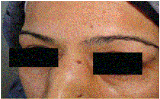
Figure 1: Enlargement of the lacrimal glands.

Imène Rachdi* Fatma Daoud Hana Zoubeidi Mehdi Somai Besma Ben Dhaou Zohra Aydi Fatma Boussema
Internal Medicine Department, Habib Thameur Hospital, Faculty of Medicine of Tunis, University of Tunis El Manar, Tunisia*Corresponding author: Rachdi I, Internal Medicine Department, Habib Thameur Hospital, Faculty of Medicine of Tunis, University of Tunis El Manar, Tunis, Tunisia, E-mail: rachdi.imene14@gmail.com
Mikulicz Syndrome is a rare chronic condition characterized by inflammatory process of lacrimal, parotid and sub-mandibular glands and lymphocytic infiltrations. We present an atypical diagnosis of sarcoidosis through a clinical presentation similar to Mikulicz syndrome in two patients aged respectively of 48 and 20 years with a good response to corticosteroids.
Mikulicz syndrome; Sarcoidosis; Salivary gland diseases; Dacryoadenitis; Granuloma
Sarcoïdosis is a systemic granulomatosis that can affect lungs, ganglions, skin and eyes. Chest manifestations reveal frequently the sarcoidosis. Ophtalmologic features reveal the disease in 7 to 20% of the cases. Mikulicz’s syndrome is characterized by symmetric lacrimal, parotidian, and submandibular gland enlargement with associated lymphocytic infiltrations. Sarcoidosis, Sjogren’s syndrome, tuberculosis and hemopathies are most common disease that can include a mikulicz syndrome.
We report two cases of Mikulicz’s syndrome revealing sarcoïdosis.
A 48-year-old patient was referred for bilateral lacrimal and parotid gland enlargement without fever. Enlargement of the lacrimal (Figure 1) and parotid (Figure 2) glands associated with nasal obstruction and sinus pain were the initial symptoms. ENT (Ear, Nose and Throat) examination showed an inflammatory nasal mucosa and the presence of 2 nodules in the oral mucosa. Skin examination showed a subcutaneous nodule on the inner side of the left arm. Routine blood examination showed hyper-gammaglobulinemia at 16.9 g/l polyclonal appearance and hypercalciuria at 7.3 mmol/24h with a high level of the conversion enzyme at 101 ECU. The calcemia and parathormone levels were normal. Serologies of toxoplasmosis were negative. The tuberculosis balance sheet was negative. The level of IgG4 was normal. The thoraco-abdominal CT scan showed interstitial pneumopathy with mediastinal adenopathies as well as moderate hepatomegaly and retroperitoneal adenopathies. Histopathological examination of nasal mucosa biopsies (Figure 3) showed non-necrotizing epithelioid and giganto-cellular granulomatous lesions of the nasal mucosa. At the examination of the biopsy of accessory salivary glands, we noted granulomatous sialadenitis. The histological examination of the subcutaneous nodule (Figure 4) noted tuberculoid inflammation with a focal pyoepithelioid appearance. The respiratory functional explorations were normal. At the broncho-alveolar lavage, we noted an inflammatory cytology. The diagnosis of sarcoidosis revealed by Mikulicz syndrome was retained. General corticosteroid therapy (0.5 mg/kg/day) was indicated. The clinical course was favourable with regression of glandular hypertrophy
Figure 1: Enlargement of the lacrimal glands.
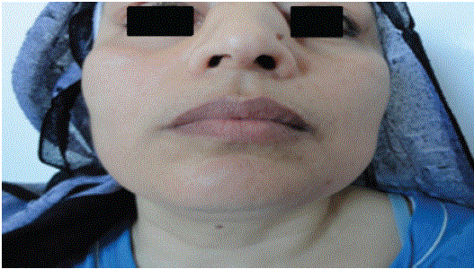
Figure 2: Enlargement of the parotid glands.
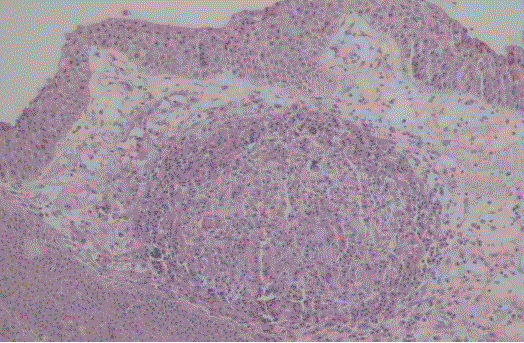
Figure 3: Histopathological examination of nasal mucosa biopsies showed non-necrotizing epithelioid and giganto-cellular granulomatous lesions of the nasal mucosa.
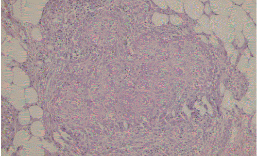
Figure 4: The histological examination of the subcutaneous nodule noted tuberculoid inflammation with a focal pyoepithelioid appearance.
A 20-year-old patient was referred for bilateral palpebral edema associated with parotid gland enlargement. She complained about intense headache at the base of the skull exaggerated by head movements associated with anosmia, nasal obstruction, palpebral oedema, hypertrophy of the parotid glands without fever. ENT examination noted the presence of nasal mucopurulent secretions in both nasal fossae with left obstruction and highly inflammatory petechial mucosa on the right side. At the routine blood examination, we noted hyper-gammaglobulinemia with a polyclonal appearance at 14.4 g/l. Phosphocalcic and liver function tests were normal. The tuberculosis balance sheet was negative. Serologies of toxoplasmosis were negative. The level of IgG4 was normal. The thoracic CT scan showed the presence of micronodular parenchymal lesions opposite the large right apico-dorsal, middle lobar, subpleural and culminal scars. Histopathological examination of the nasal mucosa and accessory salivary glands showed granulomatous lesions. Broncho-alveolar lavage: a lymphocytic alveolitis (58.3%). The diagnosis of sarcoidosis revealed by Mikulicz syndrome was retained. Systemic corticosteroid therapy (0.5 mg/kg/day) was indicated for respiratory compromise. The clinical course was favourable with regression of glandular hypertrophy after 25 days.
Sarcoidosis is a multisystemic granulomatosis affecting preferentially young, female subjects. It affects the eye in approximately 21% of cases [1]. Ophthalmologic manifestations are inaugural in 7 to 20% of cases depending on the series [1,2]. Anterior uveitis is the most frequent ocular manifestation and is noted in 21 to 70% of cases of ocular sarcoidosis depending on the series [2]. Patent orbital manifestations are rarer and are poorly described in the literature [3-5]. Among them, the lacrimal gland is the most frequent and is associated with systemic damage in the majority of cases. The lacrimal gland is affected bilaterally and asymptomatically in 60- 87% of cases [2]. Glandular enlargement is present in 8-32% of cases [2] and may be palpable as a painless bilateral superolateral mass. It is responsible for a dry syndrome in 5-42% of cases [2]. The simultaneous hypertrophy of the lacrimal and parotid glands observed in our two patients constitutes Mikulicz syndrome, mainly encountered in sarcoidosis, tuberculosis, Hodgkin’s disease and some leukemias. Anosmia is a symptom caused by various etiologies, whereas sinonasal disease is one possible reason. Patients with sinonasal disease almost always complain of nasal obstruction, rhinorrhea, and may be crusting, which also were identified to be strong indicators of sinonasal sarcoidosis such is the case of the first patient.
This mode of presentation of sarcoidosis is rarely encountered in internal medicine; its exact frequency is unknown and probably underestimated by internal medicine studies since most cases are diagnosed in ophthalmology or stomatology. Mikulicz disease is now considered part of the IgG4-related autoimmune disease spectrum, which can include inflammatory disorders of the pancreas, thyroid gland, pachymeninges, pituitary gland and infundibulum [6].
Among the differential diagnoses to consider when faced with bilateral tear gland enlargement, orbital lymphoma with involvement of the lacrimal glands should be mentioned; however, orbital lymphoma usually affects the older subject. It is most often unilateral. The biopsy of the lacrimal glands eliminates this diagnosis by identifying a giganto-epithelioid granuloma without caseous necrosis, especially since the association between systemic sarcoidosis and orbital lymphoma has already been described [7]. The other causes of Mikulicz syndrome were eliminated by the normality of the rest of the clinical examination and further investigations. If the lacrimal gland is affected, sarcoidosis is systemic in the majority of cases [8]. A positive diagnosis of sarcoidosis is obtained by histological examination of the biopsy of the lacrimal gland showing a giganto-epithelioid granuloma without caseous necrosis. Rapid regression is the rule under systemic corticosteroid treatment [9].
Granumatosis of lacrymal and parotid glands can be encountred with lymphoma or tuberculosis. It rarely reveals the diagnosis of sarcoidosis. Our cases confirm the good response to corticosteroids.
Download Provisional PDF Here
Article Type: CASE SERIES
Citation: Rachdi I, Daoud F, Zoubeidi H, Somai M, Dhaou BB, et al. (2020) Mikulicz Syndrome Revealing Sarcoidosis: About Two Observations. J Clin Case Stu 5(3): dx.doi.org/10.16966/2471-4925.203
Copyright: © 2020 Rachdi I, et al. This is an open-access article distributed under the terms of the Creative Commons Attribution License, which permits unrestricted use, distribution, and reproduction in any medium, provided the original author and source are credited.
Publication history:
All Sci Forschen Journals are Open Access