Abstract
Macroadenomas in cushing’s disease represent 10% of cases, empty sella syndrome is relatively rare, it is defined as the extension of the subarachnoid space toward the intrasellar region with displacement of the pituitary to the postero-inferior wall. The prevalence of empty sella has been reported in up to 38% of imaging studies. Some patients may present with endocrine, neurological and ophthalmological symptoms due to the aberrant anatomy of the sellar region. The association with hormonal hypersecretion and in particular Cushing’s syndrome is much rarer.
We report the case of the B.Y patient aged 65 years without any previous history hospitalized in endocrinology for further exploration and management of a left lateral pituitary macroadenoma of 11 mm in height and 14 mm in wide in iso signal T1 and T2, pituitary gland laminated facing upwards, discovered following a hypertensive peak with neurological signs such as scintillating scotoma and dizzying sensations. The patient had some hallmark signs of Cushing’s syndrome. Biological evaluation performed during the patient’s first admission showed a broken circadian rhythm of cortisol with high midnight cortisol and basal ACTH level at 176 pg/mL. Urinary free cortisol was also elevated at 29.32 ug/dL.
Two mg dexamethasone suppression test didn’t show inhibition effect with cortisol level at 4.71 ug/dL. However, an inhibitory effect was observed with 8 mg dexamethasone test. A hyperprolactinemia of disconnection was associated with normal thyroid assessment (see Table 1). The associated comorbidities presented by our patient were diabetes mellitus, high blood pressure and osteopenia. The patient benefited from a transphenoidal adenoma resection with persistence of a high level of cortisol (D12: 23.56 ug/dL).Unfortunately, the patient is lost to follow-up.
Conclusion: The singularity of our case remains in the association of two relatively rare circumstances associating a macroadenoma developed in an empty sella turcica both responsible for a hypercortisolism caused by an ACTH-secreting pituitary macroadenoma.
Keywords
Cushing disease; Empty sella turcica; Macroadenoma
Abbreviations
ESS: Empty Sella Turcica; CSF: Cerebrospinal Fluid; D: Day; MRI: Magnetic Resonance Imaging
Introduction
Macroadenomas in Cushing’s disease represent 10% of cases [1]. Empty sella syndrome is relatively rare; it is defined as the extension of the subarachnoid space toward the intrasellar region with displacement of the pituitary to the postero-inferior wall. The prevalence of empty sella has been reported in up to 38% of imaging studies [2]. The association of endocrine, neurological and ophthalmological symptoms due to the aberrant anatomy of the sellar region has been reported [1]. The association with hormonal hypersecretion and in particular Cushing’s syndrome is much rarer.
Case Report
A 63-year-old woman was referred to our endocrinology unit in 2015 following the sudden appearance of a hypertensive peak with neurological signs such as scintillating scotoma and dizzying sensations. Cushing’s disease was suspected on the basis of clinical and imaging data. The patient’s height, body weight and BMI were 154 cm, 78.5 kg and 33.12 kg/m2 respectively. The patient presented with moon face, central obesity, and atrophy of the skin without striae. The patient also complained of muscular weakness. No neurological deficits were observed, including in the visual field. Biological evaluation performed during the patient’s first admission showed a broken circadian rhythm of cortisol with high midnight cortisol at 27.73 ug/dL and basal ACTH level at 176 pg/mL.
Urinary free cortisol was also elevated at 29.32 ug/dL. Two mg dexamethasone suppression test didn’t show inhibition effect with cortisol level at 4.71 ug/dL. However, an inhibitory effect was observed with 8 mg dexamethasone test (cortisol level: 2.24 ug/dL). A hyper- prolactinemia of disconnection was associated with normal thyroid assessment and relative gonadotropine deficiency (see Table 1). The associated comorbidities presented by our patient were diabetes mellitus, high blood pressure treated by Metformin and the association of Valsartan 160 mg with Hydrochlorothiazide 12.5 mg and non-treated osteopenia.
| Parameters and kits |
Results |
Normal ranges |
| PRL cisbio RIA |
33.8 |
73-474 ui/L |
| TSH cisbio RIA |
0.52 |
0.1-0.4 mui/mL |
| FT4 cisbio RIA |
8.92 |
6.35-18.9 pg/mL |
| FSH IRMA |
7.98 |
3.5-98 ui/L |
| LH IRMA |
2.17 |
15-64 ui/L |
| Oestradiol cisbio RIA |
108 |
26-272 pmol/L |
| IGF1 cisbio RIA |
102 |
70-200 ng/mL |
| ACTH IRMA |
176 |
5-69 pg/mL |
Table 1: Showing the results of the biological evaluation of our patient.
PRL: Prolactin; TSH: Thyroid Stimulating Hormone; FT4: Free Thyroxin; FSH: Follicle Stimulating Hormone; LH: Luteinizing Hormone; ACTH: Adreno Cortico Tropic Hormone; RIA: Radioimmunoassay; IRMA: Immunoradiometric assay
The patient’s MRI at the time of admission showed an intrasellar intermediate signal formation on the two weights and barely enhancing after injection of Gadolinium salts measuring 11 × 14 mm. The pituitary stalk was in the middle position with normal size, driven backwards by an arachnoidocele. The pituitary gland was enlarged not by the adenoma, but by the arachnoidocele. The hypersignal of the post pituitary gland was lost and the pituitary gland presumably pressed against the sellar floor. The optical chiasma was without anomalies and a usual enhancement of the cavernous sinus was observed (Figure 1). The patient was diagnosed as having Cushing’s disease caused by an ACTH-secreting pituitary macroadenoma developed in an empty sella turcica, which was extirpated by trans-sphenoidal surgery complicate by cerebrospinal fluid leakage with persistence of a high level of cortisol (D12: 23.56 ug/dL). Unfortunately, the patient is lost to follow-up.
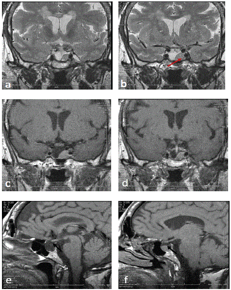
Figure 1: Highlighting of a right lateral intrasellar lesion (red arrow) of tissue signal, homogeneous in T2 (a, b) and T1 (c, d), enhanced very moderately after injection of Gadolinium salts; Left intrasellar arachnoidocele well individualized on the right para sagittal image T1 (e), in left para sagittal (f), the section passes through the adenoma (black arrow).
Discussion
Empty sella turcica is a descriptive term first used by Busch W in 1951 [3]. In a careful study of sellar morphology in a series of 788 autopsies, he found that 20% presented a total or near total absence of the sellar diaphragm and that 6% presented a complete empty sella with compression of the pituitary towards the floor of the sella turcica. In spite of the fact that the first descriptions referred to patients who had developed this sign after pituitary surgery or irradiation, empty sella has also been described as a primary pathology [4]. Our case belongs to this second group.
The association of empty sella and Cushing disease is very rare. In fact, the first reported case by Gautier D, et al. on 1980 in Hormone Res [5]. Michotte N, et al. in their retrospective analysis executed through the medical records of the university hospital of Brussels for the period from January 2007 to September 2017 included 1036 patients with ESS had observed 14.7% hormonal dysfunction; growth hormone deficiency and secondary hypogonadism were the most prevalent hormonal dysfunction in the studied population. Cushing’s disease remains rare.
Many theories have been proposed to explain the origin of ESS. Most authors believe that the main predisposing factor is the combination of a pulsatile CSF with an incompetent sellar diaphragm [6,7]. Pituitary atrophy secondary to an autoimmune process [8], glandular involution after gestational pituitary hyperplasia, or pituitary hyperplasia secondary to target gland insufficiency [8], have also been described. Infarction and posterior reabsorption of a pituitary tumour with loss of intrasellar tissue and secondary extension of the sub-arachnoid space to the sella, is another cause of this pathology [9]. The relationship of ESS and Cushing’s disease is probably fortuitous given that the frequency of ESS in the general population is similar [10].
Except for the review by Buchfelder M, et al. [11], from the reports in the literature most of the patients were cured by pituitary surgery [12-14], such as Mancini A, et al. [15] who obtained surgical cure without complications in the six patients reported. The rest of the patients were treated with bilateral adrenalectomy [16,17], radiotherapy [5,18] or medical therapy [19].
Conclusion
The singularity of our case remains in the association of two circumstances relatively rare associating a macroadenoma developed in an empty sella turcica both responsible for an ACTH dependent cortisolic hypersecretion.
Acknowledgments
The author thanks Pr Ould-Kablia, Pr Bensalah and Pr Ourad for their precious help.
Download Provisional PDF Here
Article Information
Article Type: CASE REPORT
Citation: Lachkhem A, Nouzha H, Yahi A, Heffaf L, Derraji S, et al. (2020) Cushing’s Disease on Macroadenoma Developed in an Empty Sella Turcica: About a Case. J Clin Case Stu 5(3): dx.doi.org/10.16966/2471-4925.202
Copyright: © 2020 Lachkhem A, et al. This is an open-access article distributed under the terms of the Creative Commons Attribution License, which permits unrestricted use, distribution, and reproduction in any medium, provided the original author and source are credited.
Publication history:
Received date: 06 May, 2020
Accepted date: 18 May, 2020
Published date: 22 May, 2020
