Introduction
Periodontitis (PO) concerns more than 10% of the population [1] and is associated with an increased risk of cancer [2,3]. Chronic inflammation induced by oral or small gut dysbiosis may lead to deleterious interactome and premature death [4,5].
Detection of Small Intestinal Bowel Overgrowth (SIBO) is based on breath test, which should be routinely performed “in the evaluation of common gastroenterology problems” according to a recent consensus [6]. Researchers are ongoing for early non-invasive detection of cancers using exhaled VOC; especially digestive, breast, thyroid and prostatic cancers [7-12].
The pathogenic mechanism of severe inflammation, destruction of tissues associated with PO is unknown. However, the cutting of High-Molecular-Weight-Hyaluronic acid in small fragments of LowMolecular-Weight-Hyaluronic acid (LMWHA) by bacterial proteases is established in PO [13]. We reported in a preliminary study that the severity of PO is associated with an increased level of plasmatic HA and with an increased risk of adenocarcinoma [14].
LMW-HA is known to increase endothelial permeability, to stimulate receptors of cancer stem cells and to favour cancer cells metastasis [15-18]. Hyaluronidases of oral pathogens may play a role in the occurrence of cancer associated with PO. Since bacteria may have a specific gas signature [19], we investigated whether patients with SIBO+PO and severe-associated diseases exhale specific gas. To our knowledge, no similar study has been performed yet.
Material and Methods
All data were collected during the normal course of routine gastroenterological consultations for SIBO.
There was no hypothesis testing before data collection, no data collection beyond that which is part of routine clinical practice, no scheduled data analysis before the work has already been done. This epidemiological retrospective analysis of Case Series cannot therefore be qualify as “research” and does not requires approval from ethics boards designed to protect humans involved in clinical research, according to the International Committee of Medical Journal Editors (ICMJE).
Inclusion criteria
All patients underwent a breath test and a trans abdominal ultrasound which confirmed SIBO. Patients did not take either antibiotic therapy or any medication which can modify digestive flow for at least 4 months before the breath test.
Patients presented with PO diagnosed by a periodontologist. Patients should have completed an appropriate screening for cancer (colon, stomach, breast, prostate, thyroid) according to the recommendations of the “Institute National du Cancer “(France). Plasmatic hyaluronic acid (HA) dosage had been performed. A full medical history was available, including medication intake. Patients signed a written consent for the epidemiological use of collected data; as all other patients consulting in the gastroenterological clinic.
Exclusion criteria
Ongoing tobacco abuse; lack of hyaluronic acid analysis; lack of breath test or trans abdominal ultrasound; lack of signed consent for epidemiological use of data. Intake of antibiotic therapy or of any medication which may modify digestive flow as well as surgical treatment of periodontitis within the previous 4 months.
Gases analysis
The measurement of VOC implies SPME-GC-MS with collection of gases on a PDMS/CAR, 75 µm (Supelco®) fibres chosen to trap VOCs. The patient breathes in a glass bottle with no plastic part. Condensates remain in the bottom of the bottle whilst the air is evacuated by 8mm glass tube which contains the fibre. Samples were obtained twice after fasting and twice two hours after the intake of sugars (fructose and trehalose). The air of the clinic is permanently Hepa-filtered and UVdecontaminated. Lack of any VOC contamination is checked twice each working-day with two different MX6® devices (Gazdetect® France). SPME-GC-SM analysis was performed within 24 hours. A solid phase micro extraction (SPME) holder with carboxen/polydimethylsiloxane (CAR/PDMS) fibers of 75 µm thickness was purchased from Supelco® . The SPME fiber was inserted into the glass bottle where the patients blew 10 times to collect the sample.
The analysis was performed on a GC-MS instrument (Agilent® GC 7890A/MS 5975C) equipped with a Rxi 624 Sil column (length 60m × inner diameter 250 µm × film thickness 0.25 µm) (Restek®). VOC identification was realized with mass spectra bank. Peak areas corresponding to m/z of each molecule were estimated in Areas Arbitrary Unit (AAU). The values for relative polarity are normalized according to Reichardt C, et al.
Relevant gases were selected according to previous publications on VOC in human breath. Acetonitrile, dichloromethane, ethylacetate, phenylethanol, M-xylene, 2-propanol and P-cymene were considered as environmental contaminants [19,20].
With our method which did not take into consideration the breath flow because of ambulatory easy-to-use equipment-the measurement of ethanol in breath was not considered to be reliable [21]. Acetone is reported to be a potential marker in clinical practice [22]. 2-methylbutane (isopentane) may be found in human breath [23]. 1,3 pentadiene can be produced by fungus belonging to human gut microbiota [24]. Dimethylcyclopropane (DMCP) is a well-known byproduct of gut microbiota [25]. 1-propanol is relevant to measure the impact of fasting [26] or in lung cancer [27]. Toluene is a well-known interesting gas for detection of severe human diseases [28]. High yield of various phenols are produced by gut bacteria [29]. These gases are therefore selected for statistical analysis.
Statistics
The first group includes patients with SIBO, PO and a medical history of cancer. The second group includes patients with SIBO+PO without a medical history of cancer. In order to avoid large interindividual fluctuations, gases with limited changes between T0 and T2 h were identified and ratios were calculated using VOCs with broad magnitude (broad VOC) as numerator and VOCs with mild magnitude (low VOC) as denominator. The following gazes were detected by the SPME-GC-SM analysis: ethanol, acetonitrile, dichloromethane, ethylacetate, phenylethanol, M-xylene, 2-propanol, P-cymene, acetone, 2-methylbutane (isopentane), 1,3 pentadiene, dimethylcyclopropane, 1-propanol, toluene and phenol. Only acetone, 2-methylbutane (isopentane), 1,3 pentadiene, dimethylcyclopropane, 1-propanol, toluene and phenol were further analysed.
Gases were split into two groups. The first group includes gazes with broad (broad VOC) inter-individual fluctuation at T0 or at T2 hours (standard deviation>2 means): 2-methylbutane (157,907 ± 415,878), dimethylcyclopropane (85,839 ± 307,163) and acetone (569,139 ± 1,673,014). The second group of gazes includes gazes with low (low VOC) inter-individual fluctuations at T0 or T2 hours: 1,3 pentadiene (60,356 ± 75,821), 1-propanol (10,147 ± 17,364), toluene (55,297 ± 65,925) and phenol (58,617 ± 98,145).
Ratios of broad VOC/low VOC were compared between group 1 and group 2. (Comparison of means) Calculations were performed for T0 , for T2 and for differences between T0 and T2 . Comparisons of means were performed using independent samples T tests. Since the number of patients in group1 is below 30, a Student-Fisher T-test was chosen. Specificity, false positive ratio, negative predictive value and positive predictive value and ROC curve were calculated for relevant parameters (D/TPPP).
Results
65 patients were included: 12 in group 1 (Table 1) and 53 in group 2 (Table 2). Mean of broad VOCs/low VOCs ratios were compared between group1 and group 2 (Table 3).
|
Patients |
Clinical Herpes simplex |
Plasmatic Hyaluronic acid (µg/l) |
Helicobacterpylori |
Diseases |
| 1 |
Yes |
105 |
Yes |
Thyroid cancer |
| 2 |
Yes |
75 |
No |
Colonic cancer |
| 3 |
Yes |
32 |
Yes |
Colonic cancer |
| 4 |
Yes |
58 |
Yes |
Prostatic cancer |
| 5 |
Yes |
109 |
Yes |
Breast cancer |
| 6 |
Yes |
78 |
No |
Carcinoma of the uterine cervix |
|
7 |
Yes |
68 |
No |
Thyroid cancer, carcinoma of the uterine cervix |
| 8 |
No |
35 |
No |
Thyroid carcinoma |
| 9 |
Yes |
171 |
No |
Prostatic cancer |
| 10 |
Yes |
89 |
Yes |
Colonic cancer |
| 11 |
No |
25 |
No |
Giant cell tumour of the knee |
| 12 |
No |
95 |
No |
Thyroid cancer |
Mean or
number |
9 |
78.3 |
5 |
|
| SD or % |
75% |
40.5 |
41.7% |
|
Table 1: Patients with SIBO+PO and cancer (group 1; 12 patients).
Patients with SIBO (Small Intestinal Bowel Overgrowth) + PO (Periodontitis) + medical history of cancer (mainly adenocarcinoma). Hyaluronic acid levels are high; almost twice the normal range (40 µg/l).
|
Patients |
Clinical herpes simplex |
Plasmatic Hyaluronic acid (µg/l) |
Helicobacterpylori |
Diseases |
| 1 |
No |
58 |
Yes |
Severe metabolic syndrome (myocardial infarctions, NASH) |
| 2 |
No |
95 |
No |
Severe metabolic syndrome (diabetes, NASH) |
| 3 |
No |
12 |
Yes |
Parkinson’s disease, psoriasis, thyroiditis |
| 4 |
No |
50 |
No |
Severe metabolic syndrome (diabetes, NASH) |
| 5 |
Yes |
26 |
No |
NASH |
| 6 |
Yes |
34 |
Yes |
Helicobacter pylori |
| 7 |
Yes |
12 |
No |
Esophagitis |
| 8 |
No |
65 |
Yes |
Thyroiditis |
| 9 |
Yes |
16 |
No |
Urticaria, eczema, herpes, alcohol |
| 10 |
No |
18 |
Yes |
Untreated dental cavities, oral aphtous lesions |
| 11 |
Yes |
35 |
No |
Mild COPD, eczema |
| 12 |
No |
37 |
Yes |
Thyroiditis, eczema |
| 13 |
No |
50 |
No |
Colonic diverticulosis |
| 14 |
No |
71 |
Yes |
Toxic extrapyramidal disorder |
| 15 |
Yes |
30 |
Yes |
Overweight |
| 16 |
Yes |
48 |
Yes |
Osteoporosis, gastro duodenal ulcer |
| 17 |
Yes |
65 |
Yes |
Mild liver steatosis, severe acne |
| 18 |
No |
25 |
Yes |
Vitiligo, allergy |
| 19 |
No |
22 |
No |
Zona, alcohol |
| 20 |
No |
42 |
Yes |
Severeacne (isotretinoin), psoriasis |
| 21 |
Yes |
19 |
No |
Thyroiditis |
| 22 |
No |
35 |
Yes |
Fibromyalgia, psoriasis |
| 23 |
No |
12 |
No |
Diverticulosis, thyroiditis |
| 24 |
Yes |
28 |
Yes |
Psoriasis, furonculosis |
| 25 |
Yes |
50 |
No |
Controlled HIV, urticaria |
| 26 |
Yes |
19 |
No |
Severe herpes, migraine |
| 27 |
Yes |
45 |
Yes |
Overweight, psoriasis,acne, oral aphtous lesions |
| 28 |
No |
12 |
Yes |
Vitiligo |
| 29 |
No |
56 |
Yes |
Overweight, endometriosis, chronic rhinosinusitis. |
| 30 |
No |
26 |
No |
Gougerot-Sjögren, atrophicthyroiditis, osteoporosis |
| 31 |
No |
12 |
Yes |
Severe acne (isotretinoin), chronic rhinosinusitis |
| 32 |
No |
33 |
No |
Controlled metabolic syndrome, no NASH |
| 33 |
No |
17 |
No |
Psoriasis |
| 34 |
Yes |
36 |
No |
Overweight, liver steatosis, no NASH |
| 35 |
No |
74 |
Yes |
Thyroiditis |
| 36 |
Yes |
46 |
Yes |
Liver steatosis, no NASH, mild psoriasis |
| 37 |
Yes |
39 |
No |
Atrophic thyroiditis, pollen allergy |
| 38 |
Yes |
65 |
No |
Eczema, osteopenia |
| 39 |
Yes |
14 |
Yes |
Acne, nasal polyps |
| 40 |
No |
45 |
Yes |
Asthma, steatorrhea |
| 41 |
Yes |
51 |
No |
Diarrhea |
| 42 |
No |
12 |
No |
Pollen allergy, overweight, liver steatosis |
| 43 |
No |
48 |
No |
Asthenia, acne |
| 44 |
No |
33 |
No |
Obesity, cardiac arrhythmia, glucose intolerance |
| 45 |
No |
48 |
Yes |
Dysbiosis, abdominal pain |
| 46 |
Yes |
49 |
Yes |
Mild psoriasis |
| 47 |
No |
38 |
No |
Osteopenia, controlled Hashimoto’s thyroiditis |
| 48 |
No |
9 |
Yes |
Depression, liver steatosis |
| 49 |
No |
24 |
No |
Liver steatosis, pollen allergy |
| 50 |
Yes |
56 |
No |
Asthenia, mild depression |
| 51 |
Yes |
30 |
Yes |
Eczema, severe gastro-oesophageal reflux |
| 52 |
Yes |
34 |
Yes |
Urticaria, chronic rhinosinusitis |
| 53 |
Yes |
72 |
No |
Dysbiosis, bloating, abdominal pain |
Mean or
number |
24 |
37.7 |
26 |
|
| SD or % |
45.3 |
19.6 |
49.1 |
|
Table 2: Patients with SIBO+PO, without cancer (group 2; 53 patients).
Patients with SIBO (Small Intestinal Bowel Overgrowth)+PO (Periodontitis) and no medical history of cancer. Hyaluronic acid levels are less than 40 µg/l.
| |
Group 1
(12 patients) |
Group 2
(53 patients) |
P values |
| T0 |
|
|
|
| M |
0.82 ± 0.64 |
0.60 ± 0.57 |
> 0.05 |
| D |
0.50 ± 0.26 |
0.28 ± 0.17 |
<0.01 |
| A |
3.48 ± 3.46 |
2.26 ± 2.20 |
>0.05 |
| M+D |
1.24 ± 0.85 |
0.87 ± 0.69 |
> 0.05 |
| D+A |
3.91 ± 3.58 |
2.53 ± 2.25 |
>0.05 |
| M+A |
4.30 ± 3.95 |
2.86 ± 2.52 |
>0.05 |
| M+D+A |
4.73 ± 4.07 |
3.13 ± 2.59 |
>0.05 |
| T2 hours |
|
|
|
| M |
0.89 ± 0.87 |
0.73 ± 0.71 |
>0.05 |
| D |
0.34 ± 0.20 |
0.38 ± 0.24 |
>0.05 |
| A |
2.82 ± 2.61 |
1.9 ± 1.29 |
>0.05 |
| M+D |
1.22 ± 1.05 |
1.06 ± 0.90 |
>0.05 |
| D+A |
3.15 ± 2.73 |
2.23 ± 1.47 |
>0.05 |
| M+A |
3.71 ± 3.14 |
2.62 ± 1.75 |
>0.05 |
| M+D+A |
4.04 ± 3.28 |
2.95 ± 1.95 |
>0.05 |
| T0-T2 hours |
|
|
|
| M |
-0.07 ± 0.23 |
-0.13 ± 0.14 |
>0.05 |
| D |
0.15 ± 0.21 |
-0.07 ± 0.20 |
<0.001 |
| A |
0.66 ± 0.85 |
0.37 ± 0.91 |
>0.05 |
| M+D |
0.02 ± 0.20 |
-0.19 ± 0.22 |
<0.01 |
| D+A |
0.76 ± 0.84 |
0.30 ± 0.78 |
>0.05 |
| M+A |
0.59 ± 0.81 |
0.24 ± 0.77 |
>0.05 |
| M+D+A |
0.69 ± 0.79 |
0.17 ± 0.65 |
>0.05 |
Table 3: Comparison of ratios M/TPPP, D/TPPP, A/TPPP, (M+D)/TPPP, (D+A)/TPPP and (M+D+A)/TPPP between group 1 and group 2.
The most reliable ratios to differentiate the 12 groups were dimethylcyclopentane/(toluene+phenol+1propanolol+pentadiene) (D/TPPP) either for T0 (0.50 ± 0.26 versus 0.28 ± 0.17; p<0.01) or for the difference between T0 and T2 (0.15 ± 0.21 versus -0.07 ± 0.20; p<0.001).
All 65 patients were classified according to the ratio D/TPPP. The sensitivity and the false positive rate were calculated. The figure 1 shows the ROC curve for the ratio D/TPPP. The threshold is close to 0. The sensitivity is equal to 75% and the false positive rate is equal to 31.25%. The negative predictive value is equal to 91.7% and the positive predictive value is equal to 96.4%. Patients in group1 had a higher plasmatic concentration of HA (78.3 ± 40.5 microg/l versus 37.7 ± 19.6; p<0.001). Patients in groups 1or 2 have high and similar percentage of infection with Helicobacter pylori (HP) (respectively 41.7% and 49.1%; p<0.05) or of clinical herpes simplex (58.8% versus 48%, p<0.05).
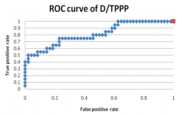
Figure 1: ROC curve for the D/TPPP ratio.
The threshold between group 1 (medical history of cancer) and group 2 (no medical history of cancer) is close to 0. The sensitivity is equal to 75% and the false positive rate is equal to 31.25%. The negative predictive value is equal to 91.7% and the positive predictive value is equal to 96.4%. The values are satisfactory (AUC=0.814), as far as simple screening is concerned.
Discussion and Conclusion
Concerning VOC detection
PO has been attributed to specific types of bacteria [30,31] and is associated with an increased risk of cancers [2,3].
Bacterial signature may be identified by breath tests focused on VOC [32-35]. Early detection of some cancers may also be detected by exhaled VOC [7-12].
In this epidemiological retrospective analysis, the ratio of DMCP/ TPPP was higher in patients with a medical history of cancer (p<0.001). The sensitivity (75%), the false positive rate (31.25%), the negative predictive value (91.7%) and the positive predictive value (96.4%) of the ratio D/TPPP are satisfactory, as far as simple screening is concerned. This is the first time that a link between DMCP and a medical history of cancer is reported.
DMCP is derived from cyclopropane ring which occurs only in organisms that synthesize specific unsaturated fatty acids (UFA). Cyclopropane Fatty Acids (CFA) are typically produced at the onset of the stationary phase in bacterial cultures [36]. The timing and extent of the UFA-to-CFA conversion and the widespread distribution of CFA synthesis among bacteria suggest an important physiological role for this phenomenon [25,36].
The following bacteria may produce CFA: i.e. Arthrobacter, Alcaligenes, Azotobacter, Bifidobacterium, Bordetella, Campylobacter, Caulobacter, Clostridium, Chlorobium, Citrobacter, Enterobacter, Helicobacter, Klebsiella, Lactobacillus, Nitrobacter, Pediococcus, Proteus, Pseudomonas, Rhizobium, Salmonella, Serratia, Streptococcus, Thiobacillus, Vibrio, Yersinia [25].
Some bacteria can be excluded since there are mainly found in soil, used in food processing or are opportunistic bacteria which develop only in severely immunosuppressed patients: Arthrobacter, Alcaligenes, Azotobacter, Bifidobacterium, Citrobacter, Lactobacillus, Nitrobacter, Pediococcus, Rhizobium, Serratia or Thiobacillus. Other bacteria can also be excluded since they induce severe acute infections and since the included patients did not have any acute infection when enrolled: i.e. Bordetella,Salmonella, Vibrio, and Yersinia.
Some bacteria are either commensal or induce acute infections or intoxications. There implication in chronic infections has never been documented: i.e. Clostridium, Enterobacter, Klebsiella, Proteus, Pseudomonas or Streptococcus [37].
Campylobacter or Helicobacter are the only remaining possible candidates.
Numerous publications have causally implicated HP or Campylobacters [38,39], in the occurrence of PO. HP synthetizes 19:0 cyclopropane [40].
97% of Campylobacter jejuni strains and 83% of Campylobacter coli strains are characterized by the presence of a 19-carbon cyclopropane fatty acid. Others Campylobacters (including Campylobacter rectus) lack 19-carbon cyclopropane [41,42].
Because the percentage of HP is similar between the two groups of patients, we hypothesize that Campylobacter jejuni or Campylobacter coli could explain the higher rate of dimethylcyclopropane in patients with a medical history of cancer.
Concerning hyaluronic acid levels
Patients in group1 have a higher plasmatic concentration of HA (78.3 ± 40.5 microg/l versus 37.7 ± 19.6; p<0.001). We reported in a preliminary study that an increased level of plasmatic HA is associated with an increased risk of adenocarcinoma in patients with severe PO [14].
Hyaluronidase activity has not been described either for Campylobacter jejunior for Helicobacter pylori. However, some strains of Campylobacter jejuni synthetize a hyaluronic acid-type capsular polysaccharide [43,44], which may modify the level of circulating HA.
Campylobacter jejuni is associated with SIBO and small gut motility decrease [45-46]. However, this bacterium has not yet been implicated in the occurrence of adenocarcinoma. [47,48]. To our knowledge Campylobacter jejuni has not been associated with PO.
Campylobacter rectus is associated with PO [30,31] and total cancer risk increase [48]. Since all Campylobacters may N-glycosylate their proteins [49], an involvement of Campylobacter rectus in the increase of plasmatic HA cannot be excluded. However, this latter bacterium does not produce dimethylcyclopropane.
To conclude, in patients with SIBO+PO and a medical history of cancer, plasmatic hyaluronic acid level is increased as well as exhaled dimethylcyclopropane concentration. The measure of VOC (especially DMCP) and of plasmatic hyaluronic acid level on a routine basis in patients with SIBO and PO could help to detect patients with a higher risk of cancer. The implication of Campylobacter jejuni or of Campylobacter rectus should be further investigated.
Acknowledgment(S) and Conflicts of Interest
No conflict of interest to disclose.
