Figure 1: A large pericardial effusion with tamponade signs-Right atrial and ventricular diastolic collapse.

Noor-ul-huda Maria* Muhammad Anwar Ch
Punjab Institute of Neurosciences, Pakistan*Corresponding author: Noor-ul-huda Maria, Punjab Institute of Neurosciences, Pakistan, E-mail: iii_glow_iii@ymail.com
Toxoplasma gondii is an obligate intracellular protozoan parasite presenting as a zoonotic infection distributed worldwide with a large proportion of cases belonging to developing countries. Toxoplasmosis is the most common central nervous system infection in patients with the acquired immunodeficiency syndrome (AIDS) because of immunodeficiency. We are presenting a case of a young boy who presented with multiple intracranial lesions where the topmost differential was of cerebral tuberculosis and how we diagnosed and managed it.
Toxoplasma gondii is an obligate intracellular protozoan parasite presenting as a zoonotic infection distributed worldwide. In HIVpositive individuals, it causes severe opportunistic infections, which is of major public health concern as it results in physical and psychological disabilities. In immunocompetent persons due to cell-mediated immunity the parasite is transformed into tissue cyst resulting in lifelong chronic infection. In HIV-infected people opportunistic infection by T. gondii occurs due to depletion of CD4 cells, decreased production of cytokines and interferon gamma and impaired cytotoxic T-lymphocyte activity which results in reactivation of latent infection [1]. Toxoplasma reactivation is frequent when CD4 T-cell count is <200cells/mm3 [2]. Toxoplasmosis is the most common central nervous system infection in patients with the acquired immunodeficiency syndrome (AIDS) who are not receiving appropriate prophylaxis [3]. A large proportion of the AIDS infected belong to developing countries and AIDS prevalence is intensified by severe poverty, malnutrition, and famine; fatal illnesses with a scorn shortage of medical amenities complemented with lack of education and development. Current Pakistani health system setting is in a dire need of improvement. Low literacy rates, high birth rates, and associated maternal mortality plus a lack of clean drinking water and appropriate sanitation system have a serious impact on general living conditions contributing to a relatively short lifespan. HIV is, therefore, becoming a growing health concern in Pakistan with a rapid rise in the reported cases. AIDS is most prevalent among injection drug users (IDUs) and unchecked deported migrant workforce [4]. Drug therapy does not eradicate T.gondii, and lifelong therapy to avoid relapse is often necessary [5]. The differential diagnosis of multiple ring-enhancing lesions depends on the age and the immune status of the patient. In the immunocompetent host, malignancies (both primary and metastatic) and pyogenic abscesses remain the most likely diagnoses in patients with large-sized lesions. Abscesses caused by atypical microorganisms and demyelinating disease should also be considered in the differential diagnosis of multiple enhancing lesions of the brain. In tropical countries, cysticercus granuloma frequently needs to be differentiated from intracranial tuberculoma. In HIVinfected patients, the leading causes of multiple enhancing lesions are toxoplasmosis and primary CNS lymphoma. Imaging characteristics that are helpful in distinguishing toxoplasmosis from CNS lymphoma include subcortical gray matter location of toxoplasmosis lesions, their multiplicity, presence of eccentric target sign and enhancing wall of the lesions thinner than that observed in lymphomas [6]. The clinical examination shows focal or diffuse signs and/or symptoms in cerebral toxoplasmosis. Neuroimaging findings may highly suggest the parenchymal infection by Toxoplasma. However, the definitive diagnosis can only be made by the protozoan demonstration in the brain tissue. Patients that do not respond clinically or on neuroimaging to toxoplasmosis treatment must be evaluated regarding other causative opportunistic agents [7]. Although a definitive diagnosis of cerebral toxoplasmosis requires the demonstration of parasites by a histopathological procedure, the clinical and radiological data could be complemented by a less invasive approach, such as molecular and immunological diagnoses [8].
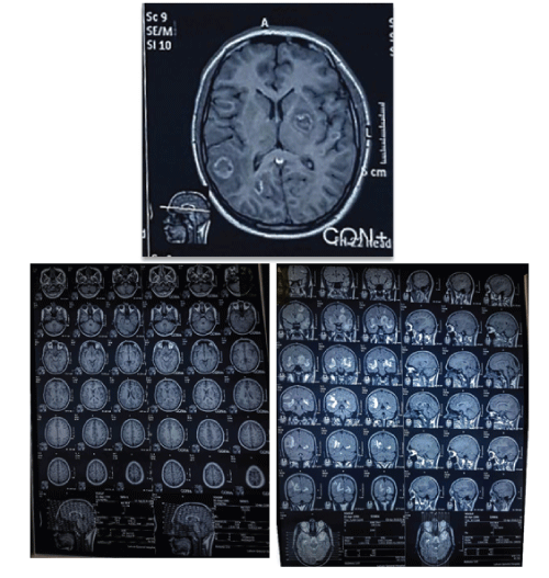
19 years old male patient, a college student belonging to middle class family was referred to us with history of seizures, right sided hemiparesis, speech difficulty, bifrontal headache, and right sided facial hypoesthesia that developed gradually over a period of the last 3 months. The patient was subjectively in the normal state of wellbeing 3 months ago when he first noticed bifrontal headache that gradually increased in intensity over the following 4 days. Patient then suffered a seizure (a tonic-clonic as described by brother) approximately 5 days after the onset of headache for which he was immediately taken to the nearby hospital emergency. An emergency computed tomography scan (CT scan) brain plain revealed multiple foci of hypodense areas that corresponded to a suspicion of multiple intracranial lesions. The patient was referred to tertiary care for further proper management. The patient presented at a specialized outdoor facility. With the conception of impaired patient care and apprehension of an impending doom, he was defaulted. He gradually developed speech difficulty, right sided hemi paresis and right sided facial numbness over a period of 10 days for which he presented to our hospital. The patient was oriented but had speech difficulty. He appeared anemic and cachectic. On further inquiry he stated past history of recurrent respiratory tract and gastrointestinal tract infections. On examination, the positive findings were power of 2/5 in the right upper and lower limb and hypoesthesia on the right side of face. There were bilateral extensor plantar reflex. Magnetic resonance imaging (MRI) was requested that showed multiple ring enhancing lesions with surrounding edema in the supratentorial region. The lesions were located in the grey white junction, periventricular region and basal ganglia. The pattern of enhancement was in the periphery and tiny nodular enhancement in the center. The largest lesion was detected at the right parietal region.
Provided the regional epidemiology of diseases (Figure 1), a diagnosis of cerebral tuberculosis was presumed and antituberculous treatment was started empirically. The recommended treatment comprised of four oral antifimic drug scheme with Rifampicin 10 mg/ kg, Isoniazid 5 mg/kg, Pyrazinamide 25 mg/kg and Ethambutol 15 mg/ kg. The complete blood count (CBC) at admission was hemoglobin 11.2 g/dl, total leukocyte count of 6500/mm3, neutrophil 80% and lymphocytes 15%. The definitive management plan included right parietal craniotomy to excise the right parietal space occupying lesion. A right sided parietal craniotomy was performed. Preoperative finding suggested abscess formation in brain tissue. Multiple tissue pieces fixed in formalin were sent for histopathological studies. After surgery patient was shifted to the recovery room. Patient started to deteriorate and was shifted to the intensive care unit. Patient showed oxygen desaturation with and suffered from fever spike of 104°C. Immediate care was started and blood sent for series of laboratory screening. It was noted that patient has a decreasing blood count as compared to the previous reports. Symptomatic treatment was started but the patient continues to follow a downward spiral curve. He stayed comatose and intubated. A tracheostomy was performed in anticipation of a prolonged airway maintenance and ventilator support. Tracheal aspirate was positive for Klebsiella pneumonia and patient had bilateral basal crepitations as well. As he continued to deteriorate with no betterment and a score of E3M3Vt, the attendants were counseled and a “do not resuscitate” agreement was signed after discussion and elaboration of predicted course of events. In the meantime the histopathological report was received. The section revealed brain tissue infiltrated lymphocytes, histiocytes and plasma cells. Extensive necrosis was also noted. Within the necrosis fragments, few cysts like structures were identified that contained the protozoan form of toxoplasma (bradyzoites). It confirmed the diagnosis of cerebral toxoplasmosis. The definite presence of cerebral toxoplasmosis in a patient >1month of age with an unknown HIV status indicates AIDS as per Centers for Disease Control’s (CDC) case definition. Given the seriousness of diseases indicative of AIDS, it is generally important to diagnose them definitively, especially when therapy that would be used may have serious side effects or when definitive diagnosis is needed for eligibility for antiretroviral therapy. Differential leukocyte count was repeated and HIV screening was requested concordantly. The lymphocyte count came out to be <200 cells/mm3 and patient was tested positive for HIV thus meeting the diagnostic criteria for Acquired Immunodeficiency Syndrome (AIDS).
Figure 1: A large pericardial effusion with tamponade signs-Right atrial and ventricular diastolic collapse.
The positive findings in our patient for AIDS were HIV positive screening, lymphocyte count <200 cells/mm3 , cerebral toxoplasmosis (confirmed by histopathological report), respiratory tract candidiasis (as per pathological investigation of tracheal aspirate) and history of recurrent lower gastrointestinal tract infection pointing to cryptosporidium infection. After confirmed diagnosis, standard treatment for cerebral toxoplasmosis was commenced with cotrimoxazole 800/160 mg TID (a total dose of 2.4 g/480g/24hrs divided in 3 doses). Patient made a miraculous recovery showing settlement of fever spikes and overall improvement .The level of consciousness also showed improvement status was deemed null and void and patient was shifted to isolation room with strict precautions and barrier nursing. By the time a multidisciplinary approach involving the AIDS center of city’s public tertiary care hospital was suggested. The relevant doctor of the specialty of infectious diseases was involved in the induction of highly active antiretroviral therapy HAART.
Cerebral toxoplasmosis is the most common cerebral focal lesion in AIDS and still accounts for high morbidity and mortality in AIDS patients. It is directly related to the prevalence of anti-Toxoplasma gondii antibodies in the population. Therefore, it is important to evaluate sensitive, less invasive, and rapid diagnostic tests. Through this case study it is emphasized that in a young patient with signs and symptoms suggestive of an infective lesion in the brain, especially in case of multiple foci on radiological examination, it is prudent to look for HIV positivity. Cerebral tuberculosis/tuberculous meningitis remain the most important differential diagnosis in the provided scenario of young patient with multiple intracranial lesions. Since there is high prevalence of tuberculosis in our country, tuberculosis is on the top of the list of differentials. The incidence of AIDS needs estimation and more research should be conducted. The burden of the disease should be measured and reassessed. Due to lack of awareness and stigma associated with HIV infection, there is insufficient data available and the level of problem is underestimated. The consequences of under reporting can perpetuate and express in the form of serious consequences with high level of transmission especially in hospital setup. There is a lack of awareness amongst the masses. It is suggested to guide and make the health personnel well awared of the probable diagnosis of AIDS. It HIV screening should be made a part of preoperative investigations. Statistical measures of the scope of the human immunodeficiency virus (HIV)/acquired immunodeficiency syndrome (AIDS) pandemic have been revised numerous times by public health agencies. At the end of 2007, the Joint United Nations Program on HIV/AIDS (UNAIDS) decreased its estimate of the numbers of people worldwide living with HIV infection by over 6 million, to 33.2 million [9]. It is proposed that a set of guidelines for a suspected case should be introduced and communicated among the healthcare providers in order not to miss any case from detection.
Cases of HIV and AIDS are being under reported that underestimates the burden of problem. This puts AIDS in the bottom of our differential lists that is always based on the incidence of disease. It is suggested that a set of guidelines should be introduced and communicated among the healthcare providers to increase their level of suspicion of AIDS cases. There should be routine screening for HIV especially if there is any high risk indicated in history and on examination. Precautions in nursing patients and in the operation theatre should always be taken in all cases to avoid inadvertent exposure of any case detected later. HIV screening kits should be made available.
In any young patient with history of recurrent/relapsing respiratory tract infections and gastrointestinal infections, HIV should be thoroughly looked for a proper history is always a key to rule out the possibility. It should be kept in mind that a negative history of any exposure is also useful if the doctor is vigilant enough to gather indirect clues from patient expression and concern of their attendants. Students from the start of their ward teaching years should be given adequate briefing about diagnostic criteria of AIDS and for self protection while working in wards. The best method to propagate the message in order to avert the possible epidemic before the condition gets out of our control is by arranging seminars and interactive programs. The importance of multidisciplinary approach towards management should be always emphasized. Proper clinical reporting should be done to point out the probable diagnosis and to highlight and tailor an up gradation of a differential that is usually not commonly considered.
A high degree of suspicion and a thorough knowledge of even the remotest possible diagnosis is the key to a proper diagnosis. The keystone of proper clinical care and saving lives especially in the most socially capable group lies in the proper care and dedication of the dealing doctor. With this case we conclude that we must always rule out HIV status in cases where manifestation related to immunosuppression are otherwise remote. In young patients without any congenital impairment in immunity, the presence of multiple intracranial foci especially with history of relapsing infections should always alert the dealing health personnel to rule out AIDS.
Download Provisional PDF Here
Article Type: CASE REPORT
Citation: Maria N, Anwar Ch M (2018) Case Report: Cerebral Toxoplasmosis Infection by T.gondii in Young Patient with AIDS. J Clin Case Stu 3(3): dx.doi.org/10.16966/2471-4925.170
Copyright: © 2018 Maria N, et al. This is an open-access article distributed under the terms of the Creative Commons Attribution License, which permits unrestricted use, distribution, and reproduction in any medium, provided the original author and source are credited.
Publication history:
All Sci Forschen Journals are Open Access