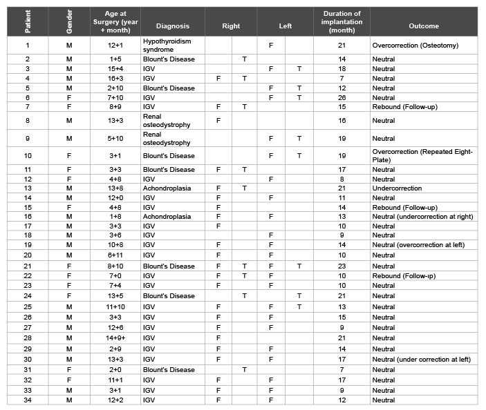
Table 1: Demographic characteristics and deformities of cases in genu varum and genu valgum groups (IGV: Idiopathic Genu Valgum; F: Femur; T: Tibia)

Mithat Oner* Bolat Mesut Karaman Ibrahim Kafadar Ibrahim Halil Guney Ahmet
Department of Orthapaedics, Erciyes University, Turkey*Corresponding author: Mithat Oner, Department of Orthapaedics, Erciyes University, Kayseri, Turkey, Tel: +90 352 4377163; Fax: +90 352 4375288; E-mail: mithatoner@mynet.com
The aim of this study was to assess the effectiveness of eight-Plate application in groups with genu varum and genu valgum. In addition, we aimed to compare the radiological measurements between groups.
We prospectively assessed 50 deformities in 34 patients who underwent surgery by the growth-controlled correction method using eight-Plate. The genu valgum group consisted of 39 deformities while the genu varum group consisted of 11 deformities. Gait patterns and range of motion in the knee joint were assessed in all patients. Again, changes in mechanical axis deviation (MAD), medial proximal tibial angle (MPTA), mechanical lateral distal femoral angle (mLDFA), and tibio-femoral angle (TFA) values were compared by obtaining pre- and post-operative orthoradiographs.
There were significant differences in MAD, MPTA, mLDFA and TFA values before and after surgery in both groups. Neutral mechanical axis was achieved in 9 deformities (81.8%) in the genu varum group and in 31 deformities (79.5%) in the genu valgum group. In terms of treatment efficiency, no significant differences were found in radiographic values between groups.
The hemiepiphysiodesis method using an eight-Plate is a reliable and effective method for the correction of deformities. Eight-Plate application provides similar improvements in the radiological values of both groups.
Genu valgum; Genu varum; Eight-Plate; Hemiepiphysiodesis
Genu valgum and genu varum are classified among angular deformities of the knee, namely coronal plane deformities of the knee. Mechanical axis shifting with gait disorder, joint instability and activity-related pain are seen as a result of the deformities. Although corrective osteotomies are considered classical treatment for the correction of these deformities, one should take care due to the need for prolonged hospitalization and immobilization, pain and wound problems [1]. Given these risks and costs, growth-controlled correction is highlighted in the treatment.
Although the classic bone block technique first described by Phemister was used initially for growth-controlled correction, it has been abandoned over time as the success of the procedure depends on timing and it is irreversible [2]. Then, temporary hemiepiphysiodesis with Blount stapling was introduced into practice, as first described by Blount [3]. In 2007, Stevens published a growth-controlled correction technique using an eight-Plate and 2 screws [4]. It gained wide acceptance in a short time due to its high success rates in trials, easy reproducibility, and simple learning curve, shorter period of hospitalization after surgery and lack of need for rehabilitation or immobilization. The eight-Plate technique has gained popularity after successful outcomes were reported in all etiologies including pathological physis such as Blount disease. The principle underlying the hemiepiphysiodesis technique using plates includes a simple, versatile extra-periosteal plate and fixation with 2 screws that serve as a hinge for physis. The procedure functions as a tension-band and a plate is required for each physis.
However, there are unclear issues such as duration of treatment or timing of implant removal in this procedure. The implant is removed when mechanical axis is corrected; thus, normal, symmetrical growth of the extremity is allowed. It is possible to repeat the eight-Plate technique in the event of rebound growth or recurrent deformities. The most important contraindications of eight-Plate application include physiological deformities and incomplete skeletal maturation [2].
In this study, we aim to assess the effectiveness of the eight-Plate technique in a clinical and radiological manner. In addition, we aim to contribute to the literature regarding complications, duration of treatment and timing of implant removal.
This study was approved by the Ethics Committee of Erciyes University on Clinical Trials (approval# 2013/276). All parents gave written informed consent before participation. We prospectively assessed 50 extremities in 34 patients who underwent surgery by the growth-controlled correction method using an eight-Plate between 2009 and 2013 (Table 1). The cases were divided into 2 groups as the genu varum (11 deformities) and genu valgum (39 deformities) groups. In all patients, pre-operative evaluation, surgery and post-operative follow-up were performed by the same surgeon.
Table 1: Demographic characteristics and deformities of cases in genu varum and genu valgum groups (IGV: Idiopathic Genu Valgum; F: Femur; T: Tibia)
In clinical assessment, gait patterns, range of motion in the knee joint and ligament instability were assessed in all patients. Leg length discrepancy was noted if present. The patients were divided into two groups as the genu varum and genu valgum groups, and full-length, weight-bearing orthoradiographs of the lower extremities with the patella facing forward were obtained. MAD, TFA, mLDFA and MTPA values were measured on the orthoradiographs.
All patients underwent surgery under general anesthesia by using a pneumatic tourniquet. After localization of the physis harboring deformity under fluoroscopy, a 3 cm incision was made, involving skin and subcutaneous tissue. The periosteum was exposed by passing through the muscle and fascia planes. Care was taken to avoid injury to the periosteum. At this point, in order to prevent injury to the perichondral ring, a Kirschner (K) wire was used to identify localization under fluoroscopy. The appropriate plate was selected under fluoroscopy. Care was taken to center the plate relative to the physis on the AP radiograph and to place the plate posterior to the mid-sagittal area on the lateral radiograph in order to prevent genu recurvatum. After localization of the plate, temporary fixation was achieved by using K wires over the screw holes. Cortical access was achieved over the K wire by using a 3.5 mm drilling bit. Then, an appropriate-sized 4.0 mm self-tapping screw was applied. One plate was applied for each physis. No post-operative fixation was used in the patients. Patients were encouraged to practice weightbearing, flexion and extension of the knee, and mobilization depending on their tolerance to pain. They were also encouraged to return to normal activities. All patients were discharged home post-operative day one.
The follow-up was scheduled at 3-month intervals. In control visits, fulllength, weight-bearing orthoradiographs of the lower extremities with the patella facing forward were obtained and MAD, TFA, mLDFA and MTAP values were measured on orthoradiographs. If the MAD and TFA values measured exceeded the neutral point due to excessive correction, they were considered as negative values. Clinically, gait patterns and ROMs were assessed in patients who attended control visits.
During the control period, the plate and screws were removed in cases with neutralization in the mechanical axis. Follow-up was scheduled at 6 month intervals in these patients, concerning rebound growth, leg length discrepancy and early closure of growth plate.
Histograms, Q-Q graphics and Shapiro-Wilk test were used to assess normal distribution of the data obtained. For intergroup comparisons, the paired-samples t test and Mann-Whitney U test were used in quantitative variables while chi-square analysis was used in qualitative variables. Data were analyzed by using SPSS for IBM version 20.0 software. p<0.05 was considered as significant.
Of the cases, 12 (35.3%) were girls while 22 (64.7%) were boys. There was genu varum in 11 cases (22%) and genu valgum in 39 cases (78%) (Table 1).
The mean age was 4 years and 5 months (one year and 5 months-13 years and 5 months) in the genu varum group while it was 9 years and 2 months (2 years and 9 months-16 years and 3 months) in the genu valgum group (p>0.05). In the genu varum group, the mean duration of treatment was 14.7 months (7-23) and mean follow-up after implantation was 22.9 months (6-50). In the genu valgum group, the mean duration of treatment was 14.4 months (7-26) and the mean follow-up after implantation was 19.8 months (6-43) (p<0.05).
There were significant differences between pre-operative and postoperative MAD, MPTA, mLDFA and TFA values in 11 deformities which underwent surgery due to genu varum (p<0.05) (Table 2) (Figure 1).
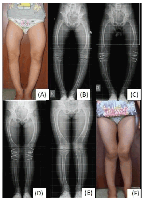
Figure 1: Genu varum; A) Clinical presentation before surgery; B) Preoperative orthoradiograph; C) Post-operative early orthoradiographs; D) Orthoradiograph at the end of treatment; E) Orthoradiograph after removal of implant; F) clinical presentation after surgery
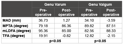
Table 2: Pre-operative and post-operative radiological values between genu varum and genu valgum groups
There were corrections to the neutral axis position in 9 cases (81.8%), insufficient axis correction in one case (9.1%) and overcorrection of axis persisted during follow-up in one case (9.1%) when the final outcomes of 11 deformities which underwent surgery due to genu varum were considered (Table 3).
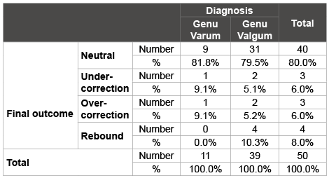
Table 3: Final outcomes between genu varum and genu valgum groups
There were significant differences between pre-operative and postoperative MAD, MPTA, mLDFA and TFA values in 39 deformities which underwent surgery due to genu valgum (p<0.05) (Table 2) (Figure 2).
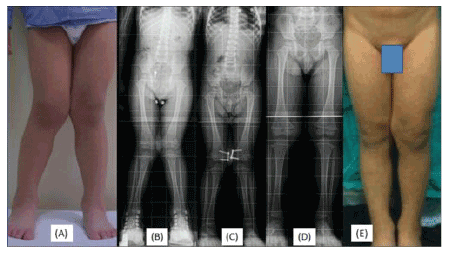
Figure 2: Genu valgum; A) Clinical presentation before surgery; B) Preoperative orthoradiograph; C) Post-operative early orthoradiographs; D) Orthoradiograph at the end of treatment; E) Orthoradiograph after removal of implant; F) clinical presentation after surgery
There were corrections to the neutral axis position in 31 cases (79.5%), insufficient axis correction in 2 cases (5.2%), overcorrection of axis persisted during follow-up in 2 cases (5.2%) and rebound deformity in 4 cases (10.3%) when the final outcomes of 39 deformities which underwent surgery due to genu valgum were considered. Of the patients with overcorrection, one patient underwent supracondylar femur osteotomy while outpatient follow-up was scheduled in the other patient (Table 3).
When the effectiveness of the treatment on MAD, MPTA, mLDFA and TFA in the genu varum and genu valgum groups was considered, no significant difference was detected between groups (p>0.05) (Table 4).
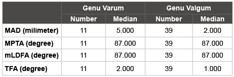
Table 4: Treatment effectiveness between genu varum and genu valgum groups by using radiological values
No mechanical problems such as permanent or early closure of the growth plaque, iatrogenic leg length discrepancy, sagittal plane deformity, implant breakage or migration were observed in any of patients during follow-up.
No loss of ROM was observed in any of our patients. None of our patients experienced problems in mobilization, nor did any of our patients require physical therapy and rehabilitation.
A wound problem was observed in one patient from the genu valgum group after the removal of the implant. The wound completely recovered after wound debridement and antibiotic therapy.
In the past century, osteotomy was used as the first choice of the correction of angular, rotational and length discrepancy deformities in children. This invasive technique was associated with common problems such as longer hospitalization, challenging rehabilitation and pain, as well as serious complications such as compartment syndrome, neurovascular injury, over correction or under correction and non-union, making it difficult to achieve the desired outcomes. These disadvantages prompted surgeons to seek for minimal invasive techniques [5,6]. As a result, the concept of epiphysiodesis was developed. After 1933, when Phemister first proposed the concept of epiphysiodesis, Dr. Walter Blount used U stapling in 1940 [3] and Metizeau et al. [7] described the use of the transphyseal screw in 1998. In 2007, a new concept, namely hemiepiphysiodesis, was introduced after the publication of the outcomes of temporary hemiepiphysiodesis using a non-locking plate and 2 cannnulated screws in children by Stevens et al. [1].
In the prospective study on 65 deformities in 34 cases by Stevens et al. [4] it was reported that the age ranged from 20 months to 17 years and the mean duration of implantation was 11 months with at least one year of follow-up (with and without implant) ranging from 14 to 26 months [1]. In a retrospective study on 54 deformities in 31 cases, Das et al. reported a mean age of 11.6 years and mean follow-up of 36 months (24-52 months) after the removal of the implant. The authors did not provide data regarding duration of implantation [8]. In a retrospective study on 50 deformities in 29 cases, Yılmaz et al. [9] reported the mean age as 10 ± 2.9 years (4 years-14 years and 4 months) and mean follow-up as 24 ± 13.4 months (7- 50). In a retrospective study on 33 deformities in 17 cases, Jelinek et al. [10] reported the mean age as 11.6 ± 3.8 (2 years and 9 months-16 years) and mean duration of implantation as 11.9 ± 6.8 months (2-30). In our study, 50 deformities in 34 cases were prospectively evaluated. The mean age was 4 years and 5 months (one year and 5 months-13 years and 5 months) in the genu varum group while it was 9 years and 2 months (2 years and 9 months-16 years and 3 months) in the genu valgum group (p>0.05). In the genu varum group, the mean duration of treatment was 14.7 months (7- 23) and mean follow-up after implantation was 22.9 months (6-50). In the genu valgum group, the mean duration of treatment was 14.4 months (7- 26) and mean follow-up after implantation was 19.8 months (6-43). Our study was found to be in agreement with the literature regarding number of cases, age distribution, duration of implantation and follow-up time.
Stevens et al. reported that neutral mechanic axis was achieved in 63 of 65 deformities, providing a 97% success rate [1]. Yılmaz et al. [9] reported the assessment criteria in detail. They found that pre- and postoperative mLDFA values in the genu varum group were 108.0 ± 8.6° and 101.8 ± 17.2° while pre- and post-operative MPTA values in the genu varum group were 82.7 ± 4.5° and 89.7 ± 4.1°, respectively. The authors also found pre- and post-operative MAD values of -34.5 ± 16.3 mm and 17.9 ± 20.5 mm in the genu varum group, respectively. Improvements in all criteria were found to be significant in the genu varum group. In addition, Yılmaz et al. found that pre- and post-operative mLDFA values in the genu valgum group were 82.1 ± 3.7° and 91.1 ± 4.9° while the pre- and post-operative MPTA values in the genu valgum group were 98.5 ± 8.0° and 87.8 ± 7.1°, respectively. The authors also found pre- and post-operative MAD values of -30.1 ± 14.8 mm and -1.7 ± 19.0 mm in the genu valgum group, respectively. Improvements in all criteria were found to be significant in the genu varum group [9]. Our study included more details than those in the literature and both the genu varum and genu valgum groups were assessed regarding changes in pre-operative and post-operative mLDFA, MPTa, TFA and MAD values, indicating significant differences. In addition, the groups were compared regarding the effectiveness of the treatment employed and no significant difference was found when radiological measurements were considered. In our study, there was correction to the neutral axis position in 9 cases (81.8%), insufficient axis correction in one case (9.1%) and overcorrection of axis persisted during follow-up in one case (9.1%) among the final outcomes of 11 deformities which underwent surgery due to genu varum; correction to neutral position was achieved in 31 cases (79.5%) among the final outcomes of 39 lower extremities which underwent surgery due to genu valgum. These rates were in agreement with the literature.
Pathological physis is not a contraindication for use of this method [11]. Boero et al. [12] reported complete correction in 22 cases (78.6%) after eight-Plate application in a series of 28 cases with pathological deformities [12]. In a study by Stevens et al. complete correction was reported in 13 of 15 cases treated with an eight-Plate [13]. As a result of their study in patients with Blount’s disease involving 18 tibia deformities, Schoerlucke et al. [14] suggested that while the eight-Plate was reasonable for hemiepiphysiodesis it was unacceptable for patients with Blount’s disease. In our study, neutral axis correction was observed in 6 (85.7%) of 7 patients with Blount disease. This was in agreement with the studies by Boero et al. [12] and Stevens et al. [11].
In the study by Stevens et al. [13] it was suggested that no intervention was required for correction of rotational deformity [1]. In our study, no intervention was performed for rotational deformity in any patient.
Insufficient correction seems to be beyond the scope of surgery. Boero et al. [12] reported partial correction in 5 cases (17.9%) and under correction in one case (3.6%) after eight-Plate application for pathological deformities in 28 cases; however, the authors reported that they did not observe under correction in the idiopathic group. They reported that osteotomy and external fixation were performed in 5 cases while internal fixation was performed following osteotomy in the remaining case.
In the study by Stevens et al. [1] no correction was observed in 2 of 15 cases treated with eight-Plate application. The authors reported that these 2 cases had Blount’s disease. In our study, it was seen that, in the final outcomes, there was insufficient axis correction in one case (9.1%) of 11 deformities which underwent surgery due to genu varum and in 2 cases (5.2%) of 39 deformities which underwent surgery due to genu valgum. Ages at surgery were 13 years and 8 months and 13 years and 3 months in the cases with insufficient axis correction in genu valgum group. This suggests that the problem could be due to advanced age. Osteotomy was planned in these cases. In the genu varum group, age at surgery was one year and 8 months in our case with achondroplasia. Follow-up at 6 months intervals was scheduled in this patient.
In their study, Yılmaz et al. [9] reported overcorrection in 7 of 50 deformities; thus, the implants were removed. It was reported that these cases achieved neutral mechanical axis during follow-up. In the literature, there is no other study reporting overcorrection. In our study, there was overcorrection in one case (9.1%) from the genu varum group. It was thought failure to attend control visits at the correct time was the reason for overcorrection in this case, in which neutral axis was achieved by the same method. In our study, there was overcorrection in 2 cases (5.2%) in the genu valgum group. Again, failure to attend control visits at the correct time was the reason for overcorrection in these cases. Supracondylar femoral osteotomy combined with external fixation was performed to correct deformity in one case since the growth plaques were closed at presentation. In the other case, implants were removed and follow-up at 3-month intervals was scheduled. To prevent overcorrection, we believe that patients should be monitored closely.
Rebound deformity can be seen after removal of the implant. In the study by Stevens et al. [1] it was reported that rebound deformity was observed in 4 cases with genu valgum and neutral mechanical axis was achieved by re-operation using the same method [1]. In our study, there were 4 cases with genu valgum which exhibited rebound deformity. No rebound deformity was observed in cases with genu varum.
In the articles by Stevens et al. [1] and Burghardt et al. [15] no early or permanent closure of growth plates was observed. In our study, no early or permanent closure of the growth plates was observed in the genu varum and genu valgum groups. We think that cases in which implants were removed should have to be followed until closure of the growth plates and skeletal maturation.
In the study on 18 tibia deformities in patients with Blount’s disease by Schroerlucke et al. implant failure was seen in 8 cases (44%). The authors reported that there were no significant differences in body mass index (BMI) and body weight between patients with or without implant failure [14]. Das et al. reported that plate and screw migration was detected in 2 cases by radiology, in which repeated plate and screw applications were performed [8]. Yılmaz et al. reported breaking a screw during implant removal in one case [9]. In their study, Stevens et al. reported no implant failure among 34 cases treated with eight-Plate application [1]. No implant failure or migration was observed in our study.
In the study by Stevens et al. superficial wound infection was observed in one case and profound infection in 2 cases, the authors reported that the case with superficial wound infection was treated with oral antibiotics while implant removal was required in those with profound infection [13]. In our study, profound infection was observed in only one patient after removal of the implant. Infection was resolved after debridement and antibiotic therapy.
Temporary, growth-controlled, partial epiphysiodesis by using an eight-Plate is a highly safe and effective treatment modality which can be applied at an early age in the treatment of coronal plane deformities of the knee. The eight-Plate technique was equally effective regarding improvement of radiological measurements in the genu varum and genu valgum groups.
None
Download Provisional PDF Here
Article Type: Case Report
Citation: Oner M, Mesut B, Ibrahim K, Halil KI, Ahmet G (2016) Comparison of Radiological Measurements in Genu Valgum and Genu Varum Deformities Treated with Eight Plate Hemiepiphysiodesis. J Clin Case Stu 1(3): doi http://dx.doi.org/10.16966/2471-4925.120
Copyright: © 2016 Oner M, et al. This is an open-access article distributed under the terms of the Creative Commons Attribution License, which permits unrestricted use, distribution, and reproduction in any medium, provided the original author and source are credited.
Publication history:
All Sci Forschen Journals are Open Access