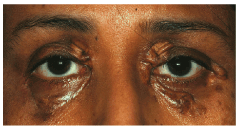
Figure 1: Persistent multiple yellowish skin lesions involving all 4 eye lids

Saif Al Dosari1 Hailah Al Hussain1 Azza Maktabi2 Hind Alkatan2,3*
1Oculoplastic Division, King Khaled Eye Specialist Hospital, Riyadh, Saudi Arabia*Corresponding author: Hind Manaa Alkatan, Chief of Ophthalmic Pathology Division, Ophthalmology Department, Director of KSU Residency, Training Program in Ophthalmology, College of Medicine, King Saud University, Riyadh, SA, Tel: +966 504492399; Fax: +966112052740; E-mail: hindkatan@yahoo.com; hkatan@ksu.edu.sa
Adult orbital xanthogranulomatous disease (AOXGD) is a heterogeneous group of syndromes that are generally rare. One of the proposed sub-classification of AOXGD includes 4 subtypes, which are differentiated based on their variable clinical features: Adult-onset xanthogranuloma (AOX), Necrobiotic xanthogranuloma (NBX), Erdheim-Chester disease (ECD), and Adult-onset asthma and periocular xanthogranuloma (AAPOX). These entities share the finding of “Hallmark” cells histopathologically, which are foamy histiocytes and Touton-type giant cells. Diagnosis of each of these can be challenging and often misleading in management. We have faced this interesting case of a 54 year-old female who presented with multiple yellowish periocular skin lesions for several years with the presumed diagnosis of simple xanthelasma based on the histopathological examination of these lesions previous excisional biopsy in another institution. The correct diagnosis of AOX was not made until the patient presented to our tertiary eye center for better cosmetic treatment. We believe that this entity is being overlooked owing to its rarity. Ophthalmologists, dermatologists, general practitioners and pathologists should consider AOXGD in their daily practice whenever applicable.
Xanthogranuloma; Histiocytes; Touton giant cell; Xanthelasma
Adult orbital xanthogranulomatous disease (AOXGD) is a heterogeneous group of syndromes that are rare and becoming more and more recognized over the last 2 decades [1,2]. These entities share a common histopathological finding of “Hallmark” xanthoma cells and Touton giant cells. They include: Adult-onset xanthogranuloma (AOX), Necrobiotic xanthogranuloma (NBX), Erdheim-Chester disease (ECD), and Adult-onset asthma and periocular xanthogranuloma (AAPOX). Their differential diagnosis is very broad and overlapping, therefore special attention to the clinical presentation is important to make the proper diagnosis, aided by the additional histopathological confirmation of tissue diagnosis. We are presenting the clinical history of a case with AOX, which has been overlooked for several years until the proper diagnosis was made in our tertiary eye care center.
A 54-year old Saudi lady presented with persistent multiple yellowish skin lesions involving all 4 eye lids over the last 4 years (Figure 1). She gave history of hyperlipidemia on treatment but was otherwise healthy. The patient had history of excisional biopsy of similar bilateral upper lid lesions done elsewhere with the histopathological diagnosis of xanthelasma. She was referred by a general plastic surgeon for better cosmetic management of her lesions. Her initial skin lesions excised at our center were also diagnosed as xanthelasma (Figure 2A). However, owing to the deeper involvement of her orbit on both sides, the extensive skin involvement and the suspicion of a more extensive pathologic condition by our oculoplastic surgeon, further excisional biopsy of the skin lesions of the right lateral canthal area and the deep orbital component was done aided by removal of the underlying periosteum. The histopathological examination of the excised orbital tissue showed sheets of foamy histiocytes with few aggregates of lymphocytes (Figures 2B and 2C) some of which are forming germinal centers. Giant cells including Touton giant cells were seen (Figure 2D). Significant fibrosis was evident but there was no evidence of necrobiosis in any of the sections (Figure 2E). The xanthoma cells showed positive staining with CD68 while they were negative to S-100 and CD1a (Figure 2F). The final diagnosis of xanthogranuloma was made. She was further evaluated medically to rule out asthma and/ or systemic associations. The areas of her skin incisions were managed by direct closure with no need for skin graft or reconstruction. The patient had good postoperative cosmetic appearance with no eyelid margin malpositioning or lagophthalmos. She was referred from a general hospital where her systemic work up was reported to be negative.
Figure 1: Persistent multiple yellowish skin lesions involving all 4 eye lids
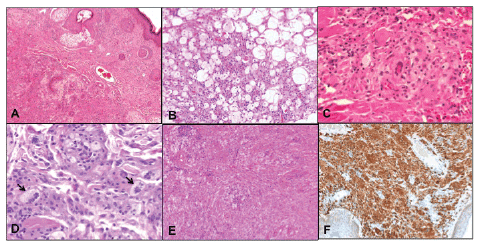
Figure 2: Histopathological examination of xanthelasma
A: Initial skin lesions diagnosed as xanthelasma
B and C: Histopathological examination of the excised orbital tissue
showed sheets of foamy histiocytes with few aggregates of lymphocytes
D: Giant cells including Touton giant cells were seen
E: Significant fibrosis was evident but there was no evidence of necrobiosis
F: Xanthoma cells showed positive staining with CD68 while negative to
S-100 and CD1a
Adult-onset xanthogranuloma (AOX) is the least common subtype of adult orbital xanthogranulomatous disease (AOXGD), which is a heterogeneous group of 4 syndromes that present with variable clinical features and are poorly understood [1,2] Among the 4 subtypes described before, Necrobiotic xanthogranuloma (NBX) is the most frequently encountered followed by Erdheim-Chester disease (ECD) and adult-onset asthma and periocular xanthogranuloma (AAPOX) [1,2]. Among the 4 syndromes, NBX is the most frequently encountered followed by ECD and AAPOX, whereas AOX is the least common subtype of AOXGDs.
Necrobiotic xanthogranuloma is characterized by the presence of subcutaneous skin lesions that tend to ulcerate and become fibrotic. It often affects adult patients aged 20 to 85 years, with no significant sex preference. Frequently associated systemic findings include internal organ involvement such as spleen, heart, lung, kidney and central nervous system, while the commonest hematologic findings are paraproteinemia and multiple myeloma [2,3,4]. ECD is an idiopathic condition of lymphohistiocytic infiltration in the orbit as well as internal organs including the heart, lungs, retro peritoneum, bones, and other tissues. This condition is often fatal, with death due to cardiomyopathy, severe lung disease, or chronic renal failure. Bilateral diffuse orbital masses should alert the clinician and/or pathologist to the possibility of this serious systemic disease [1,2,5]. AAPOX is rare, often presents with bilateral yellow-orange, elevated, indurated, and non-ulcerated xanthomatous eyelid and/or orbital masses. It typically extends into the anterior orbital fat and sometimes involves the extra ocular muscles and/or the lacrimal gland(s) [1,2,6] Most patients experience adult onset asthma within few months up to few years after the onset of the periocular lesions, the underlying mechanism of which is not understood but it is possibly related to an immunologic derangement with concomitant bronchiolar and ocular adnexal dysfunction [6].
Finally, AOX is an isolated xanthogranulomatous non-Langerhans histiocytic disease without significant systemic involvement. The pathogenesis of AOX is not clear but it seems to involve an immunemediated process based on the wide spectrum of associated immunological abnormalities and immune-related systemic diseases [7,8]. It is the least common entity among this group of diseases and affects patients with age ranging from 38 to 79 years, and no sex preference. It is often self-limited and does not require aggressive treatment unlike the other subtypes of AOXGDs [2]. Our patient did not have systemic involvement. Her hyperlipidemia possibly has aided in the misleading impression of xanthelasma initially but careful examination of additional tissue and the clinical suspicion helped us to reach the final accurate diagnosis.
The differential diagnosis is very broad with several possible entities for each category. This includes juvenile xanthogranuloma, Wegener’s granulomatosis, Langerhans histiocytosis, Rosai-Dorfman disease, orbital pseudotumer, Graves’ disease, multiple myeloma and lymphoma. The diagnosis might be challenging even within the group of AOXGDs due to overlapping presentation [9,7]. However, the histopathological findings and the immunohistochemical staining will help in the confirmation of the diagnosis. AOXGDis characterized by infiltration of the tissue by ‘‘hallmark cells’’ consisting of foamy histiocytes and Touton-type giant cells. These are non-Langerhans histiocytes thus show positive staining with CD68 and CD163 while show negative staining with S-100, Langerin and CD1a [2,9,7].
Apart from surgical excision of the periocular lesions such as in our case, intralesionalcorticosteroid injection has been recommended to avoid the use of systemic corticosteroids and cytotoxic agents [8,10]. Complications of periocular corticosteroid injection include central retinal artery occlusion, eyelid necrosis and subcutaneous fat atrophy but it was still recommended as a safe modality over partially effective methods of treatment such as radiation therapy [10].
In conclusion, it seems that the diagnosis of AOXGD in this part of the world is being overlooked possibly because of a low clinical suspicion. Surgeons and health practitioners (not only ophthalmologists) who might be involved in the surgical treatment of the periocular skin lesions should be aware of this group of diseases. Proper diagnosis is essential for early management especially in the more serious subtypes such as ECD. With enough level of suspicion, diagnosis should be made based on the clinical history and the specific histopathological findings.
The Research department at KKESH has approved this case report: RP 1502-CR. The authors have no conflict of interest and no financial disclosure in relation to this manuscript.
Download Provisional PDF Here
Article Type: Case Report
Citation: Dosari SA, Hussain HA, Maktabi A, Alkatan A (2016) Periocular Adult-Onset Xanthogranuloma (AOX) Initially Misdiagnosed as Xanthelasma: A Case Report. J Clin Case Stu 1(3): doi http://dx.doi. org/10.16966/2471-4925.117
Copyright: © 2016 Dosari SA, et al. This is an open-access article distributed under the terms of the Creative Commons Attribution License, which permits unrestricted use, distribution, and reproduction in any medium, provided the original author and source are credited.
Publication history:
All Sci Forschen Journals are Open Access