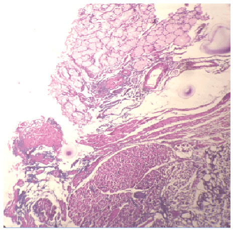
Figure 1: Lobular architecture of ectopic pancreas under intact normal duodenal mucosa and in between smooth muscle fibres of muscularispropria.(X10)

Rachana Dhakal* Ramesh Makaju
Department of Pathology, Kathmandu University School of Medical Sciences, Kavre, Nepal*Corresponding author: Rachana Dhakal, Department of Pathology, Kathmandu University School of Medical Sciences, Dhulikhel, Kavre, Nepal, E-mail: sigdelrachana@gmail.com
Heterotopic pancreas is pancreatic tissue found outside the usual anatomic location of the pancreas. It is often an incidental finding and can be found at different sites in the gastrointestinal tract. It usually remains asymptomatic, but becomes clinically evident when complicated by pathologic changes such as inflammation, bleeding, obstruction and malignant transformation. Here, we report a 33-year-old male presented at Out Patient Department with chief complaint of epigastric pain. An upper gastrointestinal endoscopy was done which revealed nodular sub mucosal lesion at first part of duodenum measuring 1.5 cm with an adjoining area showing ulcer with active bleeding. Biopsy was taken from the representative site. On gross examination, specimen consists of single piece of grey white nodular tissue. Microscopic examination revealed lobular architecture of ectopic pancreas under intact normal duodenal mucosa and in between smooth muscle fibres of muscularispropria. Pancreatic tissue consists of mixture of pancreatic acini, ducts and islets of Langerhans.
Heterotopic; Pancreas; Duodenum
Heterotopic pancreas is pancreatic tissue found outside the usual anatomic location without any vascular connection with the main pancreas [1]. It is a rare entity and a silent anomaly, but may become clinically evident when complicated by inflammation, bleeding, obstruction or malignant transformation [2]. Although echogastroscopy is helpful for diagnosis, it is difficult to distinguish from gastrointestinal stromal tumour (GIST) [3,4]. We report a case of 33-year-old male with ectopic pancreatic lesion in the duodenum.
A 33-year-old male presented at Out Patient Department with chief complaint of epigastric pain. His physical and laboratory findings were normal. An upper gastrointestinal endoscopy revealed nodular sub mucosal lesion at first part of duodenum measuring 1.5 cm with an adjoining area showing ulcer with active bleeding. Clinical diagnosis of gastrointestinal stromal tumour (GIST) was made. Biopsy was taken from representative site and sent for histopathological examination.
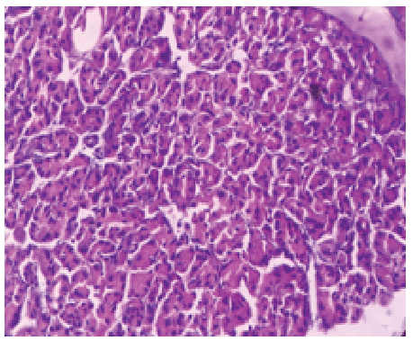
On gross examination, specimen consists of single piece of grey white nodular tissue measuring 1.5 × 1.5 × 1 cm with firm in consistency. Microscopic examination revealed lobular architecture of ectopic pancreas in the duodenum under intact normal duodenal mucosa and in between smooth muscle fibres of muscularis propria (Figure 1). Pancreatic tissue consists of mixture of pancreatic acini (Figure 2), ducts and islets of Langerhans.

Figure 1: Lobular architecture of ectopic pancreas under intact normal duodenal mucosa and in between smooth muscle fibres of muscularispropria.(X10)

Figure 2: Lobules of pancreatic tissue with acini (X40)
Karahan et al. [5] first reported heterotopic pancreas, tissue found outside the usual anatomical location of the pancreas. Heterotopic pancreas can exist at any position in the abdominal cavity. It is usually found in the upper gastrointestinal tract with >90% of the cases involving the stomach, duodenum or jejunum. Unusual locations are the colon, spleen or liver [4,5].
Three theories have been put forward regarding heterotopia. There are different theories regarding heterotopias of pancreas. The “theory of metaplasia” states, during embryological development, the metaplasia in the pancreatic endodermal tissue leads to the heterotopic pancreas [4]. The “theory of misplacement” states, there is misplacement of the pancreatic tissue during the period of rotation of the gut, due to fragmentation-before the fusion of the dorsal and ventral anlage, leading to heterotopic pancreas later in life [6]. The latest is the “theory of abnormalities of notch signaling”. This is a type of cell interaction mechanism which has been proven in mice that abnormal expression of pancreas-specific transcription factor 1a (PTF1 A) leads to formation of heterotopic pancreas [7,8].
Patients with heterotopic pancreas can be normal or present with abdominal pain and distention. In addition, it can also manifest clinically in some rare diseases of the pancreas including pancreatitis, islet cell tumour, pancreatic carcinoma and pancreatic cyst [9].
Echo gastroscope, computed tomography (CT) and gastrosopy can be helpful in diagnosis. Rate of diagnosis of this disease with the help of echo gastroscope is high. Medical treatment is not effective for heterotopic pancreas; surgical excision is the first and best choice [10-12].
The diagnosis may be sometimes difficult intraoperatively due to gross similarity of pancreatic heterotopias with gastrointestinal stromal tumour (GIST), gastrointestinal autonomic nerve tumour (GANT), carcinoid, lymphoma or even gastric carcinoma. If there is in doubt, frozen section is very helpful to establish the diagnosis intraoperatively and to avoid unnecessary extensive operations [13].
Pancreatic heterotopias are rare. It is difficult to distinguish from extramucosal gastric lesions. Preoperative diagnosis is difficult and echo gastroscope is helpful. Surgical excision provides symptomatic relief and is recommended especially if diagnostic uncertainity remains. Frozen section distinguishes heterotopic pancreas from malignant tumour and hence, avoids unnecessary radical operations.
Download Provisional PDF Here
Article Type: Case Report
Citation: Dhakal R, Makaju R (2016) Heterotopic Pancreas in Duodenum: A Case Report. J Clin Case Stu 1(3): doi http://dx.doi.org/10.16966/2471-4925.109
Copyright: © 2016 Dhakal R, et al. This is an open-access article distributed under the terms of the Creative Commons Attribution License, which permits unrestricted use, distribution, and reproduction in any medium, provided the original author and source are credited.
Publication history:
All Sci Forschen Journals are Open Access