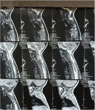
Figure 1: X Ray chest (PA And Lateral View) showed fused cervical vertebrae and thoracic kyphoscoliosis.
Ketan kataria1 Meena Shyam C2* Shivlal Soni2 Kamal kajal2
1DM Resident in Neuroanaesthesia at PGIMER, Chandigarh, India*Corresponding author: Shyam Charan Meena, Anaesthesia Department of PGIMER, Chandigarh, India, Tel: 7891669817; E-mail: drshyam.pgi@gmail.com
Article Type: Case Report
Citation: Ketan K, Meena SC, Shivlal S, Kamal K (2017) Anaesthetic Management of A Child With Klippel Feil Syndrome For Detethering of Spinal Cord: A Case Report. J Clin Anesth Manag 2(2): doi http://dx.doi.org/10.16966/2470-9956.130
Copyright: © 2017 Ketan K, et al. This is an open-access article distributed under the terms of the Creative Commons Attribution License, which permits unrestricted use, distribution, and reproduction in any medium, provided the original author and source are credited.
Publication history:
Klippel Feil syndrome is a type of bony disorder which is characterized by the abnormal fusion of two or more cervical vertebrae, which is usually present since birth. It is an autosomal dominant congenital defect. It is characterized by the classical triad of low posterior hairline, short webbed neck, restricted head and neck movements and mostly associated with a tuft of hair at lumbosacral region and torticollis (abnormal neck position). Many aspects of this syndrome are surfacing and is not clear whether it is a separate entity or if it is a part of congenital spinal deformities. Being anaesthesiologist we should be aware and ready to dealing with difficult airway management. Here we are reporting a case with same with anticipated airway difficulty and we managed the airway successfully with the help of video laryngoscope. We also kept ready fiberopetic bronchoscope as per standard institute protocol. We observed that video laryngoscope can be an alternative good option for dealing with such patients.
Klippelfeil Syndrome; Vertebral Synostosis; Cervical Vertebrae Fusion; Video Laryngoscope
Klippel Feil syndrome is a rare disease, occurring in 1 among 42,000 live births and is more common in females [1]. It has been classically described to be characterized with the triad of short neck, low hair line and restricted neck movements due to insufficiency of segmentation of two or more cervical vertebrae [2]. These abnormalities make it difficult to manage the airway. Airway management can be challenging in most of these cases mainly due to limitation in the range of neck movement due to cervical immobility. Also, cervical instability can increase the risk of neurologic damage during intubation.
We report the anaesthetic management of a two year female child who was a known case of Klippel Feil syndrome with multiple vertebral anomalies and was operated for detethering of spinal cord at C5 level.
A 2 year old female child weighing 10 kgs was brought to our hospital by the parents with chief complaint of stunted growth. On examination, the child had a short webbed neck, low lying hair line, cephalomegaly and asymmetric thorax and right hand finger flexion deformity.
X ray chest (PA and lateral view) showed fused cervical vertebrae and thoracic kyphoscoliosis. Magnetic resonance imaging (MRI) revealed split cord malformation and syrinx formation at C2-C6 with tethering of cord at cervicodorsal junction. Echocardiography revealed a normal heart. Ultrasonography for ruling out renal abnormalities revealed a normal study.
In addition to the usual anaesthetic considerations in paediatric patients undergoing surgery, airway management in this case was a special consideration. Airway examination in our patient revealed adequate mouth opening, a Mallampati grade of 2 but restricted neck extension (<20°). After reviewing the abnormalities present in our patient, we suspected that her tracheal intubation can prove to be difficult. Awake fibre optic has been described as gold standard for intubation in these patients. However, we decided to give check laryngoscope using video laryngoscope a try keeping fibreoptic as back up as this was a paediatric patient and were reasonably sure of being able to intubate her using indirect laryngoscopy. Insertion of LMA followed by fibre optic guided intubation was our plan B in case of intubation failure the patient using video laryngoscope.
Before shifting the patient to Operation theatre (OT), the difficult airway cart was prepared. In order to prevent occurrence of hypothermia, OT temperature was maintained between 33- 35°C. In addition to all the routine drugs, succinylcholine was also prepared. ENT team was kept as a follow-up for emergency tracheostomy should the need arise.
The patient was shifted to OT after premedicating her with iv midazolam 1 mg. Inside the OT, standard monitors were attached and she was induced with Inj Fentanyl 20 mcg and Inj Propofol 20 mg and a check video laryngoscopy was done, which revealed Cormac Lehane grade 2. Thereafter, 5 mg of Inj atracurium was given after checking for adequate mask ventilation. After 4 min of mask ventilation, laryngoscopy was attempted with a video laryngoscope (Figure 1) and the patient was intubated in first attempt with an uncuffed 4.5 mm ID endotracheal tube which was fixed at 10 cms. After intubation, all the monitors except saturation probe, and iv line were disconnected and the patient was made prone. Bilateral air entry was rechecked, all the monitors were attached. All the pressure points were well padded. The patient was then handed over to the surgeon. Anaesthesia was maintained with 50% nitrous oxide and oxygen with 1.5 to 2% sevoflurane, Minimum alveolar concentration (MAC) was maintained at around 1. Intraoperatively, patient was kept on Volume control ventilation mode with tidal volume of 80 ml and RR of 25/min. The surgery lasted nearly an hour and 15 mins during which the patient was administered 250 ml of normal saline. After the end of surgery, the incision site was infiltrated with 5ml of 0.25% bupivacaine for post operative analgesia. Then the child was made supine, inhalational agents were stopped and extubated when she met the standard criteria of extubation. The child made an uneventful recovery and was discharged from hospital 4 days after surgery.
Figure 1: X Ray chest (PA And Lateral View) showed fused cervical vertebrae and thoracic kyphoscoliosis.
Patients of Klippel Fiel can present with different clinical presentations and varying degrees of vertebral involvement. In addition to the typical features, these patients can also have a number of associated disorders in other body systems, which can make their anaesthetic management difficult. In addition to the upper cervical involvement, patients also present with facial asymmetry and torticollis which occurs in 21- 50% of patients. Orthopaedic neurological manifestations are seen in 20% of patients of which occipitocervical abnormalities are the most common. Scoliosis occurs in approximately 60% of patients. Some of the abnormalities include changes in atlantoaxial joint which can make the intubation difficult , scoliosis which can make the ventilation and extubation difficult, medullary canal stenosis, sprengel deformity of shoulder, rib defects are also common which can pose a challenge to the anaesthesiologist for ventilator management.
Renal anomalies are common in individuals with Klippel Feil syndrome, including a double collecting system, renal ectopia and bilateral tubular ectasia. Major renal anomalies include hydronephrosis, absence of a kidney and a horse shoe kidney, so the attending anaesthesiologist must be aware of this and plan the intraoperative management in terms of avoidance of nephrotoxic drugs and fluid management respectively. Cardiovascular anomalies occur in almost 14-29% of patients, the most common being Ventricular Septal Defect, other cardiovascular abnormalities being (Patent ductus arteriosus, mitral valve prolapse, bicuspid aortic valve, and coarctation of the aorta) [3-5]. Syncopal attacks may be precipitated by sudden rotatory movements of the neck in patients with Klippel Feil syndrome. Hence, all these conditions must be investigated before taking up a patient of Klippel Feil syndrome for surgery, and the induction, intraoperative management should be planned accordingly. It can also present with a variety of other clinical syndromes like, foetal alcohol syndrome, Goldenhar syndrome and other anomalies.
Airway management in these patients requires special care. These patients have a potentially unstable cervical spine and abnormal atlantooccipital junction and are prone to an increased risk of neurological damage. Restricted movement of cervical spine and associated anomalies can make mask ventilation and intubation difficult. Awake fibreoptic is considered the safest technique in these patients but requires a cooperative patient. Since our patient was a child, we decided to go for a check laryngoscopy with fiberoptic as a backup in case it was not possible to intubate the patient with video laryngoscopy. Fernandes et al and Ahuja et al had used similar plan to manage the airway of a klippel feil syndrome patient [6,7].
With all the modes of airway management, we feel that the optimum mode of intubation in a patient with cervical spine pathology is an awake fiberoptic intubation. The advantages are, (1) An awake spontaneously breathing patient who is maintaining his own airway (2) Spinal movement is not needed during intubation, (3) A tool for intubation that allows confirmation of tracheal tube placement, (4) It has a high rate of success (5) Complications are low and (6) Good patient acceptance. A difficult airway must be approached with caution. A comprehensive preoperative examination and ‘work up’, the availability of several alternate techniques, a willingness to call an expert help, surgeons standing by to provide a surgical airway and/or moral support and a good deal of common sense go a long way in ensuring a favorable outcome.
None
Written informed consent took for academic and research from baby’s father.
Download Provisional pdf here
All Sci Forschen Journals are Open Access