Article Information
Aritcle Type: Research Article
Citation: De Paolis M, Ali N, Romagnoli C, Romantini
M, Lana D, et al. (2016) Total Hip Arthroplasty on
Unrecognized Bone Tumors: Dramatic Outcome from
11 Cases. Int J Cancer Res Mol Mech 2(1): doi http://
dx.doi.org/10.16966/2381-3318.122
Copyright: © 2016 De Paolis M, et al. This is an
open-access article distributed under the terms
of the Creative Commons Attribution License,
which permits unrestricted use, distribution, and
reproduction in any medium, provided the original
author and source are credited.
Publication history:
Received date: 11 Sept 2015
Accepted date: 16
Feb 2016
Published date: 22 Feb 2016
Abstract
Background: Several mistakes in the diagnosis and treatment of bone tumors can be made, especially in non-specialized centers. Implanting
conventional prostheses on an unrecognized bone tumor causes contamination of the entire region with dramatic consequences for prognosis.
Purpose of our study was to try to understand which the best way to deal with these patients is. Does previous surgery affect prognosis? Does
external hemipelvectomy achieve a better overall survival and local control than limb salvage surgery?
Hypothesis: Demolitive surgery, scarifying the involved limb ensures better local control of the disease and improves life expectancy.
Patients and Methods: We retrospectively evaluated all patients with bone sarcomas at the site of a total hip arthroplasty (THA) over the years
2000-2012. After reviewing the preoperative imaging and histological slides, 11patients had a THA implanted on an unrecognized hip sarcoma.
Diagnosis was chondrosarcoma in 10 patients and osteosarcoma in one. Five patients were immediately treated with external hemipelvectomy.
Results: Five of 11 patients (45%) died of disease at a mean time of 34 months (range 2-82 months), 4 are alive with disease and only 2are
continuously disease free. Six of eleven patients (55%) had a local recurrence at a mean time of 17 months (range 3-36 months); six of these
patients had conservative treatment.
Conclusions: Although a very rare event, failure to recognize an occult malignant bone tumor during total hip arthroplasty associates with poor
survival rate. Outcome after limb saving surgery is disappointing due to a high rate of local recurrences. According to our experience external
hemipelvectomy provides better local control but this condition remains a dramatic event.
Keywords
Total hip arthroplasty; Chondrosarcoma; Pelvic tumors
Introduction
Primary sarcomas of the bone and soft tissues represent a very rare
entity. Their incidence is considered generally not more than 1% of all
malignant tumors. Specifically regarding the bone sarcomas, their
incidence is of about 0.8-1 new cases/100000 inhabitants/year. As for
other rare disease, diagnosis and treatment is complex, especially in nonspecialized
centers. Many errors in musculoskeletal oncology are due to
a misdiagnosis: lack of detection of an abnormality that would suggest
a neoplastic process, or attributing an abnormal clinical or radiological
finding to a benign etiology.
In some cases, diagnosis of a bone tumor in the femoral head or
acetabulum can be difficult and often mistaken with more common
pathologies such as osteonecrosis or osteoarthritis [1]. Implanting a
conventional prosthesis in a primary malignant bone tumor can cause
contamination of the entire region thus compromising the patient’s
life.
Surgical management of pelvic tumors is a challenge for orthopedic
surgeons [2] and even more so when previous surgery disseminated the
tumor. Literature gives few indications on how to deal with such complex
situations and consequently any recommendation and suggestion
regarding the management of these patients is difficult.
The purpose of our study was to try to understand the safest
approach to this event. Does previous surgery affect prognosis? Does
external hemipelvectomy obtain better results in achieving increased
overall survival and better local control as opposed to limb salvage
surgery?
Materials and Methods
Patients
We retrospectively studied the tumor files at our Institute of all patients
admitted for a bone lesion at the site of a THA from 2000 to 2012.We
identified 11 patients who were treated for the consequences of a THA
implanted on an unrecognized bone tumor. Five were female and 6 male,
with a mean age of 52 years (range 31-70 years). Of these we reviewed the
preoperative X-rays and saw that, although sometimes very subtle, there
was radiological evidence of the presence of tumor at the site of surgery
(Figure 1). Ten patients were treated in another institution for the first
surgery. Primary osteoarthritis was the indication in nine patients and
two were treated for femoral neck fracture. Ten patients underwent a
THA, one a hemiarthroplasty.
Diagnosis of the primary bone tumor was chondrosarcoma (CS) in 10
cases (two of these were CS dedifferentiated in high grade osteosarcoma)
and radio induced osteosarcoma in one case. In 4 cases the tumor was
located in the acetabulum, while in 7 in the femoral head. According
to Enneking’s classification [3], 9 patients were stage IIB (extracompartmental
localized disease) and two stage III (metastatic disease).
Adjuvant chemotherapy was administered in four patients: in two with CS
dedifferentiated in high grade OS, in one with OS, and in one with Gr 2-3
CS with lung metastases.
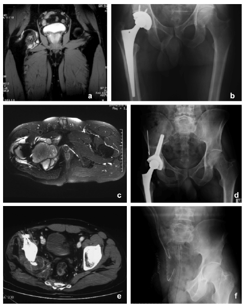
Figure 1: High grade osteosarcoma of the proximal femur a) Preoperative MRI in a 68-year-old patient presenting with pain in the right hip. Diagnosis
of aseptic necrosis of the hip was supposed. b) THA was implanted. c) 1 Month later, MRI showed a soft tissue mass around the prostheses. d)
Extra-articular resection was performed and reconstruction with allograft composite prosthesis. e) 3 Months later CT scan showed local recurrence. f)
Hindquarter amputation was performed to treat the local recurrence.
Methods of assessment
Diagnosis was confirmed at our Institution by CT-guided biopsy in 4
patients (acetabulum localization) and in 7 by review of histological slides.
Five patients were immediately treated with external hemipelvectomy and
the other six with limb salvage surgery. In the 7 patients with localization
in the proximal femur we performed 5 extra-articular resections and 2
amputations. We restored the bone defect by megaprosthesis in 3, by
allograft prosthesis composite in one (McMinn-Link prostheses was used
for the reconstruction) and by antibiotic spacer for THA infection in one.
In all cases the femoral resection was performed below the stem.
In acetabular localizations (4 cases) we performed an external
hemipelvectomy in 3 cases and a periacetabular resection and
reconstruction in one; massive bone allograft combined with stem cup
prosthesis was used. Margins were wide in 8 cases (73%), marginal in
2 and intralesional in one. All patients were staged and discussed in a
multidisciplinary setting and assessed by X-ray, MRI or CT scan of the
pelvis, chest CT and Bone Scan during preoperative staging. Follow-up
was conducted according to our protocols with chest CT scan and pelvic
MRI or CT scan with contrast medium and X-ray of the prosthesis every
3 months for the first 2 years, every 4 months for the third year and then
every 6 months.
Results
The reported results were recorded after a mean follow-up of 38
months and a minimum follow up of 6 months. Five of the 11 patients
(45%) died of disease (DOD) at a mean time of 27.4 months (range 6-40
months), 4 patients are alive with disease (AWD) at a mean time of 43
months (range 12-100 months) and only 2 patients are continuously free
of disease (CDF) with a follow up of 16 and 68 months. Among the five
patients who were immediately treated with external hemipelvectomy
only one died of disease (20%), two are CDF and the other two are AWD
(with lung metastases).
Six patients (55%) had local recurrence at a mean time of 15 months
(range 3-36 months) and of these five had had conservative treatment. In
the group of 6 patients treated conservatively, four died of disease (66.6%)
and two are alive with disease. Among the patients who died, 4 had a local
recurrence (80%). One patient treated with hemipelvectomy had a local
recurrence. The three patients treated with inadequate margins had local
recurrence.
Nine patients were free of metastases at diagnosis, 7 of these (78%)
developed lung metastases at a mean time of 14 months (range 2-36
months). The patients who developed local recurrence also developed
lung metastases, one of these also developed bone metastases. Two
patients treated conservatively developed also bone metastasis. All these
results are summarized in Table 1.
Discussion
Pelvic tumors are a big challenge for orthopedic oncologists. Wide
surgical margins are often difficult to obtain and intralesional or
debulking procedures have a higher rate of local recurrence [4] with a
worse prognosis. In our study we evaluated 11 patients who were treated
for standard THA implanted on an unknown tumor involving the
acetabulum or proximal femur. This error jeopardizes surgical treatment,
making local and systemic control of the tumor more difficult and thus
compromising the oncologic outcome. As seen in our study, 5 patients
(45%) died of disease at a mean time of 27.4 months, and only two are
CDF, in all these cases the surgeon failed to see that tumor was present.
In our cohort, chondrosarcoma was the most frequent diagnosis and it
is well known that surgical resection remains the main stay of treatment
for this tumor [5]. Primary goal of surgery was complete removal of
the tumor with limb function preservation whenever possible. The
extent of tissue contamination in these cases is very high because of the
acetabular and intramedullary reaming, and for the surgical approach
itself. Moreover, the presence of the implant makes evaluation of residual
disease or local recurrence difficult for inherent imaging artifacts. Adams
et al. [6] described a series of 8 patients treated at a tumor center after
an inadvertent internal fixation of a primary osseous sarcoma. Six of
these subsequently underwent amputation and only two limb salvage
procedures. Only four patients were alive at an average of 26.9 months.
They concluded that inadvertent surgery for high grade bone sarcomas
was associated with poor survival, in spite of the high rate of amputations.
Puri et al. [7] evaluated 14 patients with prior intervention, either as an
open biopsy or curettage/unplanned excision for a chondrosarcoma,
documenting a poorer event free survival (EFS) but a similar rate of local
recurrence. In this case though, tissue contamination is not the same as
for patients who undergo major surgery such as joint replacement. Some
of the reported survival rates [8,9] are comparable to primary treated
sarcomas, but this mainly depends on the nature of the tumor rather than
its surgical management.
Our study has the inherent limitation of being a retrospective
evaluation. The small number of patients prevents us from statistically
validating the observations. The decision for the type of surgical treatment
was not only based on the surgeon’s indications but also following the
patient’s preferences since some patients preferred conservative surgery
preserving the limb rather than an external hemipelvectomy.
Limb salvage after intralesional unplanned surgery is controversial.
According to Enneking [10] the definitive management of sarcoma
patients who underwent prior surgery involves removal of all tumor
and potentially contaminated tissue at the local site. Contamination
of surrounding tissues makes tumor resection with free margins
technically difficult. Contaminations after such surgery is comparable to
a pathological fracture, and for many authors, sarcomas accompanying
pathological fractures are considered to be a relative indication for
amputation [8]. Local recurrence rate in our study was 55% (6/11patients).
Five of these patients were treated with limb salvage surgery but only
one patient, treated immediately with external-hemipelvectomy, had a
local recurrence. Secondary amputation for local recurrence after limb
salvage procedures was performed in 4 of 6 patients. In these patients the
recurrence was very severe and re-excision was not feasible. Studies in
literature confirm that previous unplanned surgery is associated with a
higher rate of local recurrence [6-8,11,12]. Dae-Geun et al. [8] evaluated
25 patients treated with unplanned intralesional procedures, 22 had limb
salvage surgery and of these 4 had a local recurrence. Three cases had an
amputation with no local recurrence. They concluded that attempts for
limb salvage in malignancies treated intralesionally is accompanied with a
higher rate of local recurrence and extensive operative fixations represent
a relative contraindication to limb salvage procedures. This finding was
also confirmed in our study since patients treated with amputation had
less local recurrences than those treated with limb salvage.
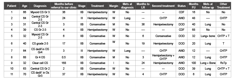
Table 1: Patient data, treatment and results at follow-up
T=Thoracotomy
W=Wide; M=Marginal; I=Intralesional; CHTP=Chemotherapy; CDF=Continuously
disease free; AWD=Alive with disease; DOD=Dead of disease; LR=Local recurrence; RxTp=Radiotherapy;
Nine patients were free of metastases at diagnosis, 7 of these (78%)
developed lung metastases at a mean time of 14 months (range 2-36
months). Patients who developed local recurrences also developed lung
metastases. Different authors sustain that improper manipulation of a
malignant tumor increases the risk of metastases [6]. Wang et al. [12]
reported 50% of lung metastases in patients who received unplanned
treatment for osteosarcma, this percentage is similar for patients with
appropriate treatment. This can be explained by the fact that this group of
patients received more aggressive chemotherapy. In our study the rate of
metastases was much higher because of the nature of the tumor. It is well
known that chondrosarcoma, the most frequent diagnosis in our series, is
resistant to chemotherapy, and this is why adequate surgical treatment is
mandatory for the oncological outcome.
Extensive surgery performed at the site of an unrecognized malignant
tumor compromises surgical resection by spreading the tumor in the
adjacent tissues. The initial diagnostic work-up is an important aspect to
avoid such mistakes. In 11 cases of our study the surgeon failed to recognize
that tumor was present, and only in one case the surgeon recognized
that there was a tumor present but it was probably misdiagnosed as a
benign lesion (case number 9: a clear cell chondrosarcoma [13] initially
diagnosed as chondromyxoid fibroma). Adequate attention to patient
history is fundamental for a correct diagnosis as can be seen with patient
number 8. Although the patient’s clinical history stated prior radiation
therapy for uterine cancer, the surgeon did not consider the possibility
of a pathological fracture from a radio induced sarcoma [14]. Another
fundamental step to avoid such errors is to perform a complete radio
graphic evaluation for patients in whom typical changes of osteoarthritis
are not present. These patients should be assessed with MRI or even CT
scan, and if there is still a doubt, a biopsy should be performed prior to
the hip reconstruction. A pathological examination of the femoral head
should always be performed or even a frozen section if an unexpected
mass is discovered during the surgery.
Conclusion
In this small series of patients it is not possible to give definitive
recommendations. The presence of any clinical or radiological sign
of pathologic bone should raise the clinical suspicion of a bone tumor.
In this case an accurate study (CT-scan; MRI or even a biopsy) should
be previously performed. Local recurrence after conservative surgery
is very high due to local contamination. Following to our experience,
independently to the method of treatment the prognosis in this group of
patients still remains poor.