Article Information
Aritcle Type: Case Report
Citation: Pai VD, Babu B, Ravindranath S, Manohar V
(2015) Sebaceous Gland Carcinoma of Upper Eyelid:
Resection and Reconstruction using Eyelid Switch and
Rotation Flap. Int J Cancer Res Mol Mech 1(4): doi
http://dx.doi.org/10.16966/2381-3318.119
Copyright: © 2015 Pai VD, et al. This is an
open-access article distributed under the terms
of the Creative Commons Attribution License,
which permits unrestricted use, distribution, and
reproduction in any medium, provided the original
author and source are credited.
Publication history:
Received date: 12 Dec 2015
Accepted date: 23
Dec 2015
Published date: 28 Dec 2015
Abstract
Sebaceous gland carcinomas are rare malignancies of the eyelid which offer significant challenge for the diagnosis and treatment. They
are frequently misdiagnosed in view of their rarity as well as resemblance to common benign pathologies like chalazion. Surgical resection
remains the mainstay of treatment. These are highly aggressive histologies with significant risk of local recurrence if not resected completely.
Reconstruction of the eyelids needs to be meticulous as protection of the cornea is of paramount importance. Although use of free flaps is
considered as a standard method of reconstruction, expertise and equipment required for the same are prohibitive for their adoption in majority
of the centers. We are reporting a case of sebaceous gland carcinoma of the upper eyelid which was resected followed by reconstruction using
lid switch of the lower eyelid and facial rotation flap as well as buccal mucosal graft.
Keywords
Sebaceous gland carcinoma; Eyelid tumors; Lid switch; Buccal mucosal graft
Introduction
Sebaceous gland carcinomas are rare malignancies of eyelid which
are challenging to diagnose and treat [1]. Significant proportion of these
tumors are misdiagnosed as they closely resemble benign pathologies
such as chalazion [2]. Hence frequently they present in advanced stage
with involvement of significant portion of the eyelid. Wide local excision
remains the mainstay of treatment although radical radiotherapy may be
used as a noninvasive treatment when surgery is not possible [3]. Surgical
resection should be wide enough to ensure complete resection with
adequate margins, at the same time not too wide to ensure a functional
eyelid. Reconstruction is the most challenging part of the management
because it is important to ensure a movable eyelid, good corneal
protection and acceptable aesthesis. We are presenting a case of sebaceous
gland carcinoma of the upper eyelid which was resected and resultant full
thickness defect was reconstructed with lid switch along with the facial
rotation flap.
Case Report
An 80 year old gentleman presented with an ulceroproliferative growth
over right upper eyelid of 6 months duration (Figure 1A). For long,
he was treated as chalazion by the local doctors before being referred to
our centre. Biopsy of the growth revealed it as sebaceous gland carcinoma
(Figure 1B). Metastatic work up revealed no evidence of regional or
distant metastasis. Clinical stage of the tumour was T3a N0 M0 [Stage II]
[4]. Tumor was resected with adequate margins all around (Figures 1C1
and 1C2). Tumor was 3 cm in diameter and closest margin was medial
margin. The resultant defect was involving full thickness of lateral 2/3rd
of the upper eyelid (Figure 1D). Lower eyelid was mobilized taking the
entire thickness and about 50% of the length of the defect of the upper
eyelid (Figure 2A). The defect in the palpebral conjunctiva over the lower
portion of the sclera was reconstructed with buccal mucosal graft (Figure
2B). Lateral canthus was recreated with facial rotation flap (Figure 2C).
Figure 2D shows the final reconstruction. Intra and post operative course
was uneventful and patient was discharged on post operative day 3. Three
weeks after the primary surgery (Figure 2E), flap division was performed
under local anesthesia (Figures 3A and 3B). Histopathological report
confirmed the pathology as high grade sebaceous gland carcinoma with
clear margins (Figures 3C and 3D). There was no pagetoid growth pattern
and there was no perineural invasion. At 6 weeks follow up, patient was
disease free with normal vision and no evidence of exposure keratitis.
Discussion
Sebaceous gland carcinoma is a high grade malignant neoplasm that
arises from the sebaceous glands. It accounts for 0.2-4.7% of the malignant
tumors of the eyelid [5]. However the incidence of sebaceous gland
carcinoma is much higher in Asian population compared to the west [6,7].
It most commonly affects upper eyelid [8]. Diagnosis is frequently delayed
not only because of its rarity but also because of the close resemblance with
the more common benign pathologies such as chalazion, conjunctivitis
and blepharitis. Surgical resection remains the gold standard treatment.
Ideally, tumors have to be excised with a 3–4 mm margin as well as
adequate depth in order to ensure long term recurrence free survival [9].
However, for tumors involving the conjunctiva, the safe margin is much
smaller than that for tumors involving the skin [10]. Role of adjuvant
radiotherapy has not been established although it is recommended for
tumors with aggressive histologic subtype, perineural invasion, or nodal
metastasis at presentation [11]. For patients who are unfit for surgery or
when the tumor is unresectable because of involvement of vital structures,
radical radiotherapy is a viable treatment option [3]. Prognosis is worse
compared to other malignancies of the eyelid with mortality and morbidity
only second to malignant melanoma [12].
In addition to protecting the eyeball, eyelids provide tear film
continuity and lacrimal pump. Eyelid defects can lead to corneal irritation,
exposure keratopathy and even loss of vision. Hence, the goals of eyelid
reconstruction are to ensure a movable eyelid and reproduce the texture of
the eyelid. The reconstruction of the eyelid is complicated by the fact that
it is composed of skin, mucosa, muscle tissue, and secretory glands unlike
other body parts. In general, partial thickness defects which are limited
to skin or conjunctiva can be reconstructed with skin grafts or local flaps.
Full-thickness defects that involve less than 25% of the eyelid’s width can
be closed primarily whereas the wider defects need more meticulous
reconstruction techniques [13]. Tissues similar to normal eyelid structure
have to be used in these reconstructions which involves both anterior and
posterior lamella. Improper reconstruction can result in significant long
term complications.

Figure 1: 1A-Patient profile showing tumor arising from the upper
eyelid; 1B-Biopsy showing high grade sebaceous gland carcinoma;
1C1 and 1C2-Resected specimen showing the tumor; 1D-Defect in the
eyelid after the resection of the tumor of the upper eyelid.
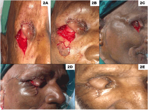
Figure 2: 2A-Defect in the upper eyelid reconstructed with the lower
eyelid switch; 2B-Buccal mucosal graft harvested to cover the defect
in the conjunctiva; 2C-Facial rotation flap planned; 2D-Completed
reconstruction with lid switch and facial rotation flap; 2E-Patient profile
3 weeks after the primary surgery.
Following techniques have been commonly used for the reconstruction
of anterior lamella: Z-plasty, the V-Y glabellar flap, median forehead
flap and the Cutler-Beard technique [14-16]. In addition, a number
of pedicled as well as free flaps have been used. Following autogenous
grafts have been used for reconstruction of the posterior lamella: labial
mucosa, hard palate mucoperiosteum, auricular and nasoseptal cartilage
[16,17]. However, each technique has its advantages and disadvantages.
In the present patient, anterior lamella was reconstructed using lid switch
with facial rotation flap whereas posterior lamella was reconstructed with
buccal mucosal graft. The choice was owing to its simplicity as well as
prior experience with using these reconstructive techniques.
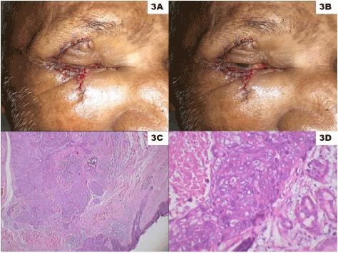
Figure 3: 3A-Results after the flap division with patient’s eye completely
closed; 3B-Results after the flap division showing the ability of the
patient to elevate the eyelid; 3C-Photomicrograph showing partially
circumscribed tumor with focally infiltrative margins, arranged in
islands, duct-like and cystic spaces. The tumor cells are polygonal to
squamoid, with high nucleus:cytoplasmic ratio (hematoxylin and eosin
stain [H&E], x40); 3D-Photomicrograph shows tumor in a duct pattern
along with central comedo necrosis in the larger duct. The cells show
pleomorphic vesicular nuclei, and overlapping with prominent nucleoli
(H&E, x400).
Conclusion
The purpose of presenting this case is to highlight the rarity of
the sebaceous gland carcinoma of the eyelid, the importance of early
diagnosis, and need for thorough understanding of the anatomy to ensure
proper reconstruction.
Download Provisional pdf here