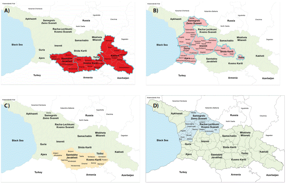
Figure 1: Risk hotspots used for FMDV serosurveillance sampling. (A) Zones with high risk villages, R1 risk hotspot. (B) Zones with low risk villages, R2 risk hotspot. (C) Migrating animal area, R3 risk hotspot. (D) Candidate Area, R4 risk hotspot.

Natela Toklikishvili1* Tamari Tchigitashvili2 Marina Turmanidze1 Tamar Tigilauri1 Eliso Mamisashvili1 Ketevan Goginashvili1 Marina Donduashvili1 Lela Kerdzevadze1 Vincenzo Lagani3
1State Laboratory of Agriculture, Tbilisi, Georgia*Corresponding author: Natela Toklikishvili, State Laboratory of Agriculture, Tbilisi, Georgia, E-mail: Natela.Toklikishvili@sla.gov.ge
Foot and Mouth Disease (FMD) is the most important economic threat to the livestock industry. Outbreaks of FMD can have a devastating impact on livestock production and trade, resulting in significant economic losses in the agricultural sector. As a result, vaccination and containment programs have been implemented internationally to minimize the spread of FMDV. The national vaccination program has been implemented in Georgia since 2012, vaccinating Large Ruminants (LR) and Small Ruminants (SR) with trivalent (A, O, Asia1) vaccine twice annually. However, active seromonitoring surveillance still shows a high seroprevalence of the disease, indicating virus circulation. In this study we attempted to estimate the prevalence of different FMDV serotypes in various risk zones within Georgia. A total of 4991 small and large ruminants were tested for the presence of FMDV nonstructural proteins (NSP) in the blood, and the exact serotypes of positive animals were further investigated through structural protein (SP) based assays. The results show that significant percentages (6.6%) of vaccinated animals were affected by FMD, and those positive animals are usually affected by more than one FMDV serotype. As such, our data call upon a stricter vaccination and monitoring program for FMDV in Georgia, especially considering that due to the geographic location of Georgia, the presence of FMD can have significant impact on transit and can be a threat for other countries as well.
Foot and Mouth Disease (FMD); Virus; Serosurveillance; Risk Zone; Georgia
Livestock production is an important economic source for countries with poor economies [1]. Animal diseases can have a significantly negative impact on the potential and security of the livestock sector, and this in turn can widely influence to nutrition, public health, stability and food availability. Foot and Mouth Disease (FMD) is the most important contagious transboundary animal disease. FMD has a great potential of causing large economic losses in case of disease outbreak because of reduced livestock productivity and trade [2,3].
FMD Virus (FMDV) has seven immunologically distinct serotypes: A, O, C, SAT1, SAT2, SAT3 and Asia 1 and is caused by picornavirus with a single-stranded ribonucleic acid (ssRNA) from genus Aphthovirus. The wide host range of FMD, its highly contagious nature and the ability of the virus to spread easily makes the disease of great importance [4], and all of this is coupled with the virus’ innate capability of easily reaching epidemic levels [3].
Furthermore, FMD is among the potential candidates for terrorist attacks, which are a big concern worldwide [5]. Disease can cause a systemic infection affecting wide range of wild and domestic animals with high mortality rate in young animals. It is endemic in many countries of Asia, the Middle East, Africa, and South America (World Organisation for Animal Health (OIE), 2019) [6].
Despite a thorough vaccination program, annual serosurveillance campaigns conducted by our institution (State Laboratory of Agriculture, Tbilisi, Georgia) have detected a relatively high seroprevalence rate of FMD in Georgia, especially in villages along the borders with Turkey, Armenia, and Azerbaijan (data not shown). This is a quite pressing issue, with a large share of Georgian GDP depending upon livestock export [7]. The last outbreak of FMD in Georgia occurred in 2002 in the Samtskhe-Javakheti region, and no clinical cases have been reported in recent years; however, if reintroduced, FMDV could easily spread throughout the country as animals are moved to live animal markets, or during seasonal migrations.
In this work we present the results of the FMDV sero-surveillance across Georgia. The purpose of the study was to determine FMD virus circulation in different risk zones for assessing FMD control measures along the estimation of virus circulation. Method used in our work is evaluation of antibody (Ab) level directed against Non-Structural Protein (NSP) of FMDV to indicate virus circulation. As NSP specific antibodies are expressed only during viral replication and are considered as a marker of infection; it is common for all 7 FMDV serotypes.
Furthermore, FMDV serotypes circulating in Georgia have not been surveyed for the last few years. Thus, we also investigated the seroprevalence of each serotype: type O, type A, and type Asia 1 among NSP-Ab positive animals, these three serotypes being the ones known to be present within the region. A detailed map of the seroprevalence of FMDV and its serotypes within Georgia will be instrumental in guiding the implementation of better practices for FMD control and eventually increasing economic productivity in animal export. Summarizing, our objectives are to: (A) Estimate the level of virus circulation; (B) Provide initial background information on the distribution of circulating FMD serotypes in different regions; and to (C) Perform analysis of the risk factors for FMD infection among various livestock sectors and geographical regions in Georgia.
The rest of the paper first presents the methods used, including sampling and testing procedures. Results follow, with discussion and conclusions at the end of the text.
During 2017, we performed an extensive serological surveillance campaign in Georgia for FMDV Non-Structural Proteins (NSP). Following extensive consultations with experts in the fields, we decided to stratify the sampling according to four risk zones (R1-R4 hotspots, Figure 1):
Figure 1: Risk hotspots used for FMDV serosurveillance sampling. (A) Zones with high risk villages, R1 risk hotspot. (B) Zones with low risk villages, R2 risk hotspot. (C) Migrating animal area, R3 risk hotspot. (D) Candidate Area, R4 risk hotspot.
• Villages with high (R1) and low (R2) risk in Georgia, excluding Candidate Area-LR are taken as indicator animals.
• Migrating animals in eastern Georgia (R3)-Only individually ear tagged SR animals are taken as indicators;
• Villages in Candidate Area (R4), where FMDV is not expected to be present-LR and SR are investigated.
A two-stage sampling design was adopted, consisting in first sampling villages and then identifying the number of animals to test within the villages.
First, a list of villages per regions and districts with approximate number of animals was created, reaching a total of 800000 animals. Following the results of a previous sero-survey carried in 2015-2016 (data not published), each Georgian village was assigned to either a “high” and “low” risk category. Particularly, the high-risk category comprised all villages with live animal markets in the S. Javakheti, K. Kartli, and Kakheti regions, as well any other village through which seasonal migration routes are lead. The low risk category comprises all other villages. The expected FMDV prevalence in the high and low risk villages was set to 15% and 10%, respectively.
Villages with less than 50 animals were excluded, resulting in 788 villages in the high risk and 1642 villages in the low risk category selected for the sero-survey. The final number of villages to sample for each category was computed using the “Estimate Percentage” function of the WinEpi online tool, www.winepi.net [8], using a 95% confidence level and a 6% accepted error. This led to selecting 117 villages for the high and 91 villages for the low risk category.
The “Detection of Disease” function of the WinEpi online tool was used to compute the number of animals to sample in each village. Setting a 95% confidence level, a population size (N) of 800000 and a 20% expected prevalence resulted in 14 samples to be tested in each village.
Thus, the total size per each risk category is 1638 samples (117 * 14) for the high risk and 1274 samples (91 * 14) for the low risk category. Both villages within each risk category and animals within each village were selected randomly.
Migration animals are considered as a different risk category. The majority of migrated animals are SR, and therefore only SR animals were included in this sampling. A two-stage sampling design was used, in which first the number of herds and then the number of samples inside each herd were identified.
The sample frame consisted of all migrating SR herds in eastern Georgia. A list of herds (assigned to villages) with an approximate number of animals was created, topping at a total of 500000SR. Herds with fewer than 50 SR were excluded from the survey for sake of statistical robustness. This led to a total of 170 herds.
The number of herds was identified using the “Sample size/estimate percentage” function of WinEpi, adopting a 95% confidence level, an expected prevalence of 11% and an acceptable error of 6%, resulting in 65 herds to be tested. The “Detection of disease” function of WinEpi was used for computing the number of animals to test within each herd. Setting a 95% confidence level, an expected prevalence of 25% and a population size of 500000 (N) resulted in 11 samples for each herd. Thus, the total sample was set to be 715 (65 * 11) animals. Both herds and animals with each herd were selected randomly.
The Racha, Lechkhum-Kvemo, Svaneti, and Mestia regions are considered as Candidate Areas of Progressive Control Pathway stage 3 [http://www.fao.org/ag/againfo/commissions/docs/pcp/pcp26012011.pdf], i.e., zones where aggressive strategies to eradicate FMD are not needed anymore.
Also in this case a two-stage sampling design was used, by first identifying the number of villages and then the number of samples within each village to be tested. A list of villages per districts with an approximate number of animals was created, with 26170 LR and 1786 SR in total. Villages with less than 50 animals were excluded from the survey to ensure more reliable statistics. The WinEpi “Sample size” function was used to compute the final number of villages to include. This resulted in 98 villages, by setting a 95% confidence level, a 5% acceptable error, an expected prevalence of 16% and an initial number of villages of 184. Similarly, the WinEpi “Detection of disease” function determined that 14 samples should have selected from each village, by considering a 95% confidence level, a 21000 total population size and an expected prevalence of 20%. Thus, the total sample size was set to 1372 animals (98 * 14). Villages and animals within villages were selected randomly, with LR and SR tested according to their proportion in each area/village/farm.
As the most important economic threat to the livestock industry, reliable tests against FMDV are needed for granting disease-free status for animals and ensuring safe international trade. According to OIE, Virus neutralization test (VNT) is the international standard for antibody detection, however nowadays Enzyme-Linked Immunosorbent Assays (ELISA) are available and they can be performed in Bio Safety Level (BSL) 2 laboratories.
We used the ID.vet competitive ELISA for the detection of specific antibodies against FMDV 3ABC non-structural protein (NSP) in serum and plasma of all susceptible species. In this test ELISA plates are coated with the FMDV NSP to detect NSP specific antibodies. We followed the producer instructions in all our applications: 50 µl of the dilution buffer was distributed to each well, along with 30 µl of positive control, negative control and test samples. Plates were incubated for 2 hours at 37°C; after incubation content of the wells were emptied and washed 5 times with 300 µl of wash solution. Next step was dispensing of 100 µl conjugate to each well followed by incubation (30 min, at 21°C). Subsequently, repeating the washing step 100 µl of the substrate solution was dispensed and incubated (15 min, at 21°C). In order to stop the reaction 100 µl of stop solution was added and results were read at 450 nm.
If anti-NSP antibodies are present in the sample they form an antigen-antibody complex which masks the virus epitopes. Dispensed anti-NSP horseradish peroxidase (HRP) conjugate fixes to the remaining free epitopes, forming an antigen-conjugate-HRP-complex. Washing step removes all the excess conjugate and after distribution of the substrate solution (TMB) color development appears. The intensity of the color development is in an inverse correlation with the quantity of NSP specific antibodies in the test sample. In the absence of antibodies, a blue solution appears which becomes yellow after addition of the stop solution. In the presence of antibodies, there is no coloration observed.
The test is valid if the mean value of the negative control OD (ODnc) is greater than 0.7 (ODnc> 0.700), the mean value of the positive control OD (ODpc) is less than 30 % of the ODnc (ODpc/ ODnc< 0.3). Results interpretation was made by computing the competition percentage (S/N %), which is calculated for each sample with the formula:
S/N%=OD sample / ODnc × 100
Samples presenting competition percentage less than or equal to 50 % are considered positive, and negative otherwise.
The Priocheck FMDV type specific blocking ELISA is a commercially available assay for in vitro detection of antibodies against FMD virus serotypes of type Asia 1, A and O in serum of cloven-hoofed animals. The test has demonstrated excellent reliability and was evaluated by the FMD world reference laboratory. Microtiter plates are coated with non-infectious FMD virus type O, A or Asia 1 antigen separately. The reaction between specific monoclonal antibody and FMDV type specific antigen is blocked by specific antibodies presenting in the tested serum sample.
For laboratory procedures we followed the manufacturer’s instructions. We dispense 80 µl of ELISA buffer, 20 serum samples, negative control, weak positive control and positive control (for type O-reference sera 1 to 4) to the appropriate wells. The plates were incubated for 60 minutes at room temperature (22°C), and then washed 6 times with 300 µl washing solution. 100 µl of the AbHRPO conjugate was added and incubated 60 min, at 22°C. The plate was washed again 6 times with 300 µl washing solution and 100 µl of chromogen (TMB) substrate was added to all wells. Followed by incubation step (15 min, at 22°C). Color development was stopped by adding 100 µl of stop solution. The optical density (OD) was measured at 450 nm.
Results calculation and interpretation for Types A and Asia 1:
Mean value of negative controls=OD450 max
The Percentage Inhibition (PI) of the weak positive control, positive control and the test sera was calculated as:
PI=100-(OD450 test sample/OD450 max) × 100
Samples with PI ≥ 50% are considered as positives; Samples with PI <50% are considered as negatives.
Test is valid if OD450 max is at least 1.000; the average PI of the weak positive controls is>50%; the mean PI of the positive controls is >70% for type A, and >75% for type Asia 1.
For Type O, first all OD values were corrected by subtracting the average of the reference sera 1. The average of the corrected OD values for reference sera 4 was then computed (corrected OD450 max). The PI values were finally calculated as
PI=100-(corrected OD test sample / corrected OD450 max) × 100
Samples were considered positive for PI ≥ 50%, negative for PI < 50%.
Test is valid if corrected OD450 max is at least 1.000; the average PI of the reference serum 2>60%; the average PI of the reference serum 3<40%
Solid-phase competitive ELISA (SPCE) is an easy and fast serological assay. It is provided as a ready-to use test, suited for large-scale surveys, can be adopted in BSL-2 laboratories; SPCE ELISA test is serotype specific and can detects the presence of antibodies in serum or plasma samples of FMDV infected animals of any susceptible species.
Test microplates are pre-coated with the FMDV inactivated antigen (type O/type A/type Asia 1) captured by the homologous monoclonal antibody (MAb). Diluted samples (1/10, 1/30, 1/90, 1/270) are incubated for 60 min at room temperature, enabling the specific antibodies eventually present in the sample to bind to the respective antigen. After 25 µl anti-FMDV type O (A or Asia 1) MAb, conjugated with peroxidase, is distributed directly without washing step, its binding with the homologous antigen will be inhibited by antibodies of positive sera previously bound to the virus, while in case of negative sera the conjugated MAb can react with the FMDV inactivated antigen. Wells are emptied and washed 4 times with 200 µl. The excess conjugate is removed by washing and 50 µl of the substrate solution is dispensed into all wells (incubated at room temperature, for 20 minutes). A colorimetric reaction develops if the conjugated MAb has bound to the virus, i.e. if test serum is negative, while color development is inhibited by positive sera. Reaction is stopped by addition of a 50 µl/well stop solution, after blocking we read the optical density (OD) of each well at 450 nm.
The percentage inhibition (PI) of the positive control and test sera is calculated as follows:
% inhibition (PI)=100-(serum OD/reference OD *) × 100
* Reference OD=mean OD of four wells of the Negative Control
The validity of the testing procedure is assessed trough precise criteria:
• OD values in the negative control wells must be ≥ 1.0for type O and Asia 1, while they are expected to be ≥ 0.8 for type A
• The positive control are expected to give ≥ 90% inhibition at 1/10 dilution and >50% inhibition at the second dilution (1/30) for all three serotypes.
Result interpretations are considered as follows: samples are positive when producing an inhibition ≥ 70% at the 1/10 dilution, negative otherwise. The second dilution (1/30) assesses the level of antibodies: a strongly positive serum is ≥80% inhibition at both 1/10 and 1/30 dilutions, while sera showing ≥ 80% inhibition at the 1/10 dilution but ≤ 50% inhibition at the 1/30 dilutions are usually considered to be low positive. The end-point titre of a positive serum corresponds to the highest dilution producing 50% inhibition.
We made interpretations of the results for each test separately according to assay interpretation criteria. Confidence intervals for proportions were computed using a normal approximation [9] as implemented in the epiR R package. A χ2 test with Holm correction was used for comparing different proportions, as implemented in the R function pairwise. prop.test [10]. All statistical analyses were performed with the R Statistical Software, Geographical Information System (GIS) mapping was performed with ArcGIS version10.1.
Samples of large and small ruminants were collected from 4991 large and small ruminants for detection of NSP prevalence. Only 4-18 months old animals were collected, and animals older than this suitable age range were excluded from the analysis. Table 1 reports the details of the samples and data collection. A total of 4991 animals were sampled across 371 villages. This number includes 715 small ruminants from villages interested by migration routes, while the rest of the animals were ruminants from high/low risks zones and from villages in the Candidate Area.
| Risk hotspot | Expected village/ herds prevalence | Number of villages | Expected individual prevalence | Target Number of samples | Total number of samples | |
| Villages in Georgia excluding Candidate Area | High Risk categories | 15 | 117 | 20 | 14 | 1737 |
| Low Risk categories | 10 | 91 | 1191 | |||
| Migrating animals (SR) in Eastern Georgia | 11 | 65 | 25 | 11 | 715 | |
| Villages in Candidate Area | 16 | 98 | 20 | 14 | 1348 | |
| Total | 371 | 4991 | ||||
Table 1: Details of the serosurveillance data collection. For each risk hotspot the following information is reported, left to right: the expected prevalence at village level, the number of sampled villages, the expected prevalence at the individual animal level, the target number of animals to sample at each village, and the final number of sampled animals.
Out of the 4991 sampled animals, 327 were positive (6.6%). Particularly, 1737 animals were collected in high risk zones with 191 positive samples (11%), 1191 in low risk zones with 89 positive samples (7%), 715 samples of small ruminants were collected in migrating animals’ zones with 18 positive samples (3%), and 1348 samples in Candidate Areas with 29 positive samples (2%). Table 2 reports detailed results for each risk hotspot.
| Risk hotspot | Tested | Positive | Estimate% | Lower% | Upper% |
| Risk 1- High risk zone LR | 1737 | 191 | 11 | 9.6 | 12.5 |
| Risk 2- Low risk zone LR | 1191 | 89 | 7.4 | 6.1 | 9.1 |
| Risk 3- Migrating animals SR | 715 | 18 | 2.5 | 1.6 | 3.9 |
| Risk 4- Candidate Area LR/SR | 1348 | 29 | 2.1 | 1.5 | 3.1 |
| Total | 4991 | 327 | 6.6 | 5.9 | 7.3 |
Table 2: Results of seroprevalence testing: For each risk hotspot the numbers of tested and positive animals are reported, along with the estimated proportion and its 95% confidence interval.
As expected, the prevalence of positive animals is higher in the highrisk villages (Risk 1hotspot zone) than in any other zone (adjusted p-value < 0.01 in all comparisons). Also, no statistically significant difference in prevalence was detected between areas interested by migrating animals and villages in the Candidate Area (Risk 4 hotspot zones, p-value: 0.707).
Out of all animals that have emerged as positive from the NSP testing, it was decided to randomly select 140 samples for further investigating the distribution of each FMDV serotype (types O, A and Asia 1). Figure 2 shows that these animals were drafted from regions belonging to all risk hotspots except for the Candidate Area. All selected animals underwent both the Solid-phase competitive ELISA (SPCE) test and the Priocheck FMDV type specific blocking ELISA. Figure 3 reports the number of animals positive to each serotype according to the two tests. According to the Priocheck test, 81, 92 and 90 animals were positive for serotype A, Asia 1 and O, respectively. In contrast, the SPCE test detected 125, 113 and 128 positive animals for the same serotypes. Most of the samples resulted positive to more than one serotypes, thus the total does not sum up to 140. Also, the Priocheck test appears to be much more sensitive than the SPCE one.
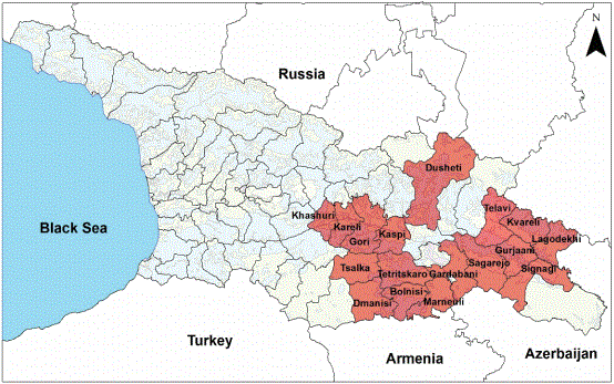
Figure 2: Georgian regions from which 140 FMDV positive samples were randomly selected for assessing the prevalence of each serotype.
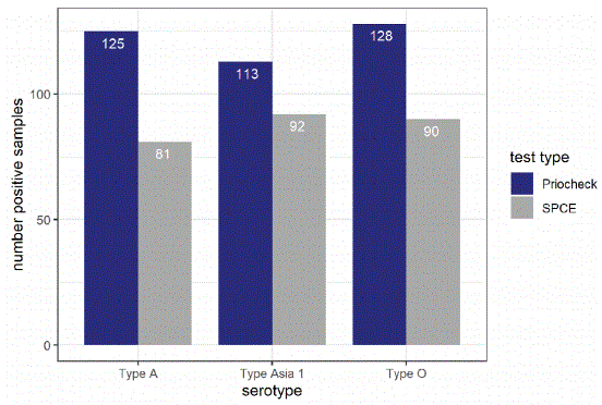
Figure 3: Results of the Structural Protein (SP) screening. For each tested serotype (O, A, Asia 1) the number of positive samples out of 140 is reported, both for the SPCE and Priocheck test.
Figure 4 reports the number of animals infected for each serotype and combination of. The majority of the animals (113 samples) were positive for all three serotypes; 10 were infected by serotypes A and O but not Asia 1; and 5 and 2 animals were positive exclusively for serotypes O and A, respectively.
Figure 4: Co-infection across different FMDV serotypes (according to PRIOCHECK test). Main bar plot (grey bars): for each serotype combination the total number of infected animals is reported. Side bar plot (blue bars): the number of positive animals for each single serotype.
In this work we investigated the seroprevalence of FMDV and three of its serotypes in Georgia. Control of FMD in Georgia has been motivated by the exportation of sheeps to Arabian countries, which is an important source of economic revenue in Georgia. There are a number of procedures for immune prevention as well as several control strategies for FMD in Georgia. First, in the summer of 2013, Georgia became a member country of the European Commission for the Control of Foot-and-Mouth Disease (EuFMD). Nowadays FMDV control plan includes FMD active surveillance, cross-border activities, use of electronic systems for animal health surveillance as well as efforts for animal identification and registration, and monitoring of migrating animals. FMD control plan includes a vaccination campaign to take protective measures against FMD. Vaccination is implemented with trivalent (A, O, Asia1) vaccine, covering the whole population of LR & SR twice annually. However, ensuring the optimal execution of all these procedures is a challenging task, and any deviation provides an edge for the spreading of the disease.
Regarding the surveillance activities performed in this study, it should be remarked that both antigen (Ag) and antibodies detection tests are available for the identification of FMD virus serotypes. Virus and its products are detectable only for relatively short period of time, in contrast to antibodies that persist for years in animals, pointing out current or past infections. Therefore, we elected antibody estimation as the preferred method for our surveillance. Particularly, we preferred Enzyme-Linked Immune Sorbent Assay (ELISA), as the ELISA test is easier, faster, suited for large scale experiments, it is performed with inactivated virus and therefore can be applied in bio safety level 2 labs (World Organisation for Animal Health (OIE), 2019) [6].
Our results underline several interesting phenomena related to the seroprevalence of FMDV in Georgia. First, despite the lack of clinical cases in recent years, seroprevalence is still relevant, with 6.6% of 4991 tested animals being FMDV positive. Zones considered at high risk do effectively show a seroprevalence (11.0%) that is statistically significantly higher than the prevalence measured in all other hotspots. At the same time, Candidate Areas, which are expected to have no or minimal FMDV presence, had the lowest seroprevalence (2.1%). Finally, all risk hotspots have a much lower prevalence that what the experts determined as expectable (reported in Table 1).
When it comes to the detection of the specific serotypes, we note that the PrioCheck test has a higher sensitivity than the SPCE test (Figure 1). Most importantly, the majority of the tested animals were positive for all of the three serotypes, indicating a very high degree of co-infection.
There are number of reasons that can explain the results of our study:
1. Annual outbreaks of disease in neighbour countries: Turkey, Armenia and Russian Federation are one of the risk factors for spreading disease in Georgia [11]. Considering the highly contagious nature of the pathogen and due to uncontrollable movement of livestock between country borders, if outbreaks take place in nearby territories the infection can easily spread across the borders. Furthermore, in conflict zones there are uncontrolled territories with animal movements as well. All these issues can explain high seroprevalence of the disease especially in villages bordering with Turkey, Armenia, and Azerbaijan.
2. Migration of animals is a very typical characteristic of Georgian farming. Animals migrate between winter and summer pastures. Free migration of animals in seasonal pastures, as well as diversity of animal maintenance (animals are not kept in closed farms) largely increases the disease spread. When migrating animals have contact with the local animal population, the risk of FMD spread is increased.
3. A further risk factor connected to animal migrations is the presence of large open, live markets for both LR and SR, where the infection can be easily transmitted between animals from different areas.
4. The vaccination campaign is a high cost and resource intensive program covered by state budget. Due to the high costs, not all animals receive appropriate booster vaccines. There is no cross-protection between the different FMDV serotypes. One more reason for high seroprevalence of FMD could be a limited vaccine effectiveness. According to governmental regulations, the choice of the vaccine is blind and largely depends on the product price, a factor that may somewhat lead to acquiring sub-optimal preparations.
The results of this study will be instrumental for future research, since information about serotype prevalence in Georgia is still a gap in the field. Particularly, the choice of vaccine in Georgia was not based on any relevant research or FMDV serotype vaccine matching, and the category of the vaccine can largely affect the efficacy of the vaccination programs. Future research should focus on a better characterization of FMDV serotypes circulating in Georgia, by using antigen detection by PCR, and virus isolation and sequencing. Antigenic matching tests are necessary to find out the proper vaccine strain. Particularly, we are considering toper form a small-scale trial for vaccine matching and for evaluation of post vaccination antibody levels within a 6 month period. This experiment would provide us with information about proper vaccine and post-vaccination immunity, as the effectiveness of vaccination campaigns largely depends on the quality and suitability of the chosen vaccine [12].
In conclusion, in this work we determined FMDV seroprevalence across different risk zones in Georgia. In particular we evaluate antibody level against non-structural protein of FMDV as a marker of infection. Three FMDV serotypes, namely A, O and Asia 1, were further investigated, being the ones known to be present within the region.
Our results contribute to the understanding of FMDV serotypes circulating in Georgia and will be useful for guiding the implementation of better practices for FMD control. A significant proportion of the Georgian Gross Domestic Product (GDP) is based on the livestock trade, and controlling FMD circulation and spread will directly affect Georgia’s economic status. Future research should focus on implementing additional laboratory diagnostic methods that will improve the quality of laboratory testing, but, more importantly, help guiding policy decisions on the FMD vaccination program, and measures to better control FMDV in Georgia.
This grant is awarded as a result of Broad Agency Announcement (BAA) HDTRA1-14-24-FRCWMD-BAA, Research and Development Enterprise, Basic and Applied Sciences Directorate, Basic Research for Combating Weapons of Mass Destruction (C-WMD), Defense Threat Reduction Agency (DTRA). We thank the National Food Agency of Georgia for their collaboration.
Download Provisional PDF Here
Article Type: RESEARCH ARTICLE
Citation: Toklikishvili N, Tchigitashvili T, Turmanidze M, Tigilauri T, Mamisashvili E, et al. (2020) Comparison Differentiation of Foot and Mouth Disease Virus Serotypes in Animals in High-Risk Zones of Georgia. J Anim Sci Res 5(1): dx.doi.org/10.16966/2576-6457.148
Copyright: © 2020 Toklikishvili N, et al. This is an open-access article distributed under the terms of the Creative Commons Attribution License, which permits unrestricted use, distribution, and reproduction in any medium, provided the original author and source are credited.
Publication history:
All Sci Forschen Journals are Open Access