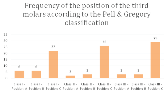
Graph 1: Frequency of the position of the third molars according to the Pell & Gregory classification.

Carlos Campos Arenas1* Sergio Campos Olivera2 Delia Olinda Huapaya3
1Dental Surgeon, Master in Stomatology, Faculty of Dentistry, National University of San Marcos, Lima, Peru*Corresponding author: Carlos Campos Arenas, Dental Surgeon, Master in Stomatology, Faculty of Dentistry, National University of San Marcos, Lima, Peru, E-mail: carlos-ky@hotmail.com
Objective: To determine the position, degree of development and level of difficulty for impacted third molar surgery in patients aged 12 to 17 years treated at the National Institute of Children’s Health.
Methodology: The sample consisted of 50 patients evaluated radiographically, who required extraction of lower third molars in the Oral Maxillofacial Surgery service, in total a total of 100 lower third molars were evaluated using the Pell & Gregory classification to evaluate their position, the Nolla stages to evaluate its development and the Pederson index to evaluate the level of difficulty for the extraction of the third molar.
Results: The age of the sample had a mean of 14.7 ± 1.53 years of age. According to the Pell & Gregory classification, there was a higher frequency of the position of the third molars in classes III with depth C with 29%, followed by class II depth C with 26%. According to the Nolla Index, the third molar development had a higher percentage in the complete Crown stage (stage 6) with 31%. And, according to the degree of difficulty in extracting the third molars, a higher percentage was observed in the moderate degree of difficulty with 59%, followed by a very difficult degree with 37%.
Conclusions: It was concluded that there is a greater frequency of position with depth C and class III of the impacted third molars, and also with a Nolla 6 stage. The level of difficulty for third molar extraction was mostly moderate, having a direct relationship with the distant position in which the third molars are located in young patients.
Third molar; Impacted tooth; Mandible; Surgical difficulty
Third molars are the most commonly extracted teeth in the practice of oral and maxillofacial surgery [1]. The difficulty of lower molar eruption is due to its late development and the phylogenetic changes that the mandible has undergone, as a result there is a lack of space for adequate eruption [2]. The intimate relationship with nearby structures such as the second molar, the inferior dental neurovascular bundle, the anterior border of the ascending ramus, the mobile lax mucosa, and the external and internal cortices can generate an alteration in the eruption of the bud [3].
The incidence of impaction of the third molars varies from 16.7% to 68.6%, without necessarily presenting any type of predilection for the gender [4]. These impacted pieces usually present pathologies such as pericoronitis, periodontitis, cystic lesions, benign tumors, root resorption and damage to the structure of the adjacent piece [5].
Although there are established indications of the removal of third molars, there is still controversy about the extraction of asymptomatic third molars [6]. Most studies indirectly report age, according to their scope, 66% are drawn between 30 and 40 years of age [7]. Some authors affirm that the early extraction of third molars is indicated as a beneficial therapy for the patient, because it will avoid complications of an infectious or cystic nature [8]. It has been reported that there is a slower recovery after third molar extraction in patients older than 24 years, along with a greater impact on their life style and oral function. Therefore, although decisions about the timing of third molar extraction are complex, the risk of morbidity associated with impaction and extraction at an older age should be taken into account [9].
It is mandatory to always carry out an accurate radiological study that shows the impacted third molar in its entirety and the structures that surround it. With a correct radiographic interpretation, the local factors that influence the complexity of the extraction can be predicted [10].
To determine the position of the impacted third molar, the Pell & Gregory classification is usually used, who classifies it according to its level of depth in relation to the occlusal surface of the second molar vertically and the relationship of the impaction with the body of the horizontally ramus [11]. As for the angulation, the classification of the Winter [12] system is used.
Taking into account the anatomical considerations and the position of the piece, better post-operative results will be obtained. For this reason, various factors have been classified and index created that help us predict the difficulty of this surgical act [13]. Pederson proposed an index of difficulty for the extraction of impacted mandibular third molars. The total scores by which difficulty is judged are mainly based on local anatomy and radiographs [14,15] (Table 1).
| Classification | Value |
| Spatial Relationship | |
| Mesionangular | 1 |
| Horizontal/Transverse | 2 |
| Vertical | 3 |
| Distoangular | 4 |
| Depth | |
| Level A: High Occlusal Level | 1 |
| Level B: Middle Occlusal Level | 2 |
| Level C: Deep Occlusal Level | 3 |
| Ramus Relationship/Space Available | |
| Class 1: Sufficient space | 1 |
| Class 2: Confined Space | 2 |
| Class 3: No space | 3 |
| Difficulty Index | |
| Very difficult | 7-10 |
| Moderately difficult | 5-6 |
| Slightly difficult | 3-4 |
Table 1: Index of difficulty for the extraction of impacted mandibular third molars, as described by Pederson [15].
Early third molar surgery requires special considerations as mentioned above. The National Children’s Institute treats patients who have not yet reached the age of majority and who frequently require this type of treatment. Therefore, this research work aims to determine the position, the degree of development and the level of difficulty for impacted third molar surgery in patients aged 12 to 17 years treated at the National Institute of Child Health.
A descriptive, cross-sectional and prospective study was carried out. A review of the medical records and panoramic radiographs of the patients treated at the Bucco-Maxillofacial Surgery Service of the Pediatric Dentistry Department of the Children’s Health Institute between 2005 and 2009 was carried out. As inclusion criteria, an age range of 12 to 17 years old, patients without systemic diseases and that the panoramic radiograph is clear enough for a correct identification of the impacted third molar. As exclusion criteria, all the histories that were not filled out correctly and the panoramic radiographs that presented some distortion or stain were discarded. The sample consisted of a total of 50 patients evaluated radiographically, who required extraction of lower third molars in the Oral Maxillofacial Surgery service, evaluating a total of 100 lower third molars.
The Pell & Gregory classification was used to record the position of the third molar. The Nolla [16] stages to establish the development of the piece and finally Peterson’s difficulty index to establish the complexity for the extraction of said piece.
The information was entered in the SPSS 17 program and presented in absolute frequencies and percentages.
It was obtained as results that the total sample 40% (n=20) was male and 60% (n=30) female. The studied age of the sample was between 12 and 17 years old, having an average of 14.7 ± 1.53.
According to the Pell & Gregory classification, it was found that, when evaluating the depth of the third molar with respect to the occlusal plane of the second molar, there was a higher frequency for depth C (77%), while, in relation to the existing space with the mandibular branch, the classes had similar frequency with a slight increase for class III (35%). In relation to the final position, there was a higher frequency in classes III with depth C with 29%, followed by class II depth C with 26%, and class I depth C with 22% (Table 2 and Graph 1).
Graph 1: Frequency of the position of the third molars according to the Pell & Gregory classification.
| Class | Frequency | |
| Class I | 34 | |
| Class II | 31 | |
| Class III | 35 | |
| Total | 100 | |
| Depth | Frequency | |
| Position A | 12 | |
| Position B | 11 | |
| Position C | 77 | |
| Total | 100 | |
| Position | Frenquency | Percentage (%) |
| Class I-Position A | 6 | 6 |
| Class I-Position B | 6 | 6 |
| Class I-Position C | 22 | 22 |
| Class II-Position A | 2 | 2 |
| Class II-Position B | 3 | 3 |
| Class II-Position C | 26 | 26 |
| Class III-Position A | 3 | 3 |
| Class III-Position B | 3 | 3 |
| Class III-Position C | 29 | 29 |
| Total | 100 | 100 |
Table 2: Frequency of the position of the third molars according to the Pell & Gregory classification.
According to the Nolla’s Index, the third molar development had a higher percentage in the complete crown stage (stage 6) with 31%, followed by a root third (stage 7) with 28% and finally the root twothirds stage with 15% (Table 3 and Graph 2).
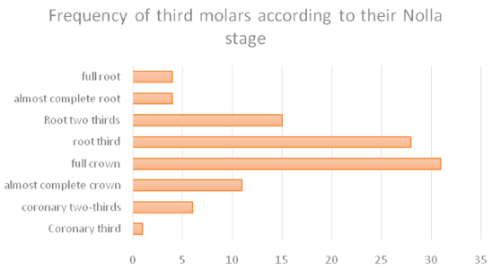
Graph 2: Frequency of third molars according to their Nolla stage.
| Nolla Stadiums | Frenquency | Percentage(%) |
| Coronary third | 1 | 1 |
| Coronary two-thirds | 6 | 6 |
| Almost complete crown | 11 | 11 |
| Full Crown | 31 | 31 |
| Root third | 28 | 28 |
| Root two thirds | 15 | 15 |
| Almost complete root | 4 | 4 |
| Full root | 4 | 4 |
| Total | 100 | 100 |
Table 3: Frequency of third molars according to their Nolla stage.
According to the degree of difficulty in extracting the third molars, a higher percentage was observed in the moderate degree of difficulty with 59%, followed by the very difficult degree with 37% (see Table 4 and Graph 3).
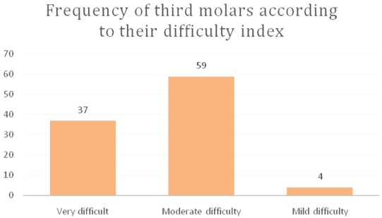
Graph 3: Frequency of third molars according to their difficulty index.
| Difficulty level | Frenquency | Percentage(%) |
| Very difficult | 37 | 37 |
| Moderate difficulty | 59 | 59 |
| Mild difficulty | 4 | 4 |
| Total | 100 | 100 |
Table 4: Frequency of third molars according to their difficulty index
The population evaluated in this study was quite young, being a pediatric institution, compared to most studies that report surgeries of symptomatic third molars after two decades of age, hence the need to establish the stage of development of the third molar as part of the characterization of the piece before the surgical procedure.
Studies on the prevalence of impaction of third molars with large samples, such as the one carried out by Hashemipour M, et al. [4] and Dias-Ribeiro E, et al. [17] had an age range of 20 to 40 years, while da Silva MC, et al. [18] and Mosquera-Valencia Y, et al. [19] had a sample between 17 and 28 years of age, and the study carried out by Kautto A, et al. [7] evaluated various age groups with a mean age of the sample of 36.4 years, a few other studies with a smaller sample such as that of Marzola C, et al. [20] and Garcia RR, et al. [21] presented age groups between 15 and 20 years of age. The study carried out by Chaparro AVC, et al. [8] was carried out with patients between 12 and 18 years of age and found that the most frequent Nolla stage was stage 7, our study took almost the same age range, however, it presented a slightly higher incidence in stage 6 followed by Nolla 7.
According to the position of the third molar based on the Pell & Gregory classification, Hashemipour M, et al. [4], Dias-Ribeiro E, et al. [17], Gatti PC, et al. [22] obtained that the highest frequency of lower third molar impaction was in position AII (43.8%, 43.4% and 28% respectively). Burgos G, et al. [3] found a higher frequency in position B II (64%), while Alfadil L, et al. [23], on the contrary, found a higher percentage in depth C (57.3%) and class I (66 .7%) in relation to the space to the mandibular ramus. As mentioned before, the first mentioned investigations had populations with higher age ranges, which implies a more advanced third molar development with the consequence of a greater approach to the occlusal plane (depth A or B), the present research work took a younger population, so it is natural that the impacted third molar is more distant (depth C). The research work of Chaparro AVC, et al. [8] worked with a population similar to ours, they coincided in presenting a higher frequency in depth C (46,7%) of the third molars, however, they found a higher frequency in class II (56.7%) while we found a slight increase in class III (35%) followed by class I (34%).
Regarding the level of difficulty for third molar surgery using Pederson’s classification; Zhang X, et al. [24] found a moderately difficult level of difficulty (37.4%) followed by slightly difficult (36.9%) more frequently, de Baranda BS, et al. [25] found a slightly difficult level of difficulty (45 .8%) followed by moderately difficult (41.5%). Unlike them, Gbotolorun OM, et al. [26] found the difficulty level very difficult more frequently (50.6%). In our study, a higher frequency of moderate difficulty level (59%) was found, similar to what was found by Zhang X, et al. [24] but not on other research works. This may be due to the degree of depth found in developing third molars in young patients, since a greater depth (depth C) implies greater difficulty for third molar extraction according to the classification of Pederson and other authors [3,8,15].
Although this classification has been widely used, it has now been shown that there are other factors than the position of the third molars to establish the level of difficulty for third molar extraction. Other researchers [20,26-29] where they take into account other local factors such as the shape of the roots, the number of roots, buccolingual position, bone density, proximity to the lower dental canal, elevation point, periodontal ligament space and other systemic patient factors such as patient age and body mass index. Sánchez-Torres A, et al. [30] carried out a systematic review including some of the new factors mentioned and incorporating some more such as the experience of the surgeon and the use of a complex surgical technique.
It was concluded that there is a greater frequency of position with depth C and class III of the impacted third molars, and also with a Nolla 6 stage. The level of difficulty for third molar extraction was mostly moderate, having a direct relationship with the distant position in which the third molars are located in young patients.
Download Provisional PDF Here
Article Type: RESEARCH ARTICLE
Citation: Arenas CC, Olivera SC, Huapaya DO (2022) Determination of the Position, Development and Level of Difficulty for Third Molar Surgery in Patients Aged 12 to 17 Years Attended in the Buccomaxillofacial Surgery Service of the National Institute of Child Health, LimaPeru. J Surg Open Access 8(1): dx.doi.org/10.16966/2470-0991.257
Copyright: © 2022 Arenas CC, et al. This is an open-access article distributed under the terms of the Creative Commons Attribution License, which permits unrestricted use, distribution, and reproduction in any medium, provided the original author and source are credited.
Publication history:
All Sci Forschen Journals are Open Access