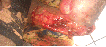
Figure 1: Important loss of substance from inside face of the elbow with telluric debris. Photo made at the arrival of the victim.

Yaovi James* KomlaSena Amouzou Messanvi Yao Akpoto Assang Dossim
Orthopeadics and Plastic surgery Department, Sylvanus Olympio Teaching Hospital, PO Box: 57, Lome, TOGO*Corresponding author: James Yaovi Edem, Orthopeadics and Plastic surgery Department, Sylvanus Olympio Teaching Hospital, PO Box: 57, Lome, TOGO, Tel: 00228 90042873; E-mail: nanoj@yahoo.fr
The elbow bone traumas are very serious lesions that can compromise the function of this joint in the future. These lesions are most severe when there is a defect of bone which affects one component of the joint.
We report a case of open trauma of the left elbow from a right-handed girl of 21 years, during an accident on the highway.
On admission, the victim had a significant defect of skin-muscle of the anterior medial aspect of the elbow. The exploration revealed a major loss of bone affecting the humeral trochlea which was completely absent. In emergency the patient has benefited from joint stabilization by an external fixation, with a filling-in of the humeral-ulnar joint space with a cement spacer. The loss of muscle substance was filled with a latissimus dorsi flap followed by a skin graft made the days that followed.
After a brief review of the literature, the authors emphasize on the need to preserve the joint space through the establishment of the spacer which is the only guarantee for maintaining the stability of the elbow, which also allows considering arthroplasty surgery later
Open trauma; Elbow; Bone defect
Complex traumatic lesions of elbow gathering the osteo-joints, vascular and mucocutaneous effects, that may occur high energy trauma waning. They are even more severe when they are associated to a loss of substances affecting the bone elements and soft parts. These lesions raise two problems that must be resolved at the time of initial support:
The aim of this work is to expose our support strategy of a case of trauma with loss of elbow musculoskeletal and skin mucous substance in a girl of 21 years in our difficult working conditions.
Miss X, aged 21 years with no particular antecedents was received in emergency for open trauma of left elbow by accident on a public highway.
At the entrance, the victim was in hypovolemic shock. The clinical examination of the left arm showed an open fracture-dislocation of the left elbow Gustillo IIIB type. This was a large wound of 17 cm long and 06 cm wide, contaminated with floor debris, laying bare the entire trauma joint cavity (Figure 1).
Figure 1: Important loss of substance from inside face of the elbow with telluric debris. Photo made at the arrival of the victim.
After correction of the hypovolemia, the victim was admitted to the surgery room for a first urgent cleaning. Examination of the wound under general anesthesia helped to highlight a significant loss of fascio cutaneous substance through which one could distinguish the jagged fibers of epitrochlear muscles. The internal ligament plan as well as the inner part of the joint capsule was completely absent.
The examination of the ulnar humeral cavity found a complete absence of the medial epicondyle and the humeral trochlea. The ulnar nerve was intact. The wound was thoroughly cleaned with hydrogen water and then with saline serum. A first trimming was performed with excision of devitalized tissue. The wound edges were brought closer without tension and closed on an aspirative Redon (Figure 2).
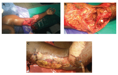
Figure 2 :
A. Aspect general of the upper limb after washing in the operation room
B. Figure showing lesion work up with full loss of external ligament plane and absence bone capitulum plane.
C. Lumb’s aspect after after the first lineage and closing.
The patient was hospitalized and an analgesic treatment with morphine and especially a triple antibiotic therapy associating gentamicin, metronidale and ceftriaxone were brought in. An X-ray assessment confirmed the bony defect touching the medial epicondyle and the vertical part of the trochlear notch of the ulna. (Figure 3).
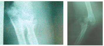
Figure 3: The X-Ray of the elbow showing
A daily dressing was achieved during three days under general anesthesia in the surgery room. During these dressings a cleaning was performed with excision and debridement of necrotic tissues. Four days later, the wound became clean (Figure 4) and the victim was readmitted to the operation-room for definite repair of lesions. After cleaning thoroughly the remnants of the joint cavity humeroulnar, a cement spacer was prepared and implemented to bridge the defect left by the bony defect and joint substance. The elbow was stabilized by an external fixator in functional position. The loss of fascio cutaneous substance was filled by a fascio-myo-cutaneous flap of the latissimus dorsi (Figure 5).
Figure 4: Picture of the elbow showing substance loss from external capitulum of humerus
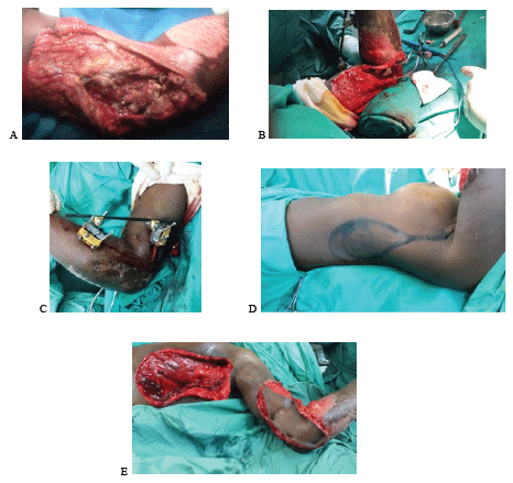
Figure 5: Definite surgical treatment
A. Elbow ’s aspect after the last washing
B. Setting up of the Spacer in acrylic Cement
C. Stabilisation by an external fixer. Simple Mount in position of elbow functioning.
D. Preparing the big dorsal tag.
E. Achievement of the big dorsal tag.
The immediate postoperative period was uneventful without occurrence of complications. At 10 months follow-up, there was complete healing of the flap. In terms of mobility, the elbow was blocked in functioning position. The active flexion was 10 degrees. The active extension 0 degrees (Figure 6).
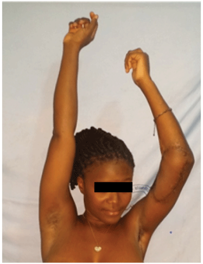
Figure 6: Functional results in10 months back.
Traumatic lesions at the elbow involving great loss of substance are uncommon, but represent a significant problem when such cases are referred to a trauma department [1]. Most of these injuries can be associated with severe bone and articular cartilage fragmentation, extensive soft tissue damage, and concomitant injuries, which may jeopardize limb integrity and cause functional impairment, particularly when treatment was delayed or inappropriate. The management of these injuries is quite challenging, and the restoration of elbow anatomy may require multiple bone and soft tissue surgical procedures. However, open intra-articular fractures of the distal humerus are frequently the result of a high-energy trauma so the first priority upon arrival in the emergency department is to stabilize the patient in time of need, because immediate reconstruction requires the patient to be in a stable general condition [1]. In addition bone defects associated with open fractures require prompt and adequate initial wound toilet, and early soft tissue cover. The bone defect is repaired secondarily usually by autologous bone grafting or the transfer of vascularised bone from the fibula or iliac crest, joint fusion, prosthetic implants or cement spacer [1-3].
Cement spacers may fill adequately the post injury defect and retain the soft tissue tension, but no biologic incorporation or long-term durability should be anticipated. The antibacterial efficacy of bone cement spacers loaded with different combinations of antibiotics makes them a good temporary or permanent treatment choice when severe wound contamination or infect is apparent [2]. The particularity in developing countries is a paucity of technical support centers with incapacity to provide adequate care and rehabilitation for this unfortunate group of patients.
There are few studies in the literature on skin reconstruction at the elbow, so there is no clear consensus. The options for skin coverage are numerous and include, from the simplest to the most complex: primary closure, grafts, local flaps, pedicled fascio cutaneous and muscular flaps, or myocutaneous flaps and free-flaps [4,5].The main factors that influence the reconstructive choice are the location and size of the loss of substance and the involvement of underlying tissues. Exposed subcutaneous tissue and muscle tissue may be covered with simple skin grafts. Structures such as tendons, nerves, vessels and bone must be covered with flaps [1]. Derderian et al. [6] uses only free flaps, whereas Choudry et al. [7] emphasises the usefulness of pedicle flaps. Hallock [8] recommends the surgeon to have in their technical armamentarium some reliable flaps that may cover all possible loss of tissue, selected on the basis of their experience. But according to Battiston et al [1], the surgeon should opt for the simplest technique, taking into account different factors, such as the size of the lesion, the quality of the coverage to restore optimal function, and the possible damage to the donating area.
Management of bone defects after severe open fractures of the distal humerus encompasses many technical difficulties. The profile of the patient and his general clinical conditions, surgical team experience and the level of multidisciplinary assistance has to be considered as well. So the decision about the kind of reconstruction should always be based upon the likelihood of salvaging a useful, functional limb, not on the prospect of local healing alone.
Download Provisional PDF Here
Article Type: Case Report
Citation: James YE, Amouzou K, Akpoto MY, Dossim A (2016) Serious Open Injury of Elbow: Support Strategy in a Developing Country. J Surg Open Access 3(1): doi http://dx.doi.org/10.16966/2470-0991.120
Copyright: © 2016 James YE, et al. This is an open-access article distributed under the terms of the Creative Commons Attribution License, which permits unrestricted use, distribution, and reproduction in any medium, provided the original author and source are credited.
Publication history:
All Sci Forschen Journals are Open Access