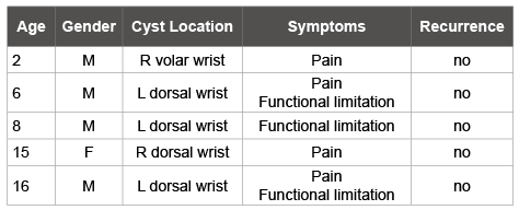
Table 1:Pediatric hand and wrist ganglia patient demographics
F=Female; M=Male; L=Left; R=Right

Adam Gendy1 Haripriya S Ayyala2 Ramazi Datiashvili2
1Temple University, 3500 N. Broad St, Philadelphia, PA 19140, USA*Corresponding author: Ramazi Datiashvili, Rutgers, New Jersey Medical School, Division of Plastic Surgery, 140 Bergen Street, Suite E1620, Newark, NJ 07103, E-mail: datiasro@njms.rutgers.edu
Background: Although there is an abundance of information about hand and wrist ganglion cysts in the adult patient population, there is limited data about these lesions in children. The purpose of this article is to present our clinical experience and review the epidemiology, etiological factors, clinical presentation, treatment, and outcomes of pediatric patients with hand and wrist ganglion cysts.
Methods: A literature review was thoroughly conducted along with a chart review of all cases of ganglion cysts operated on at a single institution, University Hospital, between May 2014 and October 2015.
Results: Five consecutive patients, between ages 2-16 years, who presented with symptomatic lesions of the hand or wrist, underwent treatment by a single surgeon (R.D.). The mean age of patients was 9.4 years, with one of the patients being female. Functional limitation was the most common indication for surgical treatment. Only one patient had a history of previous trauma. In 80% of the cases, the diagnosis was made clinically. The most common site of occurrence was the dorsal wrist (4/5), followed by the volar wrist. Surgical excision was the treatment of choice for all patients that presented with symptomatic lesions (100%). Patients were followed up on one week post-operatively and told to return if they developed any recurrences.
Conclusions: While observation has been reported to be worthwhile in the cases of the asymptomatic pediatric hand and wrist ganglia, surgical excision should be employed in those lesions that are symptomatic or do not resolve with observation alone.
Ganglion cyst; Hand; Pediatric; Wrist
Ganglion cysts are well-circumscribed, mucin-filled benign tumors that usually arise from underlying joint capsules or tendon sheaths [1]. They are the most common soft tissue lesions found within the upper extremity, accounting for approximately 33-69% of all hand masses [2]. Although they occur at all ages, they are most frequently seen in adults between the second and fourth decades of life with females affected two to three times as often as males [2,3]. Ganglion cysts are most commonly located over the dorsum of the wrist (60-70%) followed by the volar wrist (20%) and the volar retinaculum (10-20%) [4].
On gross examination, these benign growths usually have a smooth, translucent, shiny appearance and may either consist of a single main cyst or may be multi-loculated [2]. Microscopically, the walls of these cysts are primarily composed of sheets of collagen fibers that are organized within many different layers and are adorned by flattened cells that are not organized into an epithelium or synovium [1]. The capsular attachment of these structures can be shown in serial sections to contain mucin-filled clefts, which intercommunicate with each other and the underlying adjacent joint, thus forming an intricate and continuous duct between the ganglion cyst and the associated joint [5]. The clear and viscous jelly-like fluid material within the cyst consists of high concentrations of hyaluronic acid, albumin, globulin and mucopolysaccharides such as glucosamine [1].
There have been several theories postulated to explain the etiology of the ganglion cyst. However, to date, there is no single theory that fully explains the pathogenesis of this lesion. The most prevailing belief is that ganglion cysts arise as a response to repetitive minor trauma to synovial or mesenchymal cells at the synovial-capsular interface. Under the stress of repetitive stretching of the capsular and ligamentous structures that support the chronically aggravated joint, these cells appear to produce hyaluronic acid at the synovial-capsular interface which then accumulates within small channels and eventually pools together to become a ganglion cyst [5].
While a great deal of information exists for hand and wrist ganglia that occur within the adult patient population, there is a limited amount of data on pediatric ganglion cysts. The true incidence of these lesions within this patient population in all likelihood is underreported as their presence is usually painless and often does not cause functional deficiency. We present a single surgeon’s (R.D.) experience with pediatric ganglion cysts along with a review of the current literature.
In our case series, we reviewed the charts of all 5 patients between 2-16 years of age with primary ganglia of the hand and wrist that were treated consecutively by a single surgeon between May 2014 and October 2015. During this time period, five patients underwent surgical excision of their ganglion cysts, all of which were symptomatic lesions. Information was then collected on each patient, including age, gender, cyst location, symptoms, and recurrence (Table 1).

Table 1:Pediatric hand and wrist ganglia patient demographics
F=Female; M=Male; L=Left; R=Right
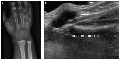
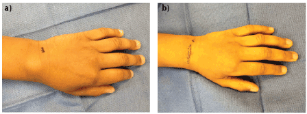
The mean age was 9.4 years old, ranging from 2 years to 16 years, with 3 of the five patients being 10 years of age or younger. There was one female and four males in the study. Functional limitation was the most common indication for surgical management of the lesion. Other indications for surgery included pain, discomfort at rest, a progressive increase in cyst size, and emotional distress to the patients and their parents. Only 1 patient reported a history of previous trauma. In 4 cases, the diagnosis was made on clinical presentation alone. In the remaining case, imaging was utilized in the investigation of the mass based on its location over the projection of the radial artery (Figure 1). The right side was affected in 40% of cases and the most common site of occurrence was the dorsal wrist (4/5), (Figure 2) followed by the volar wrist (1/5). There were no complications with any of the procedures performed in any patient. Long term follow-up was performed by means of telephone interview of the patients and their parents and ranged from 4 to 18 months. To date, there have been no recurrences or any other complications noted from any one of these patients.

Figure 1: a) Preoperative radiograph (oblique view) of the right wrist in a volar ganglion cyst in a four-year-old; b) Ultrasound of the same patient (sagittal view) showing the cyst located volar to the distal radial metaphysic

Figure 2: a) Preoperative view of the left wrist with a dorsal ganglion cyst in an eight-year-old male; b) Postoperative view
The epidemiology of pediatric hand and wrist ganglia differ than that of adults. Satku and Ganesh [6] showed that in children aged less than 10 years, volar cysts (77%) were more commonly seen than dorsal cysts (14%). Coffey et al. [7] corroborated this observation with children under the age of 12 years, with 55% of their patients suffering from volar cysts. Of note, the pediatric population classically exhibits a similar gender bias as the adult population with the proportion of females being affected by these lesions ranging in the literature from 1:6 to 4.7:1 [4,8].
The most common symptoms of ganglion cysts include pain, especially upon movement, weakness, and an aesthetically displeasing appearance. Although they are often non-tender to palpation, pain can be elicited during flexion or extension of the wrist [5].Ganglion cysts can range in size from 1-3 cm but may wax and wane with time. On physical examination, they may be firm or rubbery, mobile, and may move with associated tendons [5]. In addition, these lesions can be successfully transilluminated with light as compared to other hand and wrist lesions [2]. While radiographic imaging has not been shown to be very helpful in making the diagnosis, in children presenting with an unusual-appearing or atypical mass, further testing is warranted to rule-out underlying articular pathology or soft tissue calcifications to establish a proper diagnosis.
There are several treatment options for ganglions of the wrist and hand. Aspiration and other similar cyst puncture techniques have been shown to have variable success in adult patients presenting with dorsal ganglion cysts, with cure rates reported in the literature anywhere between 13%[9] and 85% [10]. Aspiration within the pediatric population has also been reported in the literature but has been shown to have less favorable results. Mac Collum [8] reviewed 14 children with wrist ganglia that were treated by aspiration, puncture, or indwelling suture and experienced a 43% recurrence rate with those techniques. Colon and Upton [11] reported only a 20% success rate in their case series with the aspiration of dorsal and volar ganglion cysts and a less than 20% success rate with the aspiration of retinacular cysts. Volar ganglion cysts have been shown to have higher rates of recurrence following aspiration (57-83%) as compared to dorsal ganglion cysts. The aspiration of volar ganglia further carries with it the additional risk of injuring nearby neurovascular structures [9,12]. The aspiration of any ganglion cyst is a challenging procedure to perform on younger children in the office with higher rates of recurrence and the potential to inflict damage to adjacent vital structures, therefore, it is generally not recommended for the pediatric population.
Indications for surgical excision noted in the literature as well as in our experience include associated pain, functional limitation, or gross deformity [13]. The surgical excision of hand and wrist ganglion cysts in the adult patient population has been reported to be very successful with reported recurrence rates as low as 4% for dorsal cysts and 7% for volar cysts [3]. For the pediatric patient population, however, recurrence rates after excision are much more variable, ranging between 2.8%to 35% [6,13]. In this age group, recurrence rates seem to vary depending on both patient age and the location of the excised cyst. Colon and Upton [11] had a recurrence rate of 6% for dorsal and volar ganglion cysts in mostly older teenaged patients, with no recurrences for retinacular cysts in younger patients following extirpation.
Proper management of these lesions requires complete resection of the ganglion, its stalk, and if large enough, a portion of the joint capsule to avoid recurrence [3]. As volar wrist ganglions are often located between the radial artery and the flexor carpi radial is tendon, they must be gently dissected off the radial artery to avoid the possibility of compromising blood supply to the hand. If the cyst is large enough, it can be punctured to allow for better visualization of the stalk during the dissection [2]. The potential complications of this treatment modality include vascular compromise of the hand, decreased range of motion secondary tendon injury, scapholunate dissociation, joint stiffness, and wound-healing sequel and that can include infection, neuroma, and keloid formation. Simon Cypel et al. [13] reported an 8.5% complication rate in their series, where in two cases, a segment of the radial artery was removed during the excision of a volar ganglion cyst and in one case, one patient presented post-operatively with a giant cell reaction. In light of variable recurrence rates and the potentially serious complications associated with resection, the surgical management of ganglion cysts is usually indicated for patients that present with pain, gross deformity, and/or functional limitations [2].
Only a few studies have examined the treatment of pediatric ganglia by observation alone. Wang and Hutchinson [4] found a 79% spontaneous resolution rate in a case series which included 14 children less than 10 years of age that presented with simple, asymptomatic hand and wrist ganglia. None of these lesions were accompanied by the presence of pain symptoms, neurovascular abnormalities, signs of wrist instability, or a limited range of motion. In their report, the average time to a traumatic resolution by observation alone was 6 months from the time of diagnosis, with a range between 1.5 months and 12 months, without a single recurrence [4]. They postulated that if a cyst did not resolve within 1year, it was not likely to resolve spontaneously and therefore, warranted having a conversation with the patients and their family members about the risks and benefits of continuing to observe the lesion versus undergoing surgery. They concluded that in the absence of any other symptoms, these benign lesions could be initially treated conservatively with observation with marked success prior to any surgical intervention and incurring its potential complications [4].
Although ganglion cysts are the most common soft tissues masses of the hand and wrist, there is a paucity of information about these benign lesions within the pediatric population. In contrast to previous studies, our series of patients more commonly had a ganglion cyst on the dorsum of the wrist rather than the volar side, and occurred more commonly in males than females; no patients experienced recurrence. We believe children that present with these benign growths may be managed with observation and reassurance in asymptomatic cases. However, those that present with associated pain, functional limitation, or gross deformity benefit from surgical excision.
None.
Download Provisional PDF Here
Article Type: Case Report
Citation: Gendy A, Ayyala HS, Datiashvili R (2015) Pediatric Ganglion Cysts: Case Series and Review of the Literature. J Surg Open Access 2(3): doi http://dx.doi. org/10.16966/2470-0991.119
Copyright: © 2016 Gendy A, et al. This is an open-access article distributed under the terms of the Creative Commons Attribution License, which permits unrestricted use, distribution, and reproduction in any medium, provided the original author and source are credited.
Publication history:
All Sci Forschen Journals are Open Access