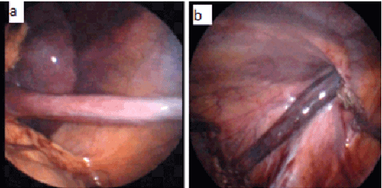
Figure 1: Laparoscopic appearance of continuous splenogonadal fusion.
a) A cord-like soft-tissue mass extended from the spleen
b) To the left inguinal ring and was attached to the testis

Mustafa Erman Dörterler1* Mehmet Emin Boleken1 Kemal Deniz2
1Harran University Medical School, Department of Pediatric Surgery, Sanlıurfa, Turkey*Corresponding author: Mustafa Erman Dorterler, Harran University, Department of Pediatric Surgery, Şanlıurfa, Turkey, Tel: +90 (414) 318 34 20; Fax: +90 (414) 318 31 92; E-mail: m.e.dorterler@hotmail.com
Splenogonadal fusion (SGF) is a rare congenital anomaly in which the spleen is connected to the gonad. Two types of SGF have been described: continuous and discontinuous. SGF is frequently associated with cryptorchidism or congenital orofacial/limb anomalies. We herein present a case involving a 7-year-old boy who underwent surgical repair of an undescended left testis. When laparoscopy was performed intra operatively because of bleeding, the patient was diagnosed with SGF and laparoscopically treated accordingly.
Spleen; Splenogonadal fusion; Testis
Splenogonadal fusion (SGF) is a rare congenital anomaly characterized by the conjunction of the splenic and gonadal structures in the intrauterine period. The involved primordial organs, the splenic anlage in the dorsal mesogastrium, and the gonadal ridge between the mesonephros and dorsal mesentery are formed between the 5th and 6th weeks of gestation. During the 5th week, as the dorsal mesogastrium rotates to the left, the splenic anlage lies in close proximity to the left gonadal ridge. Inflammation or adhesions of the peritoneal surface of the spleen could lead to fusion of these primordial organs and their subsequent caudal migration [1,2].
The condition affects young males. More than 70% of affected patients are younger than 20 years, and approximately 50% are pediatric patients [3]. SGF typically appears as an extra testicular mass and is rarely diagnosed preoperatively. Approximately 150 cases have been reported [4]. We herein describe a patient with an undescended testis combined with SGF. The diagnosis was made at laparoscopy and then treated, thereby avoiding an unnecessary orchiectomy.
A 7-year-old boy was admitted for surgery at another center with a diagnosis of an undescended left testis. When the inguinal canal was opened with a left inguinal transverse incision, a mass was identified. There was excessive intra operative bleeding, so the procedure was terminated and the patient was transferred to our clinic. The physical examination revealed that the right testis was of normal size and shape, while the left testis seemed to be larger than normal and had an irregular structure. The patient was admitted for surgery and the inguinal canal was explored through the previous left inguinal transverse incision. Because a mass and bleeding were noted in the inguinal canal, diagnostic laparoscopy was performed. A mass was observed originating from the spleen and passing through the left inguinal canal to the scrotum (Figure 1). Because the mass was considered to be SGF, a frozen biopsy was performed and the findings were consistent with spleen tissue. Therefore, the mass was separated from the spleen tissue laparoscopically and removed by excision from the left inguinal canal in a manner protecting the testis. Postoperative laboratory examination revealed that the testicular tumor markers α-fetoprotein, human chorionic gonadotropin, and lactate dehydrogenase were all within normal limits. At the 6-month follow-up, the patient had no problems and the testis was observed to have retained its normal structure.
Figure 1: Laparoscopic appearance of continuous splenogonadal fusion.
a) A cord-like soft-tissue mass extended from the spleen
b) To the left inguinal ring and was attached to the testis
SGF was first described by Boestrom [5] as a rare benign congenital anomaly characterized by fusion of the gonadal tissue with the spleen tissue in the 5th to 8th weeks of embryological development. Putschar and Manion [6] classified SGF into continuous and discontinuous types. The continuous type is characterized by connection of the spleen and gonad by a cord of splenic or fibrous tissue. Rarely, beads of splenic tissue are interspersed throughout the fibrous cord. In the discontinuous type, ectopic tissue is attached to the gonad, but has no connection to the spleen. SGF may be seen with other congenital anomalies, such as cardiac defects, cleft palate, anal atresia, microgonads, or limb anomalies [6]. Our patient had the continuous type, and no additional anomaly was found. In the discontinuous type, which is seen more often, there is no connection between the normal and ectopic spleen tissue [7].
SGF is seen primarily in young males, who present with a scrotal mass, inguinal hernia, or undescended testis. Cryptorchidism is the most common anomaly associated with SGF, and 59% of patients have bilateral undescended testes [1]. Rarely, traumatic rupture of the testis may occur, sometimes with infectious diseases such as mumps, malaria, and mononucleosis, or intestinal obstruction may be caused by the splenic tissue attached to the spermatic cord [6]. Atrophy and hypoplasia may be seen in the testis tissue adjacent to the lesion. There have been a few reports of discontinuous SGF associated with ipsilateral testicular atrophy [7,8]. One case associated with contralateral testicular aplasia has been reported in association with continuous SGF [9]. Cases with accompanying testicular aplasia have been reported. Although the diagnosis is generally made intra operatively or at autopsy, radiological methods can assist with a preoperative diagnosis [10]. The ultra sonography (USG) findings of SGF are variable and not clear, especially in discontinuous SGF. This type is usually diagnosed following orchiectomy [11]. USG may suggest continuous SGF [12]. Although rarely performed preoperatively, contrastenhanced computed tomography may reveal cord-like tissue between the spleen and left scrotal mass. If suspected, the diagnosis may be confirmed by magnetic resonance imaging (MRI) or a technetium-99m sulfur colloid liver–spleen scan. On MRI, T2-weighted imaging will show an isointense tubular structure connecting the spleen to the testis [3,12].
Laparoscopy is used for diagnosis and treatment in some cases. In our case, there was excessive bleeding during the surgical dissection for the undescended testis, and a mass was identified. The diagnosis was made at laparoscopy, and testis-sparing surgery was performed.
This rare case demonstrates the need to consider the differential diagnosis of intra scrotal or extra testicular masses. The use of laparoscopy for the diagnosis and treatment of extra testicular masses, as in this case, should be kept in mind. Laparoscopy can visualize a wide area and allow for intra operative frozen biopsy, avoiding an unnecessary orchiectomy.
Download Provisional PDF Here
Article Type: Case Report
Citation: Dörterler ME, Boleken ME, Deniz K (2015) Splenogonadal Fusion: Laparoscopic Diagnosis and Treatment. J Surg Open Access 1(2): http:// dx.doi.org/10.16966/2470-0991.106
Copyright: © 2015 Dörterler ME, et al. This is an open-access article distributed under the terms of the Creative Commons Attribution License, which permits unrestricted use, distribution, and reproduction in any medium, provided the original author and source are credited.
Publication history:
All Sci Forschen Journals are Open Access