Abstract
Background: Hip fracture is one of the most common risk factors increasing the mortality rate among the elderly population, especially in the first year after injury even when receiving operative treatment to encourage early ambulation.
Objective: To compare one year mortality rate between patients with anti-osteoporosis therapy and those without anti-osteoporotic medication after surgical intervention for hip fracture.
Method: 156 patients underwent surgical intervention for hip fractures from low energy trauma were reviewed. Demographic data, comorbidity, and anti-osteoporotic medication were collected. Patients were classified into 2 groups: Patients taking antiosteoporotic drug and those who did not receive anti-osteoporotic drug. Statistical significance was identified by P-value less than 0.05.
Results: The rate of osteoporosis treatment in patients sustained hip fracture was low (48%). Prevalence of one year mortality rate underwent surgical intervention was 15.4% (6.7% vs 23.5% in the treated and untreated groups, respectively, p=0.004).Patients with a history of anti-osteoporotic drug intake tended to receive osteoporotic drugs after treatment (P<0.001). In univariate analysis, there were several factors that significantly affected one year mortality including age (P=0.001), history of kidney disease (P=0.049), ambulatory status on the day discharged from the hospital (P=0.028) and history of osteoporotic drug intake (P=0.004). Recurrent fracture was not significantly related to one year mortality rate in these patients.
Conclusion: After surgical treatment of osteoporotic hip fracture, one year mortality rate tended to decrease among patients receiving anti-osteoporotic drugs. Physicians should promote osteoporosis therapy in all patients who have sustained hip fracture in the elderly population.
Keywords
Osteoporosis; Hip fracture; Mortality; Bisphosphonate; Treatment
Introduction
Osteoporosis represents a major public health problem because of its association with fragility or low-energy trauma fractures. Hip fracture has been recognized as the most serious complication of osteoporosis because of its consequence including disability, poor quality of life, increased risk of mortality, and health care costs [1-4]. The incidence of hip fractures has increased in many regions of the world as a result of the aging of the population, by 2050 half of hip fractures all over the world are estimated to occur in Asia [5]. In Thailand, a rising incidence of hip fracture has been clearly shown in both men and women during the past decade [6,7]. The Chiang Mai Hip fracture study from the 2006 data reported 690 hip fractures (203 men and 487 women) with a mean age of 76.7 year. The estimated cumulative incidence was 181.0 per 100,000 persons, and the adjusted incidence was 253.3 (135.9 and 367.9 in men and women, respectively), which increased by 2% per year compared with the 1997 data [7]. The first year after a hip fracture is considered to be the most critical time and the mortality rate after osteoporotic hip fracture among Thai people was 18% within the first year which was eight-fold higher than the general population [8].
An osteoporotic hip fracture generally requires surgical repair or replacement. Although surgery is the main treatment for patients with hip fracture, interventions to prevent future falls, exercise, balance training, and the treatment of osteoporosis are also important strategies for secondary prevention of hip fracture. Fortunately, a number of medications are now available and have proven to be effective in lowering the risk of future fractures [9-11]. It has been accepted that any patient with previous hip fracture is an ideal candidate for treatment due to the high risk for subsequent fractures and favorable cost-effectiveness. However, post-fracture medical treatment for osteoporosis is insufficient worldwide [12,13].
In our Orthopedic department, Phramongkutklao Army Hospital, all cases of hip fractures with surgical treatment have received at least standard medications of calcium and vitamin D supplements and focusing on secondary fracture prevention. However, the mortality rate after surgical intervention for hip fractures has not been carefully identified. The purpose of our study was to determine the rate of medical treatment for osteoporosis in patients with hip fracture and compare the 1 year mortality between treated and untreated patients, and the secondary objective was to determine subsequent fracture rate between the groups.
Methods
After study approval by the Institutional Review Board of Royal Thai Army Medical Department, a medical records investigation identified patients 65 years of age or older who sustained a hip fracture (femoral neck fracture, intertrochanteric fracture and subtrochanteric fracture). All patients underwent surgical intervention (fixation or arthroplasty) at Phramongkutklao Army Hospital between January 1, 2011 and May 31, 2012 was included in this study. Subjects were excluded from the study if they were found to have a pathological fracture (tumor, metastatic disease, etc.) or fracture caused by highenergy trauma (i.e., motor vehicle accident), peri-prosthetic fracture, previous history of secondary osteoporosis or death during hospitalization. Written informed consent was obtained before review and telephone interview was performed after verbal consent.
Data collection and outcome
A total of 176 patients with hip fracture were identified. Patient demographic information, number of comorbidities, pre-operative and postoperative status, history of prior fracture and treatment, term and cause of injury, medication at hospital admission and discharge, and radiological reports were also obtained from the medical record review. Any patient who was unable to proceed to surgery on schedule or discharged due to medical comorbidities was labeled as delayed surgery and delayed discharge, respectively
Patient’s comorbidities were reviewed by medical record: (1) Hypertension is defined as a systolic blood pressure (SBP) of 140 mm Hg or more, or a diastolic blood pressure (DBP) of 90 mm Hg or more, or taking antihypertensive medication. (2) Diabetes mellitus (or Diabetes) is a chronic, lifelong condition that affects your body’s ability to use the energy found in food, which were grossly classified into two major types (Non-Insulin Dependent Diabetes Mellitus and Insulin Dependent Diabetes Mellitus). (3) Dyslipidemia is elevation of plasma cholesterol, triglycerides (TGs), or both, or a low high-density lipoprotein level that contributes to the development of atherosclerosis. Causes may be primary (genetic) or secondary. Diagnosis is by measuring plasma levels of total cholesterol, TGs, and individual lipoproteins. (4) Heart disease refers to various types of conditions that can affect heart function, which are classified into 5 types (coronary artery disease, valvular heart disease, cardiomyopathy, arrhythmias, and heart infection). Lung disease (Pulmonary disease) is any condition causing or indicating impaired lung function. Kidney disease is a general term for any damage that reduces the functioning of the kidney. Liver disease is a general term for any damage that reduces the functioning of the liver. Dementia is usually progressive condition (such as Alzheimer’s disease) marked by the development of multiple cognitive deficits (such as memory impairment, aphasia, and the inability to plan and initiate complex behavior). Additionally, all hip fracture patients were calculated for FRAX score including 10 years probability of major bone fracture and hip fracture.
All patients received standard medications of calcium and vitamin D supplements. In this study, patients who received anti-osteoporotic agents including bisphosphonates, raloxifene, strontium ranelate, calcitonin, estrogen, and parathyroid hormone were classified as the “treated group” and those who did not receive anti-osteoporosis drugs were classified as the “untreated group”. One-year mortality rate was identified from all causes of death except non-natural cause (i.e., car accident or suicide). Any incidence of osteoporotic fracture with any sequential fracture after initial hip operation was defined as “re-fracture incidence”.
The primary outcome measurement was rate of antiosteoporotic medication and 1-year mortality rate after surgical treatment for hip fracture. This was determined by the hospital data system and reviewing the medical record in patients who still follow up. When no evidence on survival could be found in medical records or lost follow-up, patients or relatives were then contacted by telephone. Patients were classified into 2 groups as the “survival group” if they still survive, and the “deceased group” if they have died from any causes. The patients who could not be contacted by telephone were excluded in our study. The important information included post-fracture status, disability, degree of activities of daily living, history of new fractures since the first incidence of hip fracture and current medications. Other outcome measurement was to determine one year subsequent fracture rate between the treated and untreated groups.
Statistical analysis
All patients’ information was compared between the treated and untreated groups to identify any differences. Data were summarized using descriptive statistics (mean ± SD and number of patients). Information of lung disease, liver disease, time of injury, delayed surgery and recurrent fracture were compared using Fisher’s exact test. Age of patient, Charlson Comorbidity Index (CCI), and FRAX score (10 years probability of major bone fracture and hip fracture)were compared by independent T-test, monthly survival time of patients was compared by Mann-Whitney U test while the others were compared by Chi-Square test. P-value less than 0.05 were considered a significant difference. The patient characteristics including anti-osteoporotic drug intake, was compared between survival and deceased groups to identify the factors influencing 1-year mortality of the patients. Sex, lung disease, kidney disease, liver disease, cerebrovascular disease, dementia, previous drugs received, time of injury, delayed surgery, delayed discharge, and recurrent fracture were compared by Fisher’s exact test. Others were compared by Chi-Square test and P-value less than 0.05 was considered a significant difference.
Survival analysis was performed using the Kaplan-Meier test, and the cumulative survival rates (and standard error) of the groups were compared using the log-rank test. P<0.05 represented statistical significance.
Results
The 156 elderly patients with low-energy, non-pathological hip fractures treated surgically and 1-year mortality data available were included in the analysis. A summary of patient demographics is presented in Table 1.Medical records reviewed confirmed that fractures were intertrochanteric fracture (53%), femoral neck fracture (45%) and subtrochanteric fracture (2%) (Table 2); however, these were not analyzed by type of fracture because it lacked sufficient power to detect the differences. The mean age was 80 ± 6 years, 79% were female and 12.8% had previous fracture. FRAX score was comparable between treated and untreated group (14.2% vs 13.9%, p=0.675 for 10 years probability of major bone fracture; 6.0% vs 5.9%, p=0.716 for 10 years probability of hip fracture).
| Variable |
Total |
Treated Group* |
Untreated Group† |
P-value |
| n (%) |
n (%) |
n (%) |
| Age (years); Mean ± SD (Min-Max) |
80.06 ± 6.53 |
79.95 ± 6.28 |
80.16 ± 6.79 |
0.839 |
| (65-99) |
(67-92) |
(65-99) |
| Sex |
|
|
|
0.583 |
| Male |
32 (20.51) |
14 (18.67) |
18 (22.22) |
|
| Female |
124 (79.49) |
61 (81.33) |
63 (77.78) |
|
| FRAX score |
|
|
|
|
| 10 years probability of major bone fracture (percentages ± SD) |
14.03 ± 4.00% |
14.17 ± 3.86% |
13.90 ± 4.14% |
0.675 |
| 10 years probability of hip fracture (percentages ± SD) |
5.99 ± 1.89% |
6.04 ± 1.87% |
5.93 ± 1.91% |
0.716 |
| Hypertension |
|
|
|
0.936 |
| Yes |
119 (76.28) |
57 (76.00) |
62 (76.54) |
|
| No |
37 (23.72) |
18 (24.00) |
19 (23.46) |
|
| Diabetes |
|
|
|
0.904 |
| Yes |
59 (37.82) |
28 (37.33) |
31 (38.27) |
|
| No |
97 (62.18) |
47 (62.67) |
50 (61.73) |
|
| Dyslipidemia |
|
|
|
0.969 |
| Yes |
58 (37.18) |
28 (37.33) |
30 (37.04) |
|
| No |
98 (62.82) |
47 (62.27) |
51 (62.96) |
|
| Heart disease |
|
|
|
0.517 |
| Yes |
39 (25.00) |
17 (22.67) |
22 (27.16) |
|
| No |
117 (75.00) |
58 (77.33) |
59 (72.84) |
|
| Lung disease |
|
|
|
0.106 |
| Yes |
6 (3.85) |
5 (6.67) |
1 (1.23) |
|
| No |
150 (96.15) |
70 (93.33) |
80 (98.77) |
|
| Kidney disease |
|
|
|
0.100 |
| Yes |
22 (14.10) |
7 (9.33) |
15 (18.52) |
|
| No |
134 (85.90) |
68 (90.67) |
66 (81.48) |
|
| Liver disease |
|
|
|
0.051 |
| Yes |
4 (2.56) |
4 (5.33) |
- |
|
| No |
152 (97.44) |
71 (94.67) |
81 (100.00) |
|
| Cerebrovascular disease |
|
|
|
0.668 |
| Yes |
25 (16.03) |
13 (17.33) |
12 (14.81) |
|
| No |
131 (83.97) |
62 (2.67) |
69 (85.19) |
|
| Dementia |
|
|
|
0.607 |
| Yes |
21 (13.46) |
9 (12.00) |
12 (14.81) |
|
| No |
135 (86.54) |
66 (88.00) |
69 (85.190 |
|
| Pre-injury ambulatory status |
|
|
|
0.261 |
| Walking without gait aid |
112 (71.79) |
57 (76.00) |
55 (67.90) |
|
| Walking with gait aid or worse |
44 (28.21) |
18 (24.00) |
26 (32.10) |
|
| History of anti-osteoporotic drug intake |
|
|
|
< 0.001 |
| Yes |
15 (9.62) |
14 (18.67) |
1 (1.23) |
|
| No |
141 (90.38) |
61 (81.33) |
80 (98.77) |
|
| History of fracture |
|
|
|
0.105 |
| Yes |
20 (12.82) |
13 (17.33) |
7 (8.64) |
|
| No |
136 (87.18) |
62 (82.67) |
74 (91.36) |
|
| First or second time of fracture |
|
|
|
0.608 |
| First time |
153 (98.08) |
73 (97.33) |
80 (98.77) |
|
| Second time |
3 (1.92) |
2 (2.67) |
1 (1.23) |
|
| History of delayed surgery |
|
|
|
0.3689 |
| Yes |
5 (3.21) |
1 (1.33) |
4 (4.94) |
|
| No |
151 (76.79) |
74 (98.67) |
77 (95.06) |
|
| History of delayed discharge |
|
|
|
0.546 |
| Yes |
17 (10.90) |
7 (9.33) |
10 (12.35) |
|
| No |
139 (89.10) |
68 (90.67) |
71 (87.65) |
|
| Postoperative ambulatory status on discharge day |
|
|
|
0.388 |
| Walking with gait aid |
57 (36.54) |
30 (40.00) |
27 (33.33) |
|
| Wheelchair ambulatory or worse |
99 (63.46) |
45 (60.00) |
54 (66.67) |
|
| Re-fracture |
|
|
|
0.672 |
| Yes |
5 (3.21) |
3 (4.00) |
2 (2.47) |
|
| No |
151 (96.79) |
72 (96.00) |
79 (97.53) |
|
| Survival at 1 year |
|
|
|
0.004 |
| Yes (survival group) |
132 (84.62) |
70 (93.33) |
61 (76.54) |
|
| No(deceased group) |
24 (15.48) |
5 (6.67) |
19 (23.46) |
|
| *Patients who received any anti-osteoporotic agents were classified as the “treated group” while† those who did not receive anti-osteoporosis drugs were classified as the “untreated group”. |
Table 1: Demographic data comparing the “treated” and “untreated” groups
| Fracture |
n |
% |
| Intertrochanteric fracture |
83 |
53.2 |
| Femoral neck fracture |
70 |
44.9 |
| Subtrochanteric fracture |
3 |
1.9 |
| Total |
156 |
100 |
Table 2: Frequency of fracture type
No significant difference was found in medical comorbidities, pre-fracture status, previous history of osteoporotic fracture and history of delay operation between the treated and non-treated groups. However, the percentage of anti-osteoporosis drug use was significantly higher in the treated group compared with the non-treated group. Moreover, we found that a history of delayed discharge from hospital and post-operative ambulatory status on the day of discharge did not significantly differ.
In this study, 132 of 156 patients (84.6%) were surviving 1-year after surgery, while the mortality rate at 1-year was 24 patients (15.4%). All deceased patients were associated with high Charlson Comorbidity Index (CCI) (6.1 ± 1.6) and the mean survival time of this group was 8 months (range, 2-11 months). Additionally, the 1-year mortality rate was significantly higher in the untreated group (6.7 vs 23.5 in the treated and untreated groups, respectively, p = 0.004). Five patients have died from their uncontrolled medical condition (3 cases from congestive heart failure and 2 cases from pneumonia) and were associated with higher CCI (7.2 ± 0.7) even though they underwent osteoporosis therapy after surgical intervention for hip fracture (Table 3). However, the subsequent fracture (re-fracture) rate between the groups was comparable.
| Deceased patients underwent osteoporosis therapy
(n=5) |
Patient’s comorbidities |
CCI† |
Cause of death |
| Case 1 (85 years old female) |
CVA |
5 |
Pneumonia |
| Case 2 (67 years old female) |
HTN, NIDDM, DLD, CKD, Cirrhosis |
8 |
Congestive heart failure |
| Case 3 (84 years old female) |
HTN, CKD, CVA |
8 |
Congestive heart failure |
| Case 4 (81 years old male) |
HTN, CAD, COPD |
6 |
Pneumonia |
| Case 5 (80 years old male) |
HTN, NIDDM, CAD, COPD, cirrhosis |
9 |
Congestive heart failure |
| †Mean CCI was 7.2 ± 0.7; CVA= cerebrovascular accident (stroke); HTN=hypertension; NIDDM=Non-Insulin Dependent Diabetes Mellitus; DLD=dyslipidemia; CKD=chronic kidney disease; CAD=coronary artery disease; COPD=chronic obstructive pulmonary disease |
Table 3: Summarized Causes of Death in all patients taken osteoporosis therapy including comorbidities and Charlson Comorbidity Index (CCI)
A total of 75 (48%) patients received anti-osteoporotic drugs as shown in Table 4. Most of them (85%) received bisphosphonate drugs. Fifty-seven patients received only bisphosphonate. Seven patients received sequential therapy with bisphosphonate and another type of drug, five with strontium ranelate, one with teriparatide and one with nasal spray calcitonin. Four of 57 patients, receiving only bisphosphonate were treated by sequential therapy with different bisphosphonate doses. The most administered bisphosphonate drug was risedronate (55% of patients receiving bisphosphonate) followed by alendronate (34% of patients receiving bisphosphonate) and ibandronate (17% of patients receiving bisphosphonate). One patient treated with intravenous zoledronic acid. Additionally, four patients received only strontium ranelate and one patient received sequential therapy with strontium ranelate and calcitonin while six patients were treated by only teriparatide.
| Drug used in the treated group |
n |
| Biphosphonate |
|
| Only bisphosphonate |
|
| Risedronate |
28 |
| Alendronate |
17 |
| Ibandronate |
7 |
| Risedronate + Ibandronate* |
1 |
| Alendronate + Ibandronate* |
2 |
| Risedronate + Alendronate + Ibandronate* |
1 |
| Zoledronic acid (IV) |
1 |
| Sequential therapy: bisphosphonate with other agents* |
|
| Risedronate + Strontium |
4 |
| Alendronate + Strontium |
1 |
| Alendronate + Teriparatide |
1 |
| Risedronate + Calcitonin |
1 |
| Strontium |
4 |
| Strontium+ Calcitonin |
1 |
| Teriparatide |
6 |
| Total |
75 |
Table 4: Frequency of anti-osteoporotic drug intake (*sequential treatment, not given at the same time)
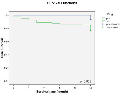
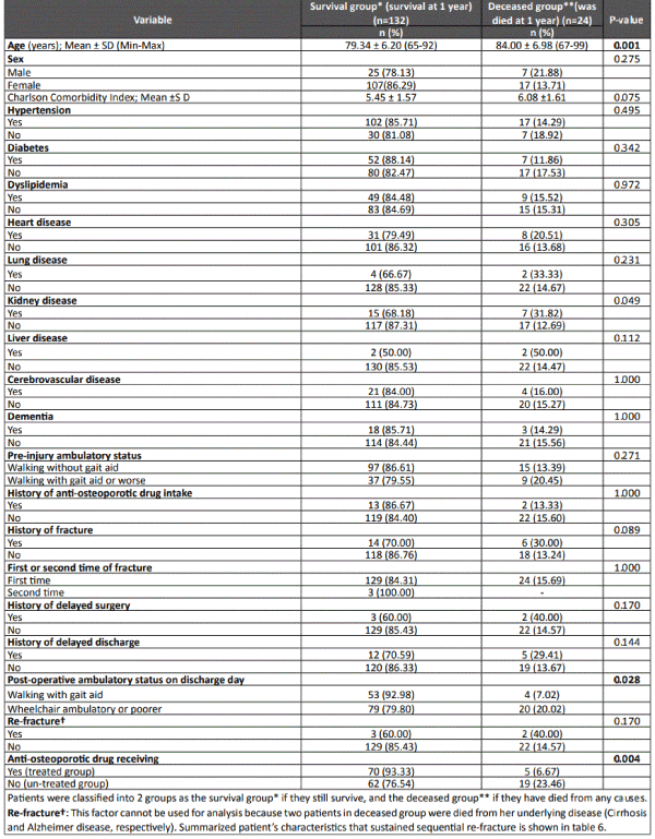
The relationship between survival rate and risk factor is shown in Table 5. Aging was significantly related to survival rate. Mean age of the survival and deceased groups was 79 ± 6 years and 84 ± 6 years, respectively. The only medical comorbidity significantly related to survival rate was kidney disease (p=0.049). All of pre- and post-injury histories were not significantly related to survival rate except ambulatory status on the day of discharge. Patients who could walk with a gait aid had significantly higher survival rates than those who were wheelchair ambulatory (p=0.028). Anti-osteoporotic drug intake significantly improved mortality rate (P-value of 0.004). However, having a history of re-fracture did not significantly correlate with survival rate of patients. Survival analysis was shown to be significantly different between the drug intake and control groups (P-value of 0.003). Kaplan-Meier survival analytic graph is shown in Figure 1.

Figure 1: Kaplan-Meier survival analytic graph. Blue line demonstrates patients with hip fracture received any antiosteoporotic drug after surgical intervention while green line defines patients with hip fracture did not receive any antiosteoporotic drug after surgical treatment

Table 5: Influencing factors compare between survival group and deceased group.
Discussion
Our populations were nearly the same baseline characteristics compare between treated and untreated group. Moreover, there was no significant difference of Charlson score between treated and untreated group (5.45 vs 6.08, p =0.075).
Many anti-osteoporotic drugs were used to prevent secondary fracture and even found that some medication reduced mortality rate in these patients. In 2006, HORIZONRFT study was found that postoperative intravenous zoledronic acid given to patient with osteoporotic hip fracture had 35% reduction of recurrent clinical fracture rate and, surprisingly, the mortality rate in this group of patient also reduced significantly by 28%. This was the first evidence about mortality benefit in bisphosphonate used [14]. For oral bisphosphonate, in 2009, oral alendronate and oral risedronate reduced mortality rate of 8% per month or about 60% per year [15]. In 2008, another study of oral bisphosphonate also showed 27% reduction of mortality rate [16].
1-year mortality rate of untreated group in our study was comparable with European countries (20%-25%) [17-19]. Moreover, the overall mortality rate was similar to other studies in Asian countries [20-22] including Thailand [8]. All deceased patients were associated with high Charlson Comorbidity Index (CCI) (6.1 ± 1.6) that correspond to the other literature. Study of Neuhaus demonstrated CCI (5 and more) predicted in-hospital mortality in patients with hip fractures [23]. Surprisingly, 1-year mortality was as low as 6.7% in the treated group which is significantly lower compared to the untreated group (6.7 vs 23.5 in treated and untreated group, respectively, p=0.004). This mean that a significant decrease in the 1-year mortality of treated versus untreated patients was demonstrated. However, five patients in the treated group had died within one year after surgical intervention for hip fracture even though they underwent osteoporotic treatment because these patients had severe medical condition and they were associated with higher CCI (7.2 ± 0.7).
The re-fracture rate in our study was also the same in both group (3.2% overall, and only 3 cases in treated group and 2 cases in un-treated group) and quite less than other studies. The HORIZON-RFT study was a secondary prevention study for treated hip fracture demonstrating zoledronic acid at a dose of 5 mg administered as a yearly infusion significantly reduced any new clinical fracture by 35%, and the rates of any new clinical fracture were 8.6% (10). Another explanation for this low re-fracture rate in this study is that the data came from interview, so only the clinical non-vertebral fracture could be detected. As we know that vertebral fracture can be silent for almost 2/3 of cases, so there are possible for very low rate of re-fracture detected in this study. Moreover, both patients in “deceased group” with sequential re-fracture have possibly died from their underlying disease (cirrhosis and moderate to severe degree of Alzheimer disease, respectively) (Table 6). Therefore, indifference of re-fracture rate between treated and un-treated group cannot be counted for the reduction of 1-year mortality rate in this study because of these confounding factors.
Re-fracture
(n=5) |
Patient’s characteristics |
Site of refracture |
Status |
|
Case 1 |
A 69 years old woman with severe osteoporosis, underlying Chronic kidney disease, treated with Strontium, calcium and vitamin D after surgical intervention with PFNA (Proximal Femoral Nail Anti-Rotation) |
Right intertrochanteric fracture followed by Left intertrochanteric fracture |
Deceased from cirrhosis (Chronic hepatitis C) |
|
Case 2 |
A 74 years old woman with severe osteoporosis, underlying Alzheimer disease (moderate to severe), treated with only Calcium and vitamin D after surgical intervention with Unipolar hemiarthroplasty |
Right femoral neck fracture followed by skull fracture |
Deceased from head injury (skull fracture) |
|
Case 3 |
An 84 years old woman with severe osteoporosis, underlying mild Alzheimer disease, treated with only Calcium and vitamin D after surgical intervention with Bipolar hemiarthroplasty |
Left femoral neck fracture followed by right femoral neck fracture |
Alive |
|
Case 4 |
An 85 years old woman with severe osteoporosis, underlying End stage renal disease and Parkinson disease, treated with Risedronate, calcium and vitamin D after surgical intervention with Dynamic hip screws fixation |
Leftstabletypeintertrochanteric fracture followed by clinical vertebral compression fracture (T10, T12, L3) |
Alive |
|
Case 5 |
An 83 years old man with severe osteoporosis, underlying Alzheimer disease, treated with Alendronate, calcium and vitamin D after surgical intervention with Bipolar hemiarthroplasty |
Left femoral neck fracture followed by periprosthetic fracture on left hip |
Alive |
Table 6: Summarized patient’s characteristics who sustained sequential re-fracture
Most of anti-osteoporotic drugs used in this study were bisphosphonates. This was because of bisphosphonates were still to be the first line of drug in Thailand. The most common bisphosphonate used in this study was risedronate. This was caused by the public health policy for universal coverage and social coverage of Thailand.
For univariate analysis, the factors significantly relate to 1-year survival rate were age, kidney disease, ambulatory status on the day discharge from the hospital. Additionally, antiosteoporotic drug intake significantly related to 1-year mortality rate in univariate analysis. Unsurprisingly, in univariate analysis, re-fracture rate did not influence 1-year mortality rate. Additionally, no benefit of anti resorptive/anti-osteoporotic agents on re-fractures rates. This may be explained by the low rate of re-fracture and re-fracture in our study correlates with patient’s comorbidities: 3 patients with Alzheimer disease and 1 patient with Parkinson’s disease. According to these reasons, no wonder why a reduced re-fracture rate may not explain the cause of improved 1-year survival rate among patient receiving anti-osteoporotic drugs in our study.
This study was limited to only 156 cases and unable to analyze which drug type affected the survival rate of patients. Some studies have demonstrated the ability to reduce risk of acute myocardial infarction using bisphosphonate [24]. However, the cause of death was not identified in this population and we could not correlate this effect with the patient survival rate. With larger and longer studies, we may be able to analyze and identify the type of anti-osteoporotic drug most related to the survival rate with a specific timing of the effect. Identifying the specific cause of death may explain the improved survival rate of patients, but this remains one limitation of this study. The second limitation was a small population of re-fracture patients with an underlying Alzheimer and Parkinson’s disease which may possibly correlate with falling [25-28]. This confounding factor may explain why no benefit of anti-osteoporotic agents on re-fracture rates, and that re-fracture rate had no effect on mortality rate in our study. Another limitation was the variable anti-osteoporotic medication which may indirectly affect to one year mortality rate even though most of treated patients (85%) received bisphosphonate postoperatively
Conclusion
1-year mortality rate tended to reduce among osteoporotic hip fracture patients, who underwent surgical intervention and received anti-osteoporotic drugs. Other factors influencing 1-year mortality rate included age, underlying liver disease and delayed surgery due to patients’ medical comorbidity. This data is useful for counseling patients and their families who sustained osteoporotic hip fracture.


