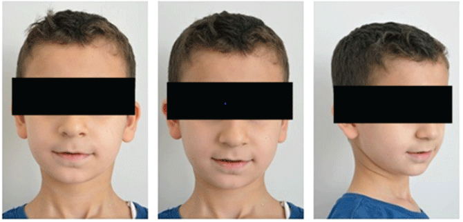
Figure 1: Extra oral photographs.


Khaled Khalaf* Zahra Seraj Hesham Hussein
Department of Orthodontics, University of Sharjah, Sharjah, United Arab Emirates*Corresponding author: Dr. Khaled Khalaf, Associate Professor and Consultant in Orthodontics, Department of Orthodontics, University of Sharjah, PO Box 27272, Sharjah, United Arab Emirates, Tel: +97165057370; E-mail: kkhalaf@sharjah.ac.ae
Hypodontia is the most common dental anomaly in humans and is defined as a condition in which there is one to six congenitally missing teeth, excluding the third molars. Genetics as well as environmental factors play important roles in the aetiology of hypodontia. The most commonly missing teeth are the mandibular second premolars followed by the maxillary lateral incisors. This report presents a rare case of agenesis of maxillary permanent canines, mandibular permanent central incisors and right mandibular permanent lateral incisor in conjunction with delayed eruption of the mandibular permanent left lateral incisor in a healthy nine-year-old male Lebanese patient without any family history of this condition.
Hypodontia; Agenesis; Non-syndromic; Delayed eruption
Hypodontia is the most common developmental anomaly in humans [1-6]. This term along with several others including: “tooth agenesis”, “congenitally missing teeth” and “aplasia of teeth” is often used to describe the condition in which there is one to six congenitally missing teeth excluding the third molars [1-3]. It can occur in conjunction with recognized genetic syndromes or as a solitary nonsyndromic anomaly, the latter of which is the most common phenotype [2]. Family studies have demonstrated that genetic mutations in some genes have been implicated in isolated forms, while mutations in other genes have been reported in syndromic forms [3].
Hypodontia most commonly affects the permanent dentition, however an association with hypodontia in the primary dentition has been reported; children with hypodontia in the primary dentition have been reported to have the corresponding successor teeth to be congenitally missing as well [2,4,5]. The overall prevalence of hypodontia varies among populations and can range from 4.4% to 13.4% [1,5,6]. Individuals with hypodontia usually have one to two congenitally missing permanent teeth, while very few have more than six missing teeth [1,5]. The mandibular second premolars have the highest incidence followed by the maxillary lateral incisors [1,7]. Therefore, the aim of the study was to report a hypodontia patient who presented with multiple congenitally missing teeth of rare occurrence so that dental health care providers are made aware of such condition to be able to form a proper diagnosis and management.
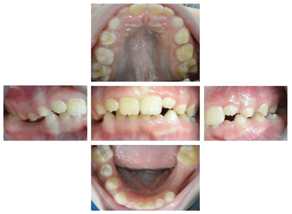
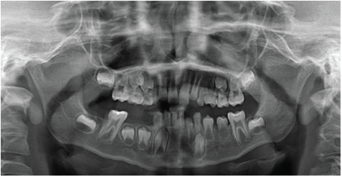
A nine-year old male Lebanese patient presented to the Pedodontic Department, University Dental Hospital Sharjah, for a routine dental examination on February 2018. The patient was healthy, with no history of systemic diseases and/or syndromes. Intraoral examination revealed a class I incisors relationship, class I molars relationship with an over jet of 4 mm and increased incomplete overbite. Clinical examination also revealed retained primary mandibular central incisors and right primary lateral incisor and a partially erupted permanent mandibular left lateral incisor. Extraoral (Figure 1) and intraoral (Figure 2) photographs were taken followed by an orthopantomogram (Figure 3). The radiograph was used to detect the presence of the permanent mandibular central and right lateral incisors and to check for any dental anomalies. The radiograph showed the absence of the permanent mandibular central incisors, right lateral incisor and upper canines bilaterally. The germs for all third molars were also not detected. Clinical history eliminated the likelihood of extractions and/or trauma incidents and there was no relevant family history for this condition. The patient and his parents were counselled regarding the prevalence, aetiology and future management options. A formal consent was sought and granted from the patient and his parents to have full records taken and disseminated for publication.

Figure 1: Extra oral photographs.

Figure 2: Intraoral photographs.

Figure 3: Orthopantomogram
Hypodontia may be classified according to the number of missing teeth as either complete and partial; or mild (two or less teeth congenitally missing), moderate (three to five teeth congenitally missing) and severe (six or more teeth congenitally missing) [8]. Severe hypodontia is usually associated with genetic disorders such as ectodermal dysplasia and Rieger syndrome [9]. However, mild to moderate hypodontia may occur due to environmental factors such as early irradiation of the germs, trauma of the dental region, and the many syndromes associated with cleft lip and palate [2].
The prevalence of congenitally missing teeth may vary with age, sampling techniques, and methods of examination, and may range from 4.4% to 13.4% [5]. According to Al-Emran, et al. [10] and in consonance with Bulk’s theory of terminal reduction [11] a missing tooth will be the most distal tooth of every morphological class; upper second incisors, second premolars and third molars. Therefore, according to Dahlberg de Ned [12], the mesial tooth; such as the central incisors and canines are considered to be the most genetically stable and are rarely absent. On the contrary, in a study conducted by Davis amongst Chinese children, the mandibular incisors were found to be the most commonly missing teeth, affecting 58.7% of children [13] According to Fukuta the prevalence of missing maxillary permanent canines was 0.13%. Furthermore, despite it being rare, the occurrence of congenitally absent permanent canines was more common on the left side of the maxilla and the right side of the mandible. Absence of a single canine was more common than multiple absences, while congenital absence of multiple permanent teeth was linked to missing maxillary permanent canines [14]. The chronology of teeth development and maturation plays an important role in diagnosing dental anomalies and treatment planning. Tooth agenesis has been associated with delayed dental development and eruption, with an estimated delay time of 0.37-0.52 years [15]. The mandibular permanent lateral incisor begins to calcify at 3-4 months and erupts at 7-8 years [16].
In our case, there were multiple permanent teeth missing associated with bilateral absence of the permanent maxillary canines and delayed eruption of the mandibular permanent lateral incisor at the age of nine years. There were however, no related dental anomalies.
Over time, many aetiological theories and concepts focusing on both genetic and environmental factors have been put forward to explain hypodontia. As a result, tooth agenesis may occur as a result of multifactorial influences. In the past, anatomical susceptibility during tooth maturation was proposed as an aetiological means, whereby specific sites in the dental lamina [17] or neural crest cells were highly susceptible to environmental stimuli such as increased oxidative stress from smoking [18]. According to evolutionary studies, the size of human jaws and number of teeth is declining [19-20]. With the advances in genetic and molecular research, the more recent studies have strived to investigate the molecular pathways and mechanisms behind tooth agenesis and have worked to identify the genes that may result in hypodontia if mutated [21-29]. The expression of many genes has been linked to tooth morphogenesis, however non-syndromic hypodontia has been most frequently related to disturbances in PAX9 (paired box gene 9), MSX1 (muscle segment homeobox 1), AXIN2 (axis inhibition protein 2), WNT10A (wingless-related integration site 10A) and EDA (ectodysplasin A) expression [24-29]. In particular, MSX1 mutations has been recognized in a family missing mandibular central incisors and all premolars [29], AXIN2 with congenitally missing lower incisors [23,30] and WNT10A with isolated hypodontia of the maxillary permanent canines [31].
In this case, there was no history of maternal smoking or significant family history for hypodontia, however, gene sequencing would help identify any causative gene mutations that may have resulted in tooth agenesis and ultimately establish whether a familiar pattern may be present.
Hypodontia patients may present with disturbances in facial growth pattern, midline deviations and deep overbites, therefore the management requires an interdisciplinary approach, careful treatment planning, long-term maintenance and family counseling [32,33]. The treatment may include strategic extraction of primary teeth, uprighting and aligning teeth and management of deep overbites.
In this case, the patient will be followed-up to monitor the eruption of the mandibular permanent left lateral incisor and referred to an orthodontist to liaise with other health care providers for the optimal management of these patients.
Download Provisional PDF Here
Article Type: CASE REPORT
Citation: Khalaf K, Seraj Z, Hussein H (2018) A Rare Hypodontia and Delayed Eruption of a Mandibular Permanent Incisor: A Case Report and Literature Review. Int J Dent Oral Health 4(5): dx.doi.org/10.16966/2378-7090.276
Copyright: © 2018 Khalaf K, et al. This is an open-access article distributed under the terms of the Creative Commons Attribution License, which permits unrestricted use, distribution, and reproduction in any medium, provided the original author and source are credited.
Publication history:
All Sci Forschen Journals are Open Access