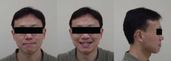
Figure 1: Facial photo before active treatment.

Chun-Shuo Huang1 Jian-Hong Yu1,2*
1Department of Orthodontics, China Medical University Hospital, Taichung, Taiwan*Corresponding author: Jian-Hong YU, Professor, School of Dentistry, College of Dentistry, and Dean, Department of Orthodontics, China Medical University Hospital, Taichung, 40402, Taiwan; E-mail: kenkoyu@mail.cmu.edu.tw
ISW (Improved Super-elastic Ti-Ni alloy wire, developed by Tokyo Medical and Dental University) for the treatment of an adult anterior functional crossbite non-extraction case will be discussed.
This patient was 37 years old adult male whose chief complaint was irregular dentition. Clinical examination revealed a bilateral Class III molar relationship with anterior functional crossbite, severe crowding over upper anterior teeth and mild crowding over lower anterior with lower midline deviated to the right side. Active treatment included ISW leveling combined with crossbite arch performed to relieve crowding and to correct anterior crossbite. MEAW (Multi-bend Edgewise Archwire) technique for the lower arch combined with Class III intermaxillary elastics (IME) was also performed. Treatment was completed within 25 months and adequate overbite and overjet were accomplished after the active treatment.
ISW; Skeletal Class III; Non-surgical orthodontics; Non-extraction orthodontics; Crossbite arch, MEAW technique; Intermaxillary Elastics (IME)
Differentially diagnosing a Class III malocclusion by dental, functional and skeletal is critical in orthodontic field [1-4]. For pseudo-class III malocclusion, also known as functional anterior crossbite case, the increase for the Vertical Dimension of Occlusion (VDO) should be the essential solution to correct the occlusal interference caused by the anterior region. In order to correct the functional anterior crossbite, the control of crown labially-tipped for the upper anterior teeth and the Class III intermaxillary elastics (IME) for the correction of occlusal interference and better jaw relationship should be performed simultaneously. To achieve a normal occlusion and a desirable cusp interdigitation, the correction of the long axis of lower dentition and better jaw relationship can also be achieved by ISW MEAW technique combined with Class III elastics [5-8].
ISW MEAW (Multi-bend Edgewise Archwire) technique with intermaxillary elastics were helpful in overjet (by class III elastics) and open bite (by up and down elastics) correction. It makes the teeth tip back and intrusion, facilitating in class I canine relationship and better long axis of lower anterior teeth.
The diagnosis of functional class III malocclusion differs from that of skeletal Class III malocclusion because it is defined as functional forward displacement of the mandible as a result of retroclined maxillary incisors. Because the major underlying cause of pseudo Class III malocclusion is the inclination of the maxillary incisors, the treatment objectives aim to change the inclination of those incisors [9-11].
The 37-year-old male complained of irregular dentition and difficulty in biting food. Before active treatment, the facial photo (Figure 1) and intraoral photo (Figure 2) of the patient was recorded on 2010.03.18.
Figure 1: Facial photo before active treatment.
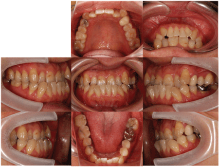
Figure 2: Intraoral photo before active treatment.
Radiographically, panoramic film (Figure 3) was also taken and absence of all third molars was shown. Simultaneously, TMJ X-ray (Figure 4) showed no deformity of condylar head before active treatment. Lateral cephalometric film (Figure 5) and, regarding the polygon before active treatment (Figure 6) was taken and measured.
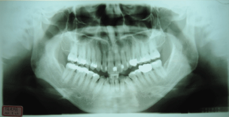
Figure 3: Panoramic film before active treatment.
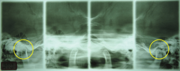
Figure 4: TMJ film before active treatment.
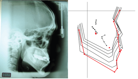
Figure 5: Lateral cephalometric film before active treatment.
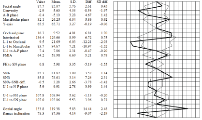
Figure 6: Polygon before active treatment.
Therefore, based on the statistics above, summary of diagnosis was listed below:
Summary of diagnosis
And treatment plan accordingly includes:
At 2010.05.23, about the start of active treatment, upper arch DBS (Direct bonding system) and leveling with 0.016 × 0.022 ISW Crossbite Arch was performed. Crossbite Arch was set between #13 and #23 for correction of anterior crossbite (Figure 7).
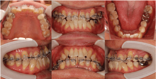
Figure 7: Start of active treatment.
Approximately after a month of treatment (2010.06.07), the anterior teeth have reached an edge-to-edge relationship. In the meantime lower arch DBS and leveling with 0.016 × 0.022 ISW wire was performed. Class III IME was used for bite raising and adjustment of jaw relationship simultaneously (Figure 8).
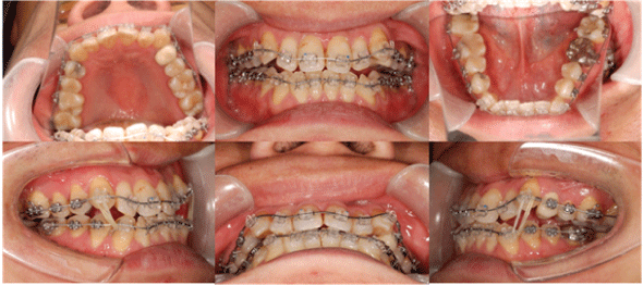
Figure 8: One month after active treatment.
Five months after active treatment, for the correction of bilateral anterior lower lateral incisor, stripping of lower anterior teeth for space creation and elastic chain for #32, #42 de-rotation was performed. Consequently, #32, #42 de-rotation was achieved by stripping, elastic chain traction, and ISW leveling (Figure 9).
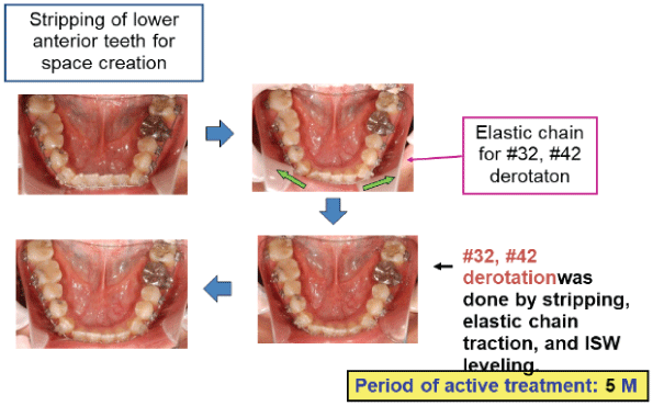
Figure 9: Five months after active treatment.
Six months after active treatment (2010.11.22), ISW MEAW and up and down IME were applied in the lower dentition. And R(3-3) and L(3-3) IME (intermaxillary elastics) was used to reinforce anchorage so as to prevent anterior teeth from flaring out (Figure 10).
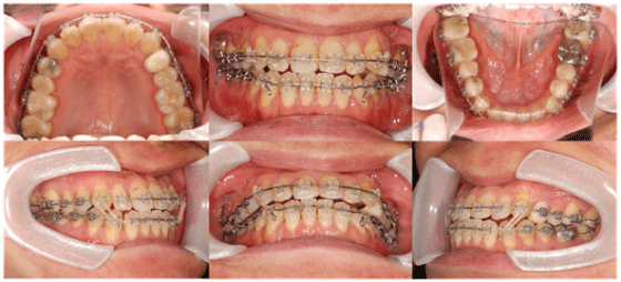
Figure 10: Six months after active treatment.
The process of IME for midline correction and finishing and detailing-interdigitation was organized and shown in figure 11 and 12.
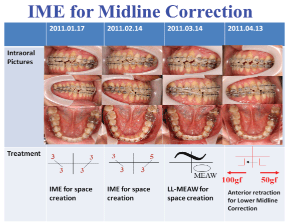
Figure 11: Process of IME for midline correction.
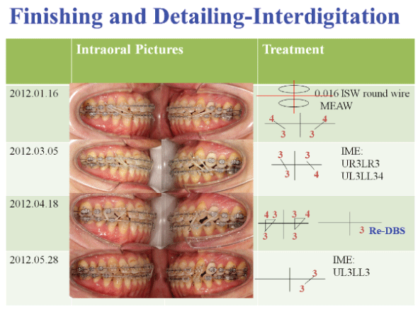
Figure 12: Process of finishing and detailing-interdigitation.
After 25 months (2012.06.27) of active treatment, de-bonding was performed and circumferential retainer was delivered for the upper arch and lower arch (Figure 13).
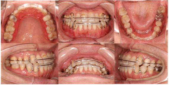
Figure 13: After 25 months of active treatment.
After active treatment, the facial photo (Figure 14) and intraoral photo (Figure 15) of the patient was recorded on 2012.06.26.
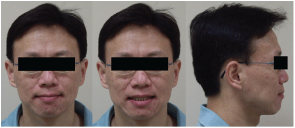
Figure 14: Facial photo after active treatment.
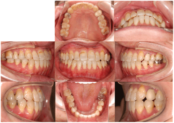
Figure 15: Intraoral photo after active treatment.
Lateral cephalometric and polygon after active treatment was shown in figure 16 and 17.
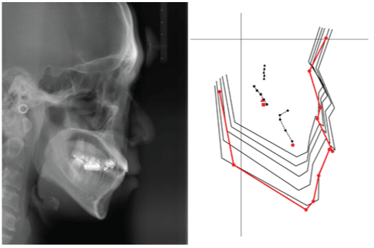
Figure 16: Lateral cephalometric film after active treatment.
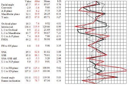
Figure 17: Polygon after active treatment.
Superimposition superimposed on SN at S was shown in figure 18. U1 to FH plane changed from 107.8° to 125.4°.
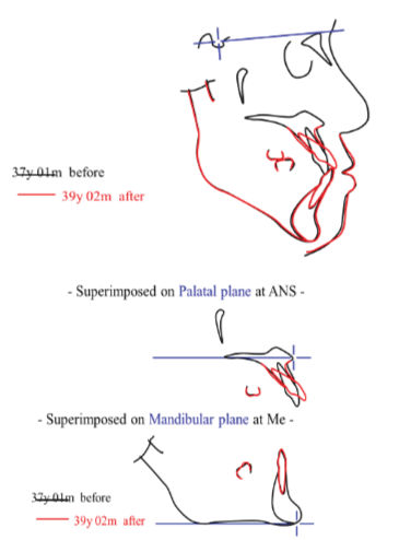
Figure 18: Superimposition after active treatment.
Differentially diagnosing a Class III case by dental, functional, or skeletal is very important before the active treatment. This case shows functional interference at the anterior portion; combine with skeletal problem, which easily induced a situation which we used to call Class III protrusion. We chose non-extraction treatment strategy using Crossbite Arch and MEAW technique for this case. After 25 months of active treatment, a desirable dentition and profile change were achieved and the patient was pleased with the treatment result.
Two crimpable stoppers and ISW to form a loop over the upper anterior area called “Crossbite Arch” was performed (Figure 19). The property of shape memory for ISW caused upper anterior teeth to flare out effectively [12-14].
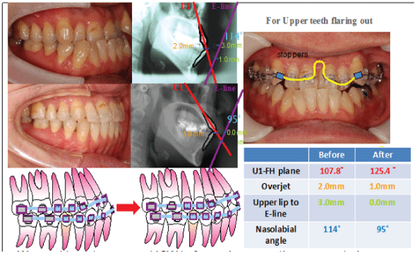
Figure 19: Crossbite Arch.
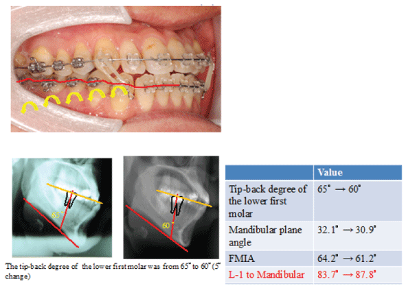
Figure 20: MEAW (Multi-bend Edgewise Archwire) technique.
Generally, ISW MEAW makes the posterior teeth tip back and intrude. This effect can create more space for the tipping of lower anterior teeth, and thus facilitate the correction of reverse overjet; in this case, the amount of lower anterior teeth flare-out is greater than the MEAW effect. Nevertheless, MEAW effect outperforms the amount of L-1 to Mandibular in some aspects listed above [15-17].
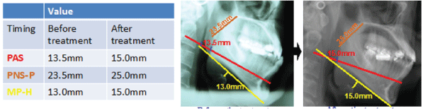
Figure 21: Tongue position and pharyngeal airway.
Three elements were discussed in this case that highly effected MFT (Myofunctional Therapy) was duration, intensity and frequency. In this case, the PAS (posterior airway space) distance before and after orthodontic treatment increased from 13.5 mm to 15.0 mm; while the PNS-P (soft palate length) increased from 23.5mm to 25.0mm and MP-H (distance from the mandibular plane to the most anterior point of the hyoid bone) increased from 13.0mm to 15.0mm. According to the statistic figure, we suspected that this patient has a high tendency of low tongue resting posture before the treatment. One can consider relevant tongue training method for better and long-term retention.
In this case, we had #33 torqued to the buccal side by means of bracket-upside-down (b-u-d) method (Figure 22). But the effect of #33 de-rotation was not favorable due to the interference from #33 distal cusp ridges with #34. Horizontally, #34 interfered by #33 distal cusp ridges. Therefore, stripping of #33 distal cusp ridges was suggested for better interdigitation [18-20].
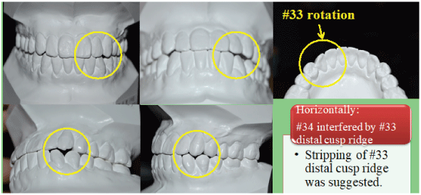
Figure 22: Torque correction by bracket-upside- down (b-u-d) method.
Download Provisional PDF Here
Article Type: CASE REPORT
Citation: Huang C-S, Yu J-H (2018) ISW for the Treatment of Functional Class III Malocclusion by Crossbite Arch and MEAW Technique in an Adult. Int J Dent Oral Health 4(2): dx.doi. org/10.16966/2378-7090.259
Copyright: © 2018 Huang C-S, et al. This is an open-access article distributed under the terms of the Creative Commons Attribution License, which permits unrestricted use, distribution, and reproduction in any medium, provided the original author and source are credited.
Publication history:
All Sci Forschen Journals are Open Access