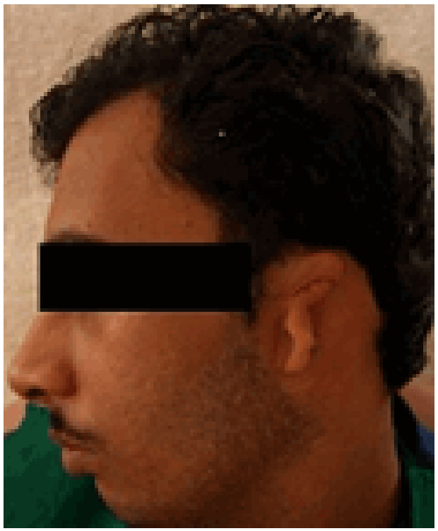
Figure 1: Auricular defect

Gita Rani1 Amarjeet Gambhir2,*
1Department of Prosthodontics, Christian Dental College, Ludhiana, Punjab, India*Corresponding author: Amarjeet Gambhir, House No. 39, New Staff Line, CMC Campus, CMC & Hospital, Ludhiana, Punjab, India, Tel: 08427543060, E-mail: amarjeetgambhir@gmail.com
Auricular defects resulting from congenital anomalies or ablative surgery pose a major reconstructive challenge for the clinician. Various treatment modalities for correction of such defects include surgical reconstruction or prosthetic rehabilitation with extraoral implants or adhesives. Sometimes, however, the patient declines surgical reconstruction; in such cases, adhesive-retained prostheses provide a cost-effective and cosmetically acceptable means of camouflage. This case report describes the successful rehabilitation of a patient with congenitally malformed ear using adhesive retained silicone auricular prosthesis. It was acceptable to the patient because of excellent support & retention and satisfactory appearance.
Auricular defect; Auricular prosthesis; Congenitally missing ear; Rehabilitation
Causes of facial tissue loss are often known to be congenital malformations, tumoral lesions or accidents. Facial defects not only cause functional problems, but also some serious psychological problems that force the individual to avoid social contact [1]. Rehabilitation of these defects presents a challenge for the clinician. Correction of auricular defects can be accomplished surgically, prosthetically or through a combination of these approaches; the choice of treatment depends on a variety of factors such as the site, size and duration of the defect as well as the patients’ preferences [2,3].
Reconstructive surgery is limited by the age and medical condition of the patient, insufficient residual tissue, compromised vascularity due to irradiation and patient’s unwillingness to undergo another surgical procedure [4]. Secondly, resemblance of the reconstructed ear to the normal one may be poor. In contrast, prosthetic treatment can produce an anatomically accurate and aesthetically pleasing replacement [5,6]. It involves the fabrication of silicone auricular prosthesis which can be retained by mechanical means or implants. Implant retained prosthesis even though more retentive, is not cost effective for economically weak patients. For such patients adhesive retained prosthesis provides a viable treatment option.
This case report presents a step by step technique for fabrication of adhesive retained silicone auricular prosthesis for a patient who had congenitally malformed ear. This patient was eager to have his defect corrected but could not afford the expensive implant retained auricular prosthesis.
A 24 year old male patient reported to the Department of Prosthodontics with chief complaint of missing left ear. Extraoral examination showed deformed/ rudimentary left auricle with a healthy tissue bed while the right ear was normal (Figure 1). Various treatment options including implant retained/adhesive retained silicone prosthesis or surgical reconstruction were presented to the patient. Due to financial constraints, he opted for adhesive retained silicone auricular prosthesis.
Horizontal markings were made with an indelible pencil in the defect area at specific places to aid in correct alignment of the prosthesis with the natural ear. These include:
A primary impression of defect area was made with irreversible hydrocolloid impression material. Hair adjacent to the area were coated with petroleum jelly (Vaseline; Chesebrough-Pond’s USA Co, Greenwich, Conn) and then irreversible hydrocolloid was bulk filled onto the defect area. Wet gauze pieces were placed over the irreversible hydrocolloid and the plaster backing was done in order to increase the strength and easy pouring of the impression. Secondary impression using custom made tray was made with light body polyvinylsiloxane followed by backing with regular body (Affinis Precious; Coltene Whaledent) (Figure 2) for more strength. Impressions were boxed, poured in Type 3 die stone (kalabhai kalstone) and working casts were obtained (Figure 3). Wax pattern was made by the sculpting technique in which ear is carved from a mirror image of patient’s natural ear using the normal side cast as reference. The prepared wax pattern was then adapted to the stone cast and corrected to match the contralateral ear. After modifications, the wax pattern was tried to finalize its position superoinferiorly and anteroposteriorly (Figure 4). The pattern was then invested & mold was formed. Conventional procedures for wax elimination of the mold were followed. After the complete removal of wax, intrinsic pigments (the colour of which was determined from the patient’s skin colour) were added to the silicone elastomer (Cosmesil; Principality Medical Ltd., South Wales) and then bulk packed. After processing, the prosthesis was removed from the mold; excess flash from the anterior margin of the prosthesis was cut and borders were thinned. The remaining excess was trimmed after the prosthesis was evaluated on the patient. The final corrections were made and the silicone prosthesis was then adapted to the defect area. After polymerisation, the auricular prosthesis was checked for adaptation. External staining with roomtemperature-vulcanised (RTV) silicone (Cosmesil; Principality Medical Ltd.) was done and then re-packed in the same mold for polymerisation to occur. The auricular prosthesis was delivered to the patient after demonstrating the path of placement. He was instructed about the use and maintenance of the prosthesis including application of the adhesive and how to clean the silicone prosthesis with a soft toothbrush and mild soap. The patient was monitored at three months interval for one year and then once a year for check up with satisfactory results (Figure 5).
Figure 1: Auricular defect
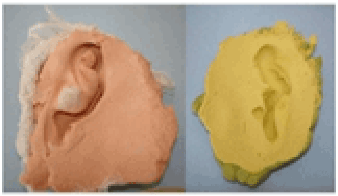
Figure 2: Making of impression
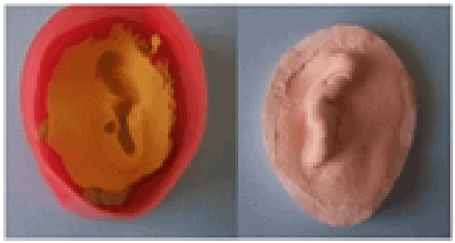
Figure 3: Beading and boxing, working cast
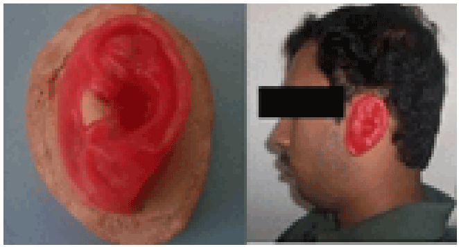
Figure 4: Try-in of the wax pattern
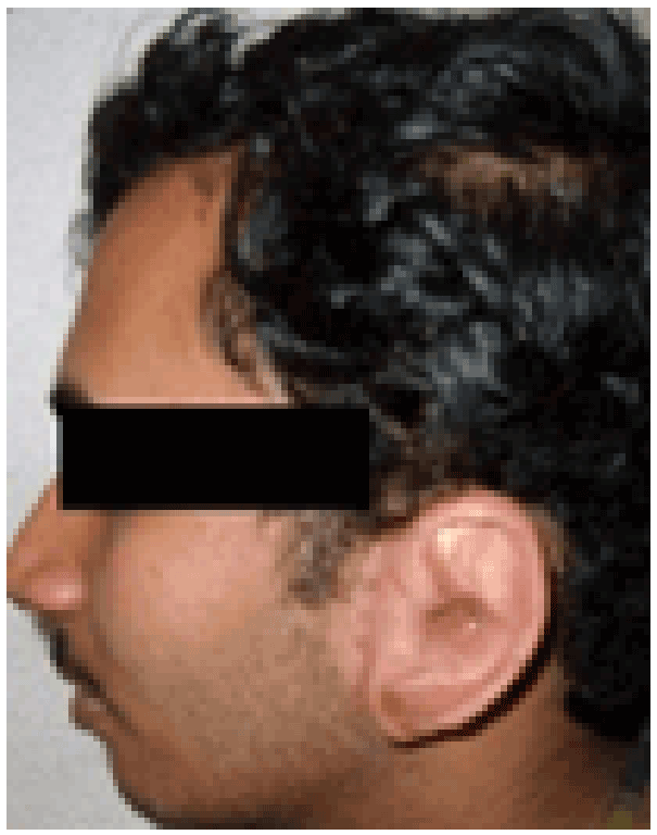
Figure 5: Post-insertion view of the prosthesis
Rehabilitation of patients with congenital or acquired auricular defects is a challenging process. The existing treatment modalities for replacement of missing ear are surgical and prosthetic. However, staged surgical ear reconstruction has not given consistently good results and prosthetic restoration is the treatment of choice [7].
Implant retained auricular prosthesis has become a valid therapeutic alternative because of improved retention and esthetics & enhanced longevity [8-10]. However, it requires sufficient healthy bone at the site of the defect for implant placement, as well as a time interval between placement and final rehabilitation, and is generally unacceptable to the patients because of financial constraints.
Adhesive retained silicone prosthesis is worn by specially formulated facial prosthetic adhesives (Daro, Pros-Aide, and Secure). This method of rehabilitation is a conservative and reversible treatment option with numerous advantages. The prosthesis can be placed immediately on a healthy tissue bed, does not require surgery, and is cost effective to the patient [11]. It is biologically inert and can be made to match any skin color using intrinsic or extrinsic color system to produce a more life-like appearance. The disadvantages include limited retention, potential for tissue irritation and difficulty in orientation of the prosthesis specially in patients with compromised manual dexterity [12]. Despite these limitations, adhesive retained silicone auricular prostheses provide a simple and cost effective but esthetically acceptable mode of rehabilitation. For patients who refuse surgery or are otherwise not good candidates for reconstruction because of underlying medical problems, silicone auricular prosthesis should be the preferred choice of treatment.
The ultimate aim of any maxillofacial rehabilitation should be to satisfy the needs & expectations of the patient with the best possible mode of treatment. This article describes the successful rehabilitation of a patient with congenitally malformed ear. Amongst the various treatment options available, the adhesive retained silicone auricular prosthesis was chosen owing to the patient’s socioeconomic status. It was acceptable to the patient because of excellent support & retention and satisfactory appearance.
Download Provisional PDF Here
Aritcle Type: Case Report
Citation: Rani G , Gambhir A (2015) Reconstruction of Auricular Defect with Adhesive - Retained Silicone Prosthesis – A Case Report. Int J Dent Oral Health 1(6): doi http://dx.doi.org/10.16966/2378-7090.156
Copyright: © 2015 Rani G, et al. This is an open-access article distributed under the terms of the Creative Commons Attribution License, which permits unrestricted use, distribution, and reproduction in any medium, provided the original author and source are credited.
Publication history:
All Sci Forschen Journals are Open Access