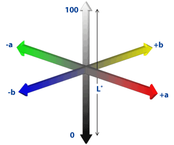
Figure 1: Visual perception of the color spaces

Cleidy Arboleda-Lopez Robert J. Manasse* Grace Viana Ana B. Bedran-Russo Carla A. Evans
Department of Orthodontics, College of Dentistry, University of Illinois at Chicago, USA*Corresponding author: Robert J. Manasse, Clinical Associate Professor, College of Dentistry, Department of Orthodontics, University of Illinois at Chicago, Chicago, IL 60612-7211, USA. E-mail: drrjm@uic.edu
Not only are today’s orthodontic patients concerned about the alignment and shape of teeth but also the color; many wish to have their teeth whitened before the end of orthodontic treatment. Common questions that orthodontists face from patients are, “When can I whiten my teeth? Can I whiten my teeth with braces?” The answer often given is based on the assumption that ideally tooth whitening should be done after the debonding of brackets at the completion of orthodontic treatment. Currently, no evidence-based protocol exists for the optimal time to whiten teeth for patients undergoing orthodontic treatment.
A recent nationwide survey of whitening protocols conducted among 3,601 active AAO members in the United States [1] showed that almost all orthodontists encounter patients seeking tooth whitening during active orthodontic treatment. Also, only about a third of those patients receive whitening treatment in the orthodontist’s office while most patients are referred to another dental practitioner for the tooth whitening procedure.
Jadad et al. [2] assessed the effectiveness of 8% hydrogen peroxide-based whitening agent in patients wearing fixed orthodontic appliances. The conclusion drawn by Jadad et al. was that Treswhite Ortho (Opalescence®) is an effective whitening agent for teeth with or without brackets.
However, there are no other studies on dental whitening in patients wearing braces to compare and validate the findings of this study and the percentage concentration of hydrogen peroxide used was low compared to other dental whitening products that have been widely studied. The lack of information makes it difficult for the practitioner to determine the right time to provide tooth whitening procedures for their patients [1,3]. Therefore, further research is needed on the effectiveness of the whitening procedure during wearing braces and how it affects the color of the tooth enamel.
The Commission Internationale de I’Eclairage (CIE L*a*b*) on Illumination, which sets standards in colorimetry and color difference equations, is commonly used in perceptual assessment of the color of teeth because of its visually uniform coverage of the color space. The L* direction in color space provides a measure of sample lightness (0 is black and 100 is white); the a* direction provides a measure of red/green (+a is a shift toward red, -a is a shift towards green); and the b* direction provides a measure of yellow/blue (+b is a shift toward yellow, -b is a shift towards blue) [4]. The visual perception of the color spaces are shown in Figure 1.
The efficacy and safety of night guard vital bleaching technique (NGVB) using 10% carbamide peroxide (CP) has been determined in previous studies and, according to the ADA, is considered as the gold standard of vital whitening procedures [5,6]. The purpose of this study was to investigate whether whitening on human teeth with brackets is effective as whitening on human teeth without brackets.
A sample size of 20 teeth in each group was needed in this study to detect the mean differences in terms of color difference, (∆E) with at least 80% power and an error of 5%. Eighty human premolar teeth extracted for orthodontic reasons were collected in accordance with an Internal Review Board approved protocol. In an attempt to standardize the initial tooth shade, only teeth in the A and B VITA shade according to the Vitapan classical shade guide (VIDENT™, California, USA) were included. The exclusion criteria included teeth with visible cracks, enamel defects, hypocalcifications, fluorosis, caries or restorations on the buccal surface. The specimens were randomly assigned to groups and prepared for the color assessment according to the experimental protocol (Figure 2).
Eighty specimen teeth were randomly divided into four groups of 20 teeth each, namely:
Group 1: Controls - No whitening/ no brackets
Group 2: Teeth without brackets whitened with 10% carbamide peroxide (Opalescence® Tooth Whitening Systems).
Group 3: Teeth with brackets with no whitening treatment
Group 4: Teeth with brackets whitened with 10% carbamide peroxide (Opalescence® Tooth Whitening Systems)

Figure 1: Visual perception of the color spaces
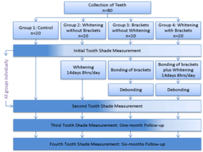
Figure 2: Flow diagram with details of the study design
The teeth were stored in 0.1% thymol solution at room temperature until they were mounted and tested. Throughout this study, the teeth were stored in artificial saliva at 37°C. The artificial saliva was formulated from 4.766 g Hepes, 0.331 g calcium chloride dehydrate, 0.184 g potassium phosphate and 9.692 g potassium chloride kept at a pH of 7.4. The anatomical roots were separated from the anatomical crowns using a slow speed water-cooled saw (Isomet 1000, Precision Saw, USA). Then the crowns were sectioned longitudinally between the buccal and lingual surfaces. A cylindrical garolite (fiberglass) mounting jig was used for orienting the buccal surface of each individual tooth. A key slot was machined on one side of each cylinder allowing the jig to be adapted only in one configuration into the spectrophotometer device (Figure 3a).
The spectrophotometer set-up (Figure 3b) included a micrometer stage, which can be adjusted vertically to position the sample accurately. After the teeth were mounted in the garolite cylinder, they were pumiced using fluoride-free paste with rubber prophylaxis cups. Then, the teeth were washed for 30 seconds with tap water and air dried in preparation for the initial tooth color assessment as per the experimental protocol.
The same protocol was followed for bonding brackets on the teeth in Groups 3 and 4 irrespective of whether whitening was going to be performed. The teeth were cleaned with tap water prior to bonding procedures. Then, each enamel surface was etched with 37% phosphoric acid for 10 seconds. The teeth were then rinsed with water for 20 seconds and air dried. A thin coating of Transbond™ XT primer was applied on the surface and lightly air dried. The mesh pads of standard stainless steel premolar brackets (American Orthodontics, Sheboygan, WI, USA) were coated with a single coating of TransbondTM XT (3M Unitek, USA) adhesive. Brackets were then placed in the middle of the tooth in occlusalgingival and mesial-distal directions on each tooth using bracket tweezers. Excess adhesive resin was carefully removed with an explorer. The bracket was cured with an Ortholux™ LED (3M Unitek, USA) curing light for 5 seconds mesially, 5 seconds distally, 5 seconds occlusally, and 5 seconds gingivally for a total of 20 seconds.
Forty customized whitening trays were made from 1mm soft clear Essix® Bleach Tray and Model Duplication Material (Dentsply Raintree Essix, Glenroe, USA) for whitening the teeth in Groups 2 and 4 with 10% carbamide peroxide (Opalescence® Tooth Whitening Systems, USA).
The whitening gel was applied to the buccal surfaces in the customized whitening trays (Figure 3c), and the teeth were treated for 8 hours daily for 14 consecutive days following the manufacturer’s specifications. The teeth were kept at 37°C while undergoing the whitening process. Each time, after tooth whitening, the gel was removed; the surface was washed with tap water and dried with compressed air. Then the specimens were stored again in artificial saliva at 37°C until the next whitening session.
The teeth in Groups 3 and 4 were debonded following the same protocol. A bracket removing plier (Orthopli, Philadelphia, PA, USA) was placed against the wings of the bracket and squeezed. Squeezing the bracket wings caused distortion of the bracket pad and induced bond failure between the pad and the adhesive resin. This method has been described as the safest way to remove metal brackets [7,8].
The remaining adhesive on the enamel surfaces of the teeth in Groups 3 and 4 was removed using protocol two-step polishing procedure. After the adhesive was removed with a 12 fluted (universal) carbide bur on a highspeed hand-piece manually operated under a lighted 5X magnifying glass (Figure 3d), a 20 fluted (universal) carbide bur was utilized to remove the remaining adhesive and produce a smooth surface [9].
Objective tooth color measurements of all the teeth were recorded using a spectrophotometer (CM-2600d, Konica Minolta Sensing, Inc., Japan), (Figure 3d). Before the initial measurement at each time point, the spectrophotometer was calibrated as outlined by the manufacturer. During each time point, the color of a 0.8 mm square area at the center of the crown of each premolar tooth was measured three times with the spectrophotometer (Figure 3e). The average of the three measurements was considered as the measured value for determination of specimen color. Differences in individual color components (∆L, ∆a, and ∆b) were calculated and the total color differences(∆E) were calculated by means of the equation ∆E = √ ∆L2 + ∆a2 + ∆b2 . 4 Tooth shade measurements of 80 premolar teeth were taken at the following time points:
T1: Initial shade measurement taken on day 1 (baseline).
T2: Two weeks shade measurement after the initial measurement.
T3: Four weeks shade measurement after the initial measurement.
T4: Six months shade measurement after the initial measurement.
The spectrophotometer was positioned at the same measurement site
for all recordings to ensure that all the measurements were consistent.
The three color differences (∆E) measurements calculated in this study were:
∆E1: The difference between baseline and two weeks shade measurements after whitening.
∆E2: The difference between baseline and four weeks shade measurements after whitening
∆E3: The difference between baseline and six months shade measurements after whitening
Descriptive statistics were prepared. Normality assumptions and homogeneity of the variances were tested. One-way analysis of variance (ANOVA) and Student paired t-tests hypotheses at 5% significance level were tested. Corresponding non-parametric hypotheses, when necessary, were also tested. The data analysis was processed using SPSS version 22.0 (Chicago, IL., USA).
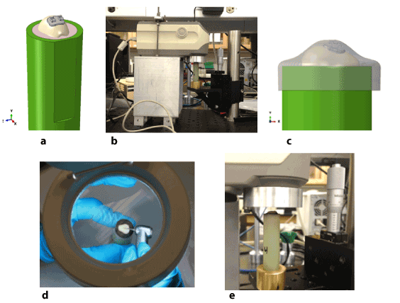
Figure 3: Set up of study: a)Orthodontic bracket bonded on premolar mounted in the garolite cylinder, b) Spectrophometric set-up c) Customized whitening tray, d) Adhesive removal under 5X magnification, e) Close up view of garolite in spectrophotometer.
The initial untreated mean color measurements for all four groups were similar. The values of color differences from the shade measurements: ΔE1,ΔE2 and ΔE3 with respect to baseline are shown (Table 1).
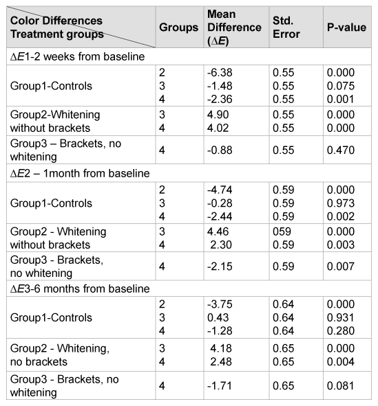
One-way ANOVA showed statistically significant mean differences among the effects of the four treatment groups on ΔE color differences: ΔE1, F (3,76)=49.04; ΔE2, F (3, 76)=27.46; ΔE3, F (3, 76)= 16.96 and P values <0.001. The post-hoc Scheffé test was used to compare the mean difference of each pair on the four treatment groups (Table 2). Post-hoc Scheffé tests indicated that the comparison between treatment Group 2 (whitening without brackets) and each of the other treatment groups: 1 (control), 3 (brackets without whitening) and 4 (whitening with brackets) showed color mean difference values: ΔE1, ΔE2 and ΔE3 were statistically significant, (p values <0.001). There were also statistically significant mean differences on comparison of treatment Group 4 (whitening with brackets) with the treatment Group 3 (brackets without whitening) but only for ΔE2 at–1 month from baseline, (p values= 0.007) (Table 2). The ΔE3 value for Group 1 (controls) is not significantly different from Groups 3 and 4, but is different from Group 2.
Additional results using Student paired t-tests did not show statistically significant mean differences for Group 4 (whitening with brackets) for each pair of the mean difference values ΔE1,ΔE2 and ΔE3 (Table 3).
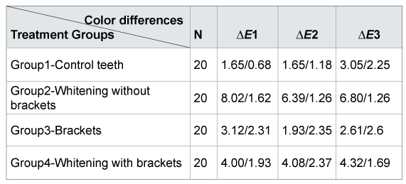
∆E1: 2 week post whitening; E2: 1 month from baseline; E3: 6 months from baseline
Table 1: Values of the color mean differences (∆E): N, mean and standard deviation
Groups: 1-Control, 2 - Whitening without brackets, 3-Brackets, no whitening and 4-whitening with brackets
Table 2: Results of the Post Hoc Scheffé test for (∆E) by treatment groups
Presently a high percentage of patients undergoing orthodontic treatment are seeking tooth whitening procedures [1], but there is limited evidence supporting the effectiveness of tooth whitening while wearing braces. The tooth color measurements were based on the CIE L* a* b* system and a three-dimensional color space system, which is an objective and accurate method [4].The results of this in vitro study show significant changes in the color of natural teeth when whitening is done in a system mimicking active orthodontic treatment (Group 2 (whitening without brackets) and Group 4 (whitening with brackets). However, the mean ∆E in the group that was bleached without brackets (Group 2, whitening without brackets) was significantly higher than Group 4 (whitening with brackets). These results stand in agreement to those found by Jadad et al. [2]. In their study, it was concluded that Treswhite Ortho (Opalescence®, 8% hydrogen peroxide) was an effective way to conduct whitening procedures during active orthodontic treatment with the same rate of improvement seen between both bracketed and non-bracketed groups. The present findings also show that both groups have a positive whitening outcome. However, Group 2’s (whitening without brackets) was far more effective. Group 3 (brackets without whitening) also demonstrated a color change probably due to the bonding process.
The major change in Group 2 (whitening without brackets) took place during the first two weeks and then was stable for the next six months. This color stability has been shown in previous studies in which the tooth shade was evaluated after whitening teeth without brackets [10]. Interestingly, Group 4 (whitening with brackets) did not show as much change within the first two weeks as did Group 2 (whitening without brackets) and this value did not change for the other time points. Many factors could be involved in the regression of tooth whitening such as extrinsic staining from food and beverages and the natural yellowing that occurs as a result of the continuous deposition of secondary dentin by the pulp [10-14]. In the present in vitro study, discoloration from dietary intake can be eliminated. Significant regression was not found in both groups 2 (whitening without brackets) and 4 (whitening with brackets). The bracket bonding process does not seem to interfere negatively with the color measurements.
When effectiveness and stability are desired, tooth whitening should be recommended after completion of orthodontic treatment. If patients in active orthodontic treatment request tooth whitening and are seeking immediate results or do not wish to wait until bracket removal, whitening procedures may provide some increase in whitening quality
The results of the present in vitro study showed that bleaching technique using10% carbamide peroxide in a customized tray was effective and change the shade of teeth both without and with orthodontic brackets. The change in color of teeth whitened without brackets was significantly higher than the teeth with bonded brackets. Long-term stability seems not to be an issue in either group.
Download Provisional PDF Here
Article Type: Research Article
Citation: Arboleda-Lopez C, Manasse RJ, Viana G, Bedran-Russo AB, Evans CA (2015) Tooth Whitening during Orthodontic Treatment: a SixMonth in vitro Assessment of Effectiveness and Stability. Int J Dent Oral Health 1(4): doi http://dx.doi. org/10.16966/2378-7090.120
Copyright: © 2015 Arboleda-Lopez C, et al. This is an open-access article distributed under the terms of the Creative Commons Attribution License, which permits unrestricted use, distribution, and reproduction in any medium, provided the original author and source are credited.
Publication history:
All Sci Forschen Journals are Open Access